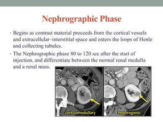
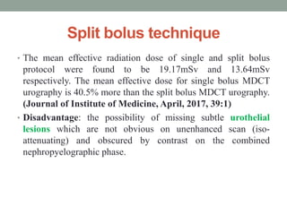
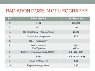
CT urography provides a comprehensive non-invasive evaluation of the urinary tract. It is performed as a multiphase exam, including an unenhanced phase followed by corticomedullary, nephrographic and excretory phases after intravenous contrast administration. The nephrographic phase provides the highest sensitivity for detecting renal masses and abnormalities. Various dose reduction techniques can be used, including lower dose protocols, split-bolus contrast injection, dual-energy CT, and iterative reconstruction. These allow radiation doses to be reduced by up to 80% compared to traditional triple-phase CT urography exams.
This presentation include biliary anatomy ,indication, contraindication post op care of percutaneus transhepatic biliary drainage with important technique. and advantage and disadvantage of different technique. This is important for radiologist, radiographers, intervention radiologist radiology resident. Thanks
A presentation about Intravenous Urography (Also known as Intravenous Pyeography).
The presentation contains 41 slides, and is divided into 4 parts :
1 - Introduction.
2 - The procedure.
3 - Examples for abnormal findings.
4 - Studies comparing IVU accuracy with KUB & USG with CT Scan.
This presentation was prepared and presented by me in the tutorials of the Radiology Department of Sebha Medical Center.
Purpose of this presentation is to educate non radiologist about basic CT anatomy of abdominal viscera and basic interpretation of very common diseases
This is about Introduction To renal CT scan Protocol what are the indication and tailoring how to optimize the the right protocol for the patient according to the indication .
Hopping you like it and helping you in daily practice .
Dr Hisham AlKhatib
Consultant Radiologist
Triphasic CT (TPCT) Scan of the liver is essential in view of the dual blood supply of the liver. TPCT allows characterisaiton of all liver lesions and close to pathological correlaiton by non invasive imaging alone. Additionally providing segmental vascular analysis as a surgicical guide.
computed tomography intravenous urography protocol and advancements ,,, slides coves urinary system anatomy glance ,, contrast media used in procedure , radiation doses and some pathological findings
NVBDCP.pptx Nation vector borne disease control programSapna Thakur
NVBDCP was launched in 2003-2004 . Vector-Borne Disease: Disease that results from an infection transmitted to humans and other animals by blood-feeding arthropods, such as mosquitoes, ticks, and fleas. Examples of vector-borne diseases include Dengue fever, West Nile Virus, Lyme disease, and malaria.
ARTIFICIAL INTELLIGENCE IN HEALTHCARE.pdfAnujkumaranit
Artificial intelligence (AI) refers to the simulation of human intelligence processes by machines, especially computer systems. It encompasses tasks such as learning, reasoning, problem-solving, perception, and language understanding. AI technologies are revolutionizing various fields, from healthcare to finance, by enabling machines to perform tasks that typically require human intelligence.
The prostate is an exocrine gland of the male mammalian reproductive system
It is a walnut-sized gland that forms part of the male reproductive system and is located in front of the rectum and just below the urinary bladder
Function is to store and secrete a clear, slightly alkaline fluid that constitutes 10-30% of the volume of the seminal fluid that along with the spermatozoa, constitutes semen
A healthy human prostate measures (4cm-vertical, by 3cm-horizontal, 2cm ant-post ).
It surrounds the urethra just below the urinary bladder. It has anterior, median, posterior and two lateral lobes
It’s work is regulated by androgens which are responsible for male sex characteristics
Generalised disease of the prostate due to hormonal derangement which leads to non malignant enlargement of the gland (increase in the number of epithelial cells and stromal tissue)to cause compression of the urethra leading to symptoms (LUTS
New Directions in Targeted Therapeutic Approaches for Older Adults With Mantl...i3 Health
i3 Health is pleased to make the speaker slides from this activity available for use as a non-accredited self-study or teaching resource.
This slide deck presented by Dr. Kami Maddocks, Professor-Clinical in the Division of Hematology and
Associate Division Director for Ambulatory Operations
The Ohio State University Comprehensive Cancer Center, will provide insight into new directions in targeted therapeutic approaches for older adults with mantle cell lymphoma.
STATEMENT OF NEED
Mantle cell lymphoma (MCL) is a rare, aggressive B-cell non-Hodgkin lymphoma (NHL) accounting for 5% to 7% of all lymphomas. Its prognosis ranges from indolent disease that does not require treatment for years to very aggressive disease, which is associated with poor survival (Silkenstedt et al, 2021). Typically, MCL is diagnosed at advanced stage and in older patients who cannot tolerate intensive therapy (NCCN, 2022). Although recent advances have slightly increased remission rates, recurrence and relapse remain very common, leading to a median overall survival between 3 and 6 years (LLS, 2021). Though there are several effective options, progress is still needed towards establishing an accepted frontline approach for MCL (Castellino et al, 2022). Treatment selection and management of MCL are complicated by the heterogeneity of prognosis, advanced age and comorbidities of patients, and lack of an established standard approach for treatment, making it vital that clinicians be familiar with the latest research and advances in this area. In this activity chaired by Michael Wang, MD, Professor in the Department of Lymphoma & Myeloma at MD Anderson Cancer Center, expert faculty will discuss prognostic factors informing treatment, the promising results of recent trials in new therapeutic approaches, and the implications of treatment resistance in therapeutic selection for MCL.
Target Audience
Hematology/oncology fellows, attending faculty, and other health care professionals involved in the treatment of patients with mantle cell lymphoma (MCL).
Learning Objectives
1.) Identify clinical and biological prognostic factors that can guide treatment decision making for older adults with MCL
2.) Evaluate emerging data on targeted therapeutic approaches for treatment-naive and relapsed/refractory MCL and their applicability to older adults
3.) Assess mechanisms of resistance to targeted therapies for MCL and their implications for treatment selection
These simplified slides by Dr. Sidra Arshad present an overview of the non-respiratory functions of the respiratory tract.
Learning objectives:
1. Enlist the non-respiratory functions of the respiratory tract
2. Briefly explain how these functions are carried out
3. Discuss the significance of dead space
4. Differentiate between minute ventilation and alveolar ventilation
5. Describe the cough and sneeze reflexes
Study Resources:
1. Chapter 39, Guyton and Hall Textbook of Medical Physiology, 14th edition
2. Chapter 34, Ganong’s Review of Medical Physiology, 26th edition
3. Chapter 17, Human Physiology by Lauralee Sherwood, 9th edition
4. Non-respiratory functions of the lungs https://academic.oup.com/bjaed/article/13/3/98/278874
Acute scrotum is a general term referring to an emergency condition affecting the contents or the wall of the scrotum.
There are a number of conditions that present acutely, predominantly with pain and/or swelling
A careful and detailed history and examination, and in some cases, investigations allow differentiation between these diagnoses. A prompt diagnosis is essential as the patient may require urgent surgical intervention
Testicular torsion refers to twisting of the spermatic cord, causing ischaemia of the testicle.
Testicular torsion results from inadequate fixation of the testis to the tunica vaginalis producing ischemia from reduced arterial inflow and venous outflow obstruction.
The prevalence of testicular torsion in adult patients hospitalized with acute scrotal pain is approximately 25 to 50 percent
Prix Galien International 2024 Forum ProgramLevi Shapiro
June 20, 2024, Prix Galien International and Jerusalem Ethics Forum in ROME. Detailed agenda including panels:
- ADVANCES IN CARDIOLOGY: A NEW PARADIGM IS COMING
- WOMEN’S HEALTH: FERTILITY PRESERVATION
- WHAT’S NEW IN THE TREATMENT OF INFECTIOUS,
ONCOLOGICAL AND INFLAMMATORY SKIN DISEASES?
- ARTIFICIAL INTELLIGENCE AND ETHICS
- GENE THERAPY
- BEYOND BORDERS: GLOBAL INITIATIVES FOR DEMOCRATIZING LIFE SCIENCE TECHNOLOGIES AND PROMOTING ACCESS TO HEALTHCARE
- ETHICAL CHALLENGES IN LIFE SCIENCES
- Prix Galien International Awards Ceremony
- Video recording of this lecture in English language: https://youtu.be/lK81BzxMqdo
- Video recording of this lecture in Arabic language: https://youtu.be/Ve4P0COk9OI
- Link to download the book free: https://nephrotube.blogspot.com/p/nephrotube-nephrology-books.html
- Link to NephroTube website: www.NephroTube.com
- Link to NephroTube social media accounts: https://nephrotube.blogspot.com/p/join-nephrotube-on-social-media.html
Flu Vaccine Alert in Bangalore Karnatakaaddon Scans
As flu season approaches, health officials in Bangalore, Karnataka, are urging residents to get their flu vaccinations. The seasonal flu, while common, can lead to severe health complications, particularly for vulnerable populations such as young children, the elderly, and those with underlying health conditions.
Dr. Vidisha Kumari, a leading epidemiologist in Bangalore, emphasizes the importance of getting vaccinated. "The flu vaccine is our best defense against the influenza virus. It not only protects individuals but also helps prevent the spread of the virus in our communities," he says.
This year, the flu season is expected to coincide with a potential increase in other respiratory illnesses. The Karnataka Health Department has launched an awareness campaign highlighting the significance of flu vaccinations. They have set up multiple vaccination centers across Bangalore, making it convenient for residents to receive their shots.
To encourage widespread vaccination, the government is also collaborating with local schools, workplaces, and community centers to facilitate vaccination drives. Special attention is being given to ensuring that the vaccine is accessible to all, including marginalized communities who may have limited access to healthcare.
Residents are reminded that the flu vaccine is safe and effective. Common side effects are mild and may include soreness at the injection site, mild fever, or muscle aches. These side effects are generally short-lived and far less severe than the flu itself.
Healthcare providers are also stressing the importance of continuing COVID-19 precautions. Wearing masks, practicing good hand hygiene, and maintaining social distancing are still crucial, especially in crowded places.
Protect yourself and your loved ones by getting vaccinated. Together, we can help keep Bangalore healthy and safe this flu season. For more information on vaccination centers and schedules, residents can visit the Karnataka Health Department’s official website or follow their social media pages.
Stay informed, stay safe, and get your flu shot today!
TEST BANK for Operations Management, 14th Edition by William J. Stevenson, Ve...kevinkariuki227
TEST BANK for Operations Management, 14th Edition by William J. Stevenson, Verified Chapters 1 - 19, Complete Newest Version.pdf
3. Anatomy
• Urinary system consists of
1. Kidneys (2)
2. Ureter (2)
3. Urinary bladder (1)
4. Urethra (1)
• Function: filters blood and
create urine as a waste by-
product.
4. Kidneys
• Bean shaped
retroperitoneal structure
• Located: T12-L3
• Right kidney is 2 cm lower
than left kidney
• Long axis of kidney is
directed downwards,
outward and parallel to long
axis of psoas muscle.
10. Ureter
• Ureter are narrow tubes that carry
urine from the kidneys to the
bladder.
• The ureters are constricted at 5
places
Ureteropelvic junction
At the crossing of iliac artery
At juxta position of vas deferens or
broad ligament
As the ureter pass through the wall
of the bladder
At ureteric orifice
11. Urinary Bladder
• Triangle-shaped, hollow muscular
organ
• Lies in pelvic cavity as a reservoir
of urine.
• Its size and position vary,
depending on the amount of urine
it contains.
• Bladder capacity: 300-600 ml
• First urge to void is felt at a
bladder volume of 150ml
12. Vascular supply of Urinary System
• Kidneys have an
extensive blood
supply via the renal
arteries which leave
the kidneys via the
renal vein.
13. How contrast media reaches to Kidney ?
Median antecubital vein- Internal jugular vein
– SVC – RA- RV- PA – LA – LV – Arch of aorta
– Thoracic aorta – Abdominal Aorta – Renal
Arteries – micro vasculature of kidney – Renal
Vein – Iliac vein - IVC
14.
15.
16.
17. Imaging Modalities
• Conventional radiography (KUB)
• Special radiography (IVU-Intravenous Urography)
• Ultrasound
• CT (KUB and Urography)
• Magnetic Resonance Urography
• Nuclear medicine ( DTPA , DMSA renal scan)
• PET CT
18. History
KUB (Kidney, Ureter, Bladder)
• KUB is typically used to investigate
gastrointestinal conditions such as a
bowel obstruction and gallstones, and
can detect the presence of
kidney stones.
• Despite its name, a KUB is not
typically used to investigate pathology
of the kidneys, ureters, or bladder,
since these structures are difficult to
assess (the kidneys may not be visible
due to overlying bowel gas.)
19. History
Intravenous urography (IVU) was the
gold standard in the imaging evaluation of
ureteral stones and urinary tract obstruction.
Advantage
• estimation of physiologic function
• estimation of degree of obstruction
• detection of anatomical abnormalities of
the urinary tract.
Disadvantages
• Difficulty in radiolucent calculi
identification;
• contrast-related adverse reactions.
• a long and tedious study time to find the
exact size and location of the calculi.
20. History
CT KUB/NECT KUB is an excellent cross sectional imaging
modality of choice for the diagnosis of urinary tract.
IVU has largely been replaced by NCCT, due to its high sensitivity
and specificity and the ease of performing the study.
ADVANTAGES
• higher detection rate for the number of calculi and related
obstructions.
• rapid image acquisition time & advanced image quality.
• no requirement for contrast material eliminating risk of
nephrotoxicity of the contrast material.
21. CT Urography (CTU)
• Although all imaging modalities play an important role in
imaging the urinary tract, CT urography represents the most
comprehensive imaging examination of the urinary tract.
• The ESUR defines CT urography (CTU) as a diagnostic
examination optimized for imaging the kidneys, ureters and
bladder with thin-slice Multidetector CT, administration of
intravenous contrast medium, acquisition of images in the
excretory phase with multiplanar imaging of the urinary
system.
• CT urography (CTU) has become the primary diagnostic
modality used in the evaluation of patients with hematuria.
• However it remains limited in evaluation of urothelium as
compared with IVU because of its lower spatial resolution.
22. CT Urography (CTU)
• CTU is a multiphase CT examination in which the clinical
presentation will largely determine the protocol used.
• Because of the multiphasic and functional nature of CTU,
the radiation dose can reach high levels, so suggestions for
optimized use of CTU with reduced dose have been
published by scientific societies.
23. Indications
• signs of obstruction including hydronephrosis, ipsilateral
renal enlargement, PUJ, VUJ.
• First-line technique in the evaluation of hematuria.
• To evaluate patients with Calculi, Renal masses, urothelial
tumors.
• Carcinoma (RCC, TCC).
• Congenital anomalies like retrocaval ureter, Ureteral
duplication, Crossed fused kidneys, Ectopic kidneys.
24. Contraindications
• A positive pregnancy test
• Hypersensitivity to iodinated contrast material
• Impaired/ insufficient renal function
• The risk versus benefits
25. Patient Preparation
• NPO for 4-6 hours
• Proper hydration of patient & full bladder
• RFT ,USG
• Consent
• H/o Regarding recent exams
• Check for allergy/ hypersensitivity to Iodine.
• Remove metals and give patient a hospital gown.
• Open Vein cannulation
26. Patient Positioning and parameters
• Supine with feet first
• Hands above the head
• Laser light positioned to center
abdomen in the Iso-center of
gantry/
• Topogram position/ landmark:
AP, from 2” above the
xiphisternum to 2” below the
symphysis pubis.
• Scan orientation: craniocaudal
27. Parameters
• FOV: variable
• Slice thickness: 3-5 mm
• Slice interval : 1.5-2.5 mm
• Recon algorithm/ Kernel: Smooth medium
• 3D recon: MPR
28. ContrastAdministration
Two different approaches;
1. Single-bolus injection
2. Split-bolus injection .
• A single- bolus injection administers 100 to 150 mL of
LOCM 300-370 mg/ml
• At rate of 2 to 3 mL/sec
• Injection site –Preferably anticubital vein with 18-20 G
cannulation
29. Approaches for CT urography
1. Hybrid CTU
2. Only CT Urography
Hybrid CT urography
CT and IVU are done together.
Disadv:- Imaging at different location
30. CTU phases
• Unenhanced phase
• Corticomedullary phase
• Nephrographic phase
• Excretory phase
CT urography protocols are being refined, and
efforts are being focused on optimization of radiation
exposure and urothelial imaging
31. Unenhanced phase
Scan of entire abdomen region to
• Detect renal parenchymal calcifications
• radiopaque urinary tract calculi
• to help in characterization of renal lesions (by providing
baseline unenhanced attenuation values , presence of
fat/calcium)
32. CECT Phases
Phase Timing
Corticomedullary 30-70 sec
Nephrogram 80-120 sec
Excretory 3-15 mins or longer
CECT Phases are required for renal mass characterization,
visualization of ureteric obstructions and strictures, bladder
wall abnormalities and infections and inflammations.
33. Cortico Medullary Phase
• Begins as contrast material enters the cortical capillaries and
peritubular spaces and filters into the proximal cortical tubules.
• During this stage, Renal cortex can be differentiated from renal
medulla at this stage because
(1) the vascularity of the cortex is greater than that of the medulla
(the renal veins and other abdominal visceral parenchyma are
better evaluated)
(2) contrast material has not yet reached the distal aspect of the
renal tubules
34. Cortico Medullary Phase
• The acquisition of Corticomedullary phase images is not
routine, since it is well established that renal masses are
more visible in the Nephrographic phase compared to the
Corticomedullary phase.
• Szolar et al. showed the Nephrographic phase is superior
to Corticomedullary phase in depiction of small renal
masses, due to statistically significant larger attenuation
difference between lesion and renal medulla in
Nephrographic phase compared to Corticomedullary phase.
• Apart from potentially missing lesions, another major
pitfall of the Corticomedullary phase is the detection of
pseudo lesions (false positives), due to inhomogeneous
enhancement of renal medulla or the appearance of normal
renal medulla during this phase
35. Nephrographic Phase
• Begins as contrast material proceeds from the cortical vessels
and extracellular–interstitial space and enters the loops of Henle
and collecting tubules.
• The Nephrographic phase 80 to 120 sec after the start of
injection, and differentiate between the normal renal medulla
and a renal mass.
36. Nephrographic phase
• It has the highest sensitivity in the detection of renal
masses, and correlation with unenhanced images is
required to show unequivocal enhancement.
37. Pyelographic phase/Excretory Phase
• 3–15 minutes after contrast administration
• useful for evaluating urothelial lesions from kidneys to the
bladder.
• The contrast excretes into the collecting system decreasing the
attenuation of the nephrogram
• Longer delays are beneficial for opacification of the distal
ureters.
39. PatientAftercare
• Needle wound site dressed and checked for extravasation.
• Check patient understands how to receive the results.
• Ensure patient understands any preparation instructions are
finished
• Escort to changing rooms.
40. Image improvement techniques
• use of abdominal compression bands.
• Saline Infusion (approx. 250 mL of 0.9%)
• Diuretic Administration( low-dose furosemide10 mg 2–3
minutes Prior CM inj. injection allows less dense,
homogeneous opacification of the collecting system )
• Alternatively, oral water (1000 mL within 15 to 20 minutes)
before the examination can cause sufficient opacification of
the calyces and ureters in most instances.
• Patient Positioning in prone to better visualize lower ureter.
• Preprocedure bowel preparation.
• Ten-minute decompressed/ release images help to visualize
almost the entire ureters.
• Twenty-minute and post voiding images are useful for bladder
evaluation.
41. Radiation dose
• The biggest drawback with CTU is the exposure to a
larger amount of radiation dose, which accompanies
multiple CT acquisitions.
• Several techniques have been developed to reduce the
overall radiation dose that the patient receives.
• In the early days of CTU, articles reported that a three-
phase CTU protocol was used, with effective dose levels
of 25–35 mSv compared to excretion urography with a
mean effective dose of 3.6 mSv .
• A recent CT dose survey performed in the Netherlands
showed that, for (split-bolus two-phase) CTU,
comparable median effective doses would be 3.6 mSv
for the unenhanced phase and 6.6 mSv for the concurrent
Nephrographic and excretory phases.
42. Dose reduction technique
1 a. For the unenhanced phase, low-dose protocols have
been shown to be comparable to standard-dose acquisitions
since the large difference in attenuation between calculi
and soft tissue allows good contrast despite increased
Image noise that comes with dose reduction.
43. Dose reduction technique
b. Combined low- and normal-dose CT urography.
• Because diagnosis with CT urography is reached by combining the
information from the various phases, all three scans obtained with
this technique do not have to have optimal image quality. It is
sufficient that the image quality be excellent in one of the three.
• A reduced dose, resulting in more image noise, can be accepted in
the other phases, such as the unenhanced and excretory phases, if
these images are systematically reviewed together with normal-
dose Corticomedullary or Nephrographic phase images.
• A study showed that the Effective dose was reduced by more than
65 % using the 100-kV protocol and by more than 76 % with
introduction of 80-kV protocol for unenhanced and excretory
phases .
44. Dose reduction technique
2. The use of dual-energy CT permits virtual non-contrast
(VNC) images to be post-processed from a single-contrast
enhanced CT acquisition, which potentially removes the
need for a True Non-contrast(TNC) CT acquisition.
• Another clinical application of dual-energy CT is in the
characterization of renal calculi into five categories
(namely Calcium oxalate, struvite , uric acid, calcium
phosphate, cystine )
45. Dual-Energy CT
• Dual-energy CT essentially involves the acquisition of
images at two separate X-ray photon energy spectra
absorptions at low and high peak kV levels .
• The primary advantage of dual-energy CT is its ability to
differentiate materials based on the differences of their
attenuation at different photon energy spectra.
46. Dual-Energy CT
• During post-processing of post contrast dual-energy CT
images of the genitourinary system, detection and
subtraction of iodine (producing ‘iodine overlay’ and VNC
images respectively) create material specific images.
• The biggest drawback of virtual unenhanced imaging is
that small calculi might be missed. Even small calculi can
be important in patients with hematuria.
47. Dose reduction technique
3. A split-bolus technique is another method employed to reduce
radiation doses.
• Also called Two phase technique
• The objective is to acquire images in a combined
nephropyelographic phase.
• unenhanced acquisition is followed by IV administration of 30-50 ml
of contrast material and a second bolus of 80-100 ml of IV contrast
material is given after 8-10 min delay during which the acquisition is
made. Thus, in a single phase acquisition the renal
parenchyma(nephrographic phase) and the collecting system, ureter
and bladder (Pyelographic phase) are assessed in reduced no. of
phases at reduced radiation dose.
48. Split bolus technique
• The mean effective radiation dose of single and split bolus
protocol were found to be 19.17mSv and 13.64mSv
respectively. The mean effective dose for single bolus MDCT
urography is 40.5% more than the split bolus MDCT urography.
(Journal of Institute of Medicine, April, 2017, 39:1)
• Disadvantage: the possibility of missing subtle urothelial
lesions which are not obvious on unenhanced scan (iso-
attenuating) and obscured by contrast on the combined
nephropyelographic phase.
49. Dose reduction technique
4. There is potential in combining both VNC and split-bolus
techniques with resulting marked reduction in radiation doses,
and preliminary studies show promising results.
5. Iterative reconstruction techniques has already become
mainstream in body CT examinations. The current clinical
implementations of iterative reconstruction can improve image
quality and allow dose reductions of 40–50% in general
abdominal CT examinations, whereas for a high-contrast
examination such as CTU, this reduction may even be higher
resulting the effective dose of 6.1 mSv for a complete three-
phase CTU examination.
51. Advanced Imaging Visualization Technique
• The use of post-processing software allows more
powerful and complete interpretation of CT images.
Multiplanar reconstruction (MPR) images can be
dynamically manipulated and assessed in real-time
• Dynamic MPR evaluation greatly enhances visualisation
of anatomical structures and lesions since it allows the
images to be viewed in any slice thickness and literally
any plane, not limited to the conventional axial, coronal
and sagittal sections. This is useful not only in evaluating
the genitourinary system but also other complex bodily
systems.
52. Advanced Imaging Visualization Technique
• Volume rendering technique (VRT)
is another post-processing
technique which allows the
construction of a three-dimensional
image of the urinary collecting
system in the excretory phase,
which also resembles an IVU
image.
• Evaluation of axial CT images
(source images) with a wide
window setting is important for
accurate diagnosis. Coronal or
oblique multiplanar reconstruction
images help to define the location
and extent of the lesions shown on
axial images .
58. Coronal (A) and axial (B) non-contrast computed
tomography shows uniformly small left kidney
59. Coronal (Aand B) and axial (C and D) contrast
enhanced CT shows absent left kidney (arrowhead)
60. Computed tomography (CT) urogram showing
papillary blush with calculi within dilated collecting
tubules (arrows)
61. Advantages of IVU over CT urography
• Better spatial resolution
• Lesser radiation dose
• Cheaper
62. Ct Urography:Advantages
• Multidetector CT : Thin slices in single breath hold allow
optimal anatomic information
• Isotropic MPR reconstruction.
• Better contrast resolution.
63. Summary
• Multidetector CT is currently the imaging modality of choice in the
evaluation of urinary tract disease and has largely supplanted IVU.
• CT urography is probably the most complete imaging investigation of the
urinary tract in the present day. Higher radiation dose, however, as the
major disadvantage of CT, is a problem amplified by the multiphasic nature
of CT urography.
• Development of split-bolus techniques to combine scan phases within
single acquisitions has resulted in significant reductions in radiation doses.
• In recent years, the emergence of dual-energy CT has made feasible many
useful applications which complement CT urography and further lend
support to the reduction of radiation doses.
• Associated advanced image visualization tools not only support more
comprehensive interpretation of images, but also serve as a means to
display images in a fashion suitable for clinicians to convey findings to
patients.
64. References
• CT for Technologist, 1st edition by lois E. Romans
• How Much Dose Can Be Saved in Three-Phase CTUrography?
://www.ajronline.org/doi/10.2214/AJR.11.7209
• https://slideplayer.com/slide/12963342/
• A comparison of radiation dose in single and split bolus
multidetector computed tomography urography, Joshi BR , Jha
A
• http://www.indianjurol.com/article.asp?issn=0970-
1591;year=2014;volume=30;issue=1;spage=55;epage=59;aulast
=Cabrera
65. References fromArticles
Tze-Anns’Low K, Teh HS. CT Urography: An Update in
Imaging Technique. Current Radiology Reports. 2015 Aug
1;3(8):31.
van der Molen AJ, Miclea RL, Geleijns J, Joemai RM. A
survey of radiation doses in ct urography before and after
implementation of iterative reconstruction. American Journal
of Roentgenology. 2015 Sep;205(3):572-7.
Jinzaki M, Matsumoto K, Kikuchi E, Sato K, Horiguchi Y,
Nishiwaki Y, Silverman SG. Comparison of CT urography
and excretory urography in the detection and localization of
urothelial carcinoma of the upper urinary tract. American
Journal of Roentgenology. 2011 May;196(5):1102-9.