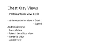
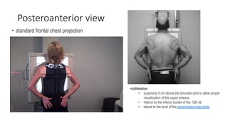
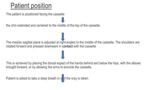
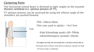
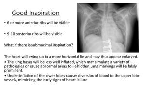
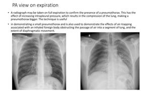
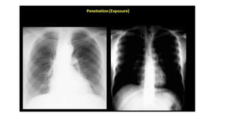
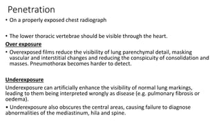
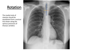
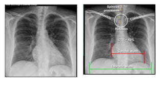
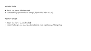
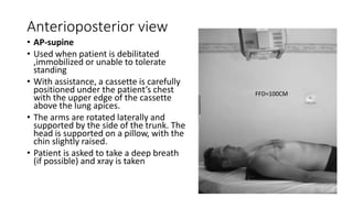
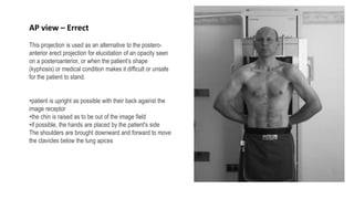
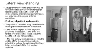
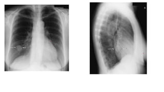
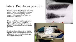
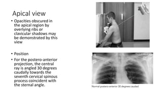
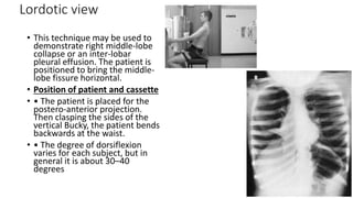
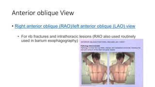
Chest radiography
positioning and Technique
Chest Xray Views
• Posteroanterior view- Erect
• Anteroposterior view – Erect
- Supine
Additional views
• Lateral view
• lateral decubitus view
• Lordotic view
• Apical view
Posteroanterior view
• standard frontal chest projection
•collimation
• superiorly 5 cm above the shoulder joint to allow proper
visualization of the upper airways
• inferior to the inferior border of the 12th rib
• lateral to the level of the acromioclavicular joints
Patient position
The patient is positioned facing the cassette
the chin extended and centered to the middle of the top of the cassette.
The median sagittal plane is adjusted at right-angles to the middle of the cassette. The shoulders are
rotated forward and pressed downward in contact with the cassette
This is achieved by placing the dorsal aspect of the hands behind and below the hips, with the elbows
brought forward, or by allowing the arms to encircle the cassette.
Patient is asked to take a deep breath in and the xray is taken.
Centering Point
The horizontal central beam is directed at right-angles to the cassette
thoracic vertebrae (i.e. spinous process of T7),
T7 spinous process can be assessed by using the inferior angle of the
shoulders are pushed forward.
FFD=180cm/6feet
Film size used in adults= 14x17cm
Film size used= 14x17cm
KVp( Kilovoltage peak)=60-70kVp
mAs(milliampere second)=20mAs
The higher the kVp, the more likely the x-ray beam will be able to
through more thicker and dense material. Based on how
of tissue kVp is selected.
Three main factors that determine the
technical quality of the radiograph
• Inspiration
• Penetration
• Rotation
Good Inspiration
• 6 or more anterior ribs will be visible
• 9-10 posterior ribs will be visible
What if there is submaximal inspiration?
The heart will swing up to a more horizontal lie and may thus appear enlarged.
• The lung bases will be less well inflated, which may simulate a variety of
pathologies or cause abnormal areas to lie hidden.Lung markings will be falsly
prominent.
• Under-inflation of the lower lobes causes diversion of blood to the upper lobe
vessels, mimicking the early signs of heart failure
PA view on expiration
• A radiograph may be taken on full expiration to confirm the presence of a pneumothorax. This has the
effect of increasing intrapleural pressure, which results in the compression of the lung, making a
pneumothorax bigger. The technique is useful
• in demonstrating a small pneumothorax and is also used to demonstrate the effects of air-trapping
associated with an inhaled foreign body obstructing the passage of air into a segment of lung, and the
extent of diaphragmatic movement.
Penetration
• On a properly exposed chest radiograph
• The lower thoracic vertebrae should be visible through the heart.
Over exposure
• Overexposed films reduce the visibility of lung parenchymal detail, masking
vascular and interstitial changes and reducing the conspicuity of consolidation and
masses. Pneumothorax becomes harder to detect.
Underexposure
Underexposure can artificially enhance the visibility of normal lung markings,
leading to them being interpreted wrongly as disease (e.g. pulmonary fibrosis or
oedema).
• Underexposure also obscures the central areas, causing failure to diagnose
abnormalities of the mediastinum, hila and spine.
Rotation
The medial ends of
clavicles should be
equidistant from a vertical
line drawn along the
spinous processes of
thoracic vertebra
Rotation to left
• Heart size maybe overestimated
• aortic arch may appear spuriously enlarged, hyperlucency of the left lung
Rotation to Right
• Heart size maybe underestimated
• rotation to the right may cause: pseudo-mediastinal mass, hyperlucency of the right lung
Anterioposterior view
• AP-supine
• Used when patient is debilitated
,immobilized or unable to tolerate
standing
• With assistance, a cassette is carefully
positioned under the patient’s chest
with the upper edge of the cassette
above the lung apices.
• The arms are rotated laterally and
supported by the side of the trunk. The
head is supported on a pillow, with the
chin slightly raised.
• Patient is asked to take a deep breath
(if possible) and xray is taken
FFD=100CM
FFD=100CM
•AP images are of inferior quality to PA images (posterior-anterior)
•This projection moves the heart away from the image receptor plane, increasing
magnification and reducing the accuracy of assessment of heart size
As a general rule the heart should not be considered large if the CTR is increased on
an AP view
• The scapula are usually visible in the lung fields because they are not rotated out of
view.
The normal biomechanics of blood flow are different from those in the erect position,
producing relative prominence of upper-lobe vessels and mimicking the signs of heart
failure.
• Pleural fluid will layer against the posterior chest wall, producing an ill-defined
increase attenuation of the affected hemithorax rather than the usual blunting of the
costophrenic angle; fluid levels are not seen.
• A pneumothorax, if present, will be located at the front of the chest in the supine
position. Unless it is large, it will be more difficult to detect if a lateral horizontal beam
image is not employed.
AP view – Errect
This projection is used as an alternative to the postero-
anterior erect projection for elucidation of an opacity seen
on a posteroanterior, or when the patient’s shape
(kyphosis) or medical condition makes it difficult or unsafe
for the patient to stand.
•patient is upright as possible with their back against the
image receptor
•the chin is raised as to be out of the image field
•if possible, the hands are placed by the patient's side
The shoulders are brought downward and forward to move
the clavicles below the lung apices
Lateral view-standing
• A supplementary lateral projection may be
useful in certain clinical circumstances for
localizing the position of a lesion and
demonstrating anterior mediastinal masses
not shown on the postero-anterior
projection.
• Position of patient and cassette
• The patient is turned to bring the side under
investigation in contact with the cassette.
• • The median sagittal plane is adjusted
parallel to the cassette. • The arms are
folded over the head or raised above the
head to rest on a horizontal bar.
• • The mid-axillary line is coincident with the
middle of the film, and the cassette is
adjusted to include the apices and the lower
lobes to the level of the first lumbar
vertebra.
Lateral Decubitus position
• Patient lies on the affected side.The
patient’s hands should be raised
to avoid superimposing on the
region of interest, legs may be
flexed for balance.
•
• Often useful in revealing a pleural
effusion that cannot be easily
observed in an upright view, since
effusion will collect in a dependent
position.
• The lateral decubitus view may be
helpful in determining the confines
of a cavity and in demonstrating a
small pneumothorax
Apical view
• Opacities obscured in
the apical region by
overlying ribs or
clavicular shadows may
be demonstrated by this
view
• Position
• For the postero-anterior
projection, the central
ray is angled 30 degrees
caudally towards the
seventh cervical spinous
process coincident with
the sternal angle.
Lordotic view
• This technique may be used to
demonstrate right middle-lobe
collapse or an inter-lobar
pleural effusion. The patient is
positioned to bring the middle-
lobe fissure horizontal.
• Position of patient and cassette
• • The patient is placed for the
postero-anterior projection.
Then clasping the sides of the
vertical Bucky, the patient bends
backwards at the waist.
• • The degree of dorsiflexion
varies for each subject, but in
general it is about 30–40
degrees
Anterior oblique View
• Right anterior oblique (RAO)/left anterior oblique (LAO) view
• For rib fractures and intrathoracic lesions (RAO also used routinely
used in barium esophagography)
References
• CLARKS POSITIONING 13edition
• Radiopedia

Chest radiography positioning and Technique.pptx

  • 1.
  • 2.
    Chest Xray Views •Posteroanterior view- Erect • Anteroposterior view – Erect - Supine Additional views • Lateral view • lateral decubitus view • Lordotic view • Apical view
  • 3.
    Posteroanterior view • standardfrontal chest projection •collimation • superiorly 5 cm above the shoulder joint to allow proper visualization of the upper airways • inferior to the inferior border of the 12th rib • lateral to the level of the acromioclavicular joints
  • 4.
    Patient position The patientis positioned facing the cassette the chin extended and centered to the middle of the top of the cassette. The median sagittal plane is adjusted at right-angles to the middle of the cassette. The shoulders are rotated forward and pressed downward in contact with the cassette This is achieved by placing the dorsal aspect of the hands behind and below the hips, with the elbows brought forward, or by allowing the arms to encircle the cassette. Patient is asked to take a deep breath in and the xray is taken.
  • 5.
    Centering Point The horizontalcentral beam is directed at right-angles to the cassette thoracic vertebrae (i.e. spinous process of T7), T7 spinous process can be assessed by using the inferior angle of the shoulders are pushed forward. FFD=180cm/6feet Film size used in adults= 14x17cm Film size used= 14x17cm KVp( Kilovoltage peak)=60-70kVp mAs(milliampere second)=20mAs The higher the kVp, the more likely the x-ray beam will be able to through more thicker and dense material. Based on how of tissue kVp is selected.
  • 6.
    Three main factorsthat determine the technical quality of the radiograph • Inspiration • Penetration • Rotation
  • 7.
    Good Inspiration • 6or more anterior ribs will be visible • 9-10 posterior ribs will be visible What if there is submaximal inspiration? The heart will swing up to a more horizontal lie and may thus appear enlarged. • The lung bases will be less well inflated, which may simulate a variety of pathologies or cause abnormal areas to lie hidden.Lung markings will be falsly prominent. • Under-inflation of the lower lobes causes diversion of blood to the upper lobe vessels, mimicking the early signs of heart failure
  • 8.
    PA view onexpiration • A radiograph may be taken on full expiration to confirm the presence of a pneumothorax. This has the effect of increasing intrapleural pressure, which results in the compression of the lung, making a pneumothorax bigger. The technique is useful • in demonstrating a small pneumothorax and is also used to demonstrate the effects of air-trapping associated with an inhaled foreign body obstructing the passage of air into a segment of lung, and the extent of diaphragmatic movement.
  • 10.
    Penetration • On aproperly exposed chest radiograph • The lower thoracic vertebrae should be visible through the heart. Over exposure • Overexposed films reduce the visibility of lung parenchymal detail, masking vascular and interstitial changes and reducing the conspicuity of consolidation and masses. Pneumothorax becomes harder to detect. Underexposure Underexposure can artificially enhance the visibility of normal lung markings, leading to them being interpreted wrongly as disease (e.g. pulmonary fibrosis or oedema). • Underexposure also obscures the central areas, causing failure to diagnose abnormalities of the mediastinum, hila and spine.
  • 11.
    Rotation The medial endsof clavicles should be equidistant from a vertical line drawn along the spinous processes of thoracic vertebra
  • 13.
    Rotation to left •Heart size maybe overestimated • aortic arch may appear spuriously enlarged, hyperlucency of the left lung Rotation to Right • Heart size maybe underestimated • rotation to the right may cause: pseudo-mediastinal mass, hyperlucency of the right lung
  • 14.
    Anterioposterior view • AP-supine •Used when patient is debilitated ,immobilized or unable to tolerate standing • With assistance, a cassette is carefully positioned under the patient’s chest with the upper edge of the cassette above the lung apices. • The arms are rotated laterally and supported by the side of the trunk. The head is supported on a pillow, with the chin slightly raised. • Patient is asked to take a deep breath (if possible) and xray is taken FFD=100CM
  • 15.
  • 16.
    •AP images areof inferior quality to PA images (posterior-anterior) •This projection moves the heart away from the image receptor plane, increasing magnification and reducing the accuracy of assessment of heart size As a general rule the heart should not be considered large if the CTR is increased on an AP view • The scapula are usually visible in the lung fields because they are not rotated out of view. The normal biomechanics of blood flow are different from those in the erect position, producing relative prominence of upper-lobe vessels and mimicking the signs of heart failure. • Pleural fluid will layer against the posterior chest wall, producing an ill-defined increase attenuation of the affected hemithorax rather than the usual blunting of the costophrenic angle; fluid levels are not seen. • A pneumothorax, if present, will be located at the front of the chest in the supine position. Unless it is large, it will be more difficult to detect if a lateral horizontal beam image is not employed.
  • 17.
    AP view –Errect This projection is used as an alternative to the postero- anterior erect projection for elucidation of an opacity seen on a posteroanterior, or when the patient’s shape (kyphosis) or medical condition makes it difficult or unsafe for the patient to stand. •patient is upright as possible with their back against the image receptor •the chin is raised as to be out of the image field •if possible, the hands are placed by the patient's side The shoulders are brought downward and forward to move the clavicles below the lung apices
  • 18.
    Lateral view-standing • Asupplementary lateral projection may be useful in certain clinical circumstances for localizing the position of a lesion and demonstrating anterior mediastinal masses not shown on the postero-anterior projection. • Position of patient and cassette • The patient is turned to bring the side under investigation in contact with the cassette. • • The median sagittal plane is adjusted parallel to the cassette. • The arms are folded over the head or raised above the head to rest on a horizontal bar. • • The mid-axillary line is coincident with the middle of the film, and the cassette is adjusted to include the apices and the lower lobes to the level of the first lumbar vertebra.
  • 21.
    Lateral Decubitus position •Patient lies on the affected side.The patient’s hands should be raised to avoid superimposing on the region of interest, legs may be flexed for balance. • • Often useful in revealing a pleural effusion that cannot be easily observed in an upright view, since effusion will collect in a dependent position. • The lateral decubitus view may be helpful in determining the confines of a cavity and in demonstrating a small pneumothorax
  • 22.
    Apical view • Opacitiesobscured in the apical region by overlying ribs or clavicular shadows may be demonstrated by this view • Position • For the postero-anterior projection, the central ray is angled 30 degrees caudally towards the seventh cervical spinous process coincident with the sternal angle.
  • 23.
    Lordotic view • Thistechnique may be used to demonstrate right middle-lobe collapse or an inter-lobar pleural effusion. The patient is positioned to bring the middle- lobe fissure horizontal. • Position of patient and cassette • • The patient is placed for the postero-anterior projection. Then clasping the sides of the vertical Bucky, the patient bends backwards at the waist. • • The degree of dorsiflexion varies for each subject, but in general it is about 30–40 degrees
  • 24.
    Anterior oblique View •Right anterior oblique (RAO)/left anterior oblique (LAO) view • For rib fractures and intrathoracic lesions (RAO also used routinely used in barium esophagography)
  • 25.
    References • CLARKS POSITIONING13edition • Radiopedia