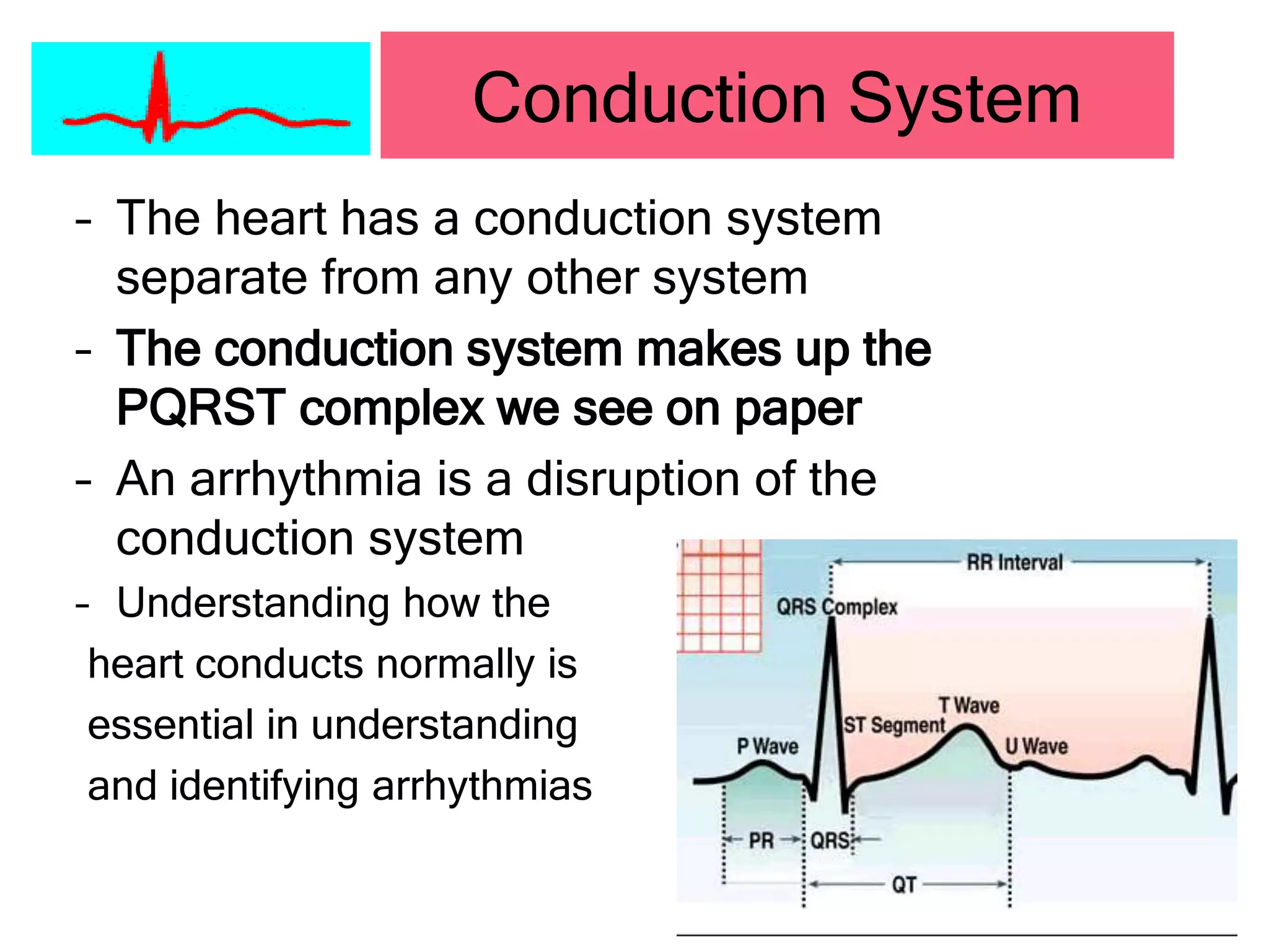
Cardiac
Arrhythmias
DR ABID NAEEM
Anatomy & Physiology
• Blood Flow through
heart
– Superior and Inferior
Vena Cava
– Right Atrium
– Right Ventricle
– Pulmonary Artery
– Lungs
– Pulmonary Vein
– Left Atrium
– Left Ventricle
– Aorta
– Body
Sinus
1-Sinus Rythem-HR-60-100/M
2-Sinus Arrythmia-HR-60-100/M
3-Sinus Arrest- Sinus arrest- sinus stop-one
beat miss
4-Sinus Bradycardia-HR-<60/M
5-Sinus Tachycardia-HR-100-160/M. >160
SVT
Atrial
1 -Wandering pace maker-HR-<60
2-Premature Atrial Contraction-HR-NA(Not
Assessable)
3-Atrial Tachycardia-HR-160-250/m
Atrial flutter-hr-250-400/m-P-regular
5-Atrial fibrillation-HR-variable=p-p-Irregular
(Junctional)-(AV-Node)
1-Junctional Rhythum-HR-40-60/M
2-Juctional escape-HR-40-60/M
3-Premature Contraction
4-Accelarated Junctional Rhythum-HR-60-
100
5-Juntional Tachycardia-HR-100-180/M
BUNDLE OF HIS/RT & LF BRANCHES/
Parkinje Fibers/ VENTRICULAR
RHYTHUM
1-Ventricular Rhythum-HR-20-40/M
2-Ventricular Muscle-HR-10-20/M
3-Primary Ventricular Standstill-No QRS-P.wave present
4-Asystole-straight line
5-Idiopathic Ventricular-hr-20-40/M
6-Ventricular tachycardia- HR-150-250/M
7-Ventricular Flutter-HR-250-300/M
8-Ventricular Fibrillation-HR-NA
Heart Blocks
FIRST DEGREE HEART BLOCK
RATE-NORMAL=P-R INTERVAL(N-0.12 0.20/SEC=0.20/SEC
2ND DEGREE HEART BLOCK (Mobitz Type I (Wenckebach)
PROGRESSIVE P-R PROLONGATION-TILL 01 BEAT DROP
2ND DEGREE HEART BLOCK (Mobitz Type 2)
P-R NORMAL=2,3,4 P.waves before each QRS-HR-60-100/M
3RD DEGREE HEART BLOCK
P AND QRS NAS ITS OWN RATE-HR-30-60/M
Conduction System
– The heart has a conduction system
separate from any other system
– The conduction system makes up the
PQRST complex we see on paper
– An arrhythmia is a disruption of the
conduction system
– Understanding how the
heart conducts normally is
essential in understanding
and identifying arrhythmias
• SA Node
• Inter-nodal and
inter-atrial pathways
• A-V Node
• Bundle of His
• Purkinje Fibers
Conduction System
SA Node
 The primary pacemaker
of the heart
 Each normal beat is
initiated by the SA node
 Inherent rate of 60-100
beats per minute
 Represents the P-wave
in the QRS complex or
atrial depolarization
(firing)
AV Node
– Located in the septum of
the heart
– Receives impulse from
inter-nodal pathways
and holds the signal
before sending on to the
Bundle of His
– Represents the PR
segment of the QRS
complex
AV Node
– Represents the PR segment of the cardiac
cycle
– Has an inherent rate of 40-60 beats per
minute
– Acts as a back up when the SA node fails
– Where all junctional rhythms originate
QRS Complex
• Represents the
ventricles
depolarizing (firing)
collectively. (Bundle
of His and Purkinje
fibers)
• Origin of all
ventricular rhythms
• Has an inherent rate
of 20-40 beats per
minute
ECG Trace
• Isoelectric
line
(baseline)
• P-wave
– Atria firing
• PR interval
– Delay at AV
ECG Trace
• QRS
– Ventricles
firing
• T-wave
– Ventricles
repolarizing
ECG Trace
• ST segment
– Ventricle
contracting
– Should be at
isoelectric line
– Elevation or
depression may
be important
• U wave
– Perkinje fiber
repolarization?
ECG- Analysis
1) RATE
2) RHYTHUM
3) AXIS
4) WAVE FORM
ECG-PAPER
1-RATE
Calculation
 300,150,100
75,60,50
1-What is the rate?
• Count the number of beats occurring in one minute
• Counting the p-waves will give the atrial rate
• Counting QRS will give ventricular rate
2-RHYTHUM
2-Is the rhythm regular?
• Verify by assessing R-R interval
• Confirm by assessing P-P interval
3-axis
LEADS- I, AVF
REULE OF THUMB
1- I,AVF= R ,UP= Normal axis
2- 1,AVF= R,WAVE DOWN =Extreme
T-RT. Axis deviation
3- I -R=UP,
AVF-R=DOWN =LT.Axis. Deviation
4-1,R =down.
AVF,R=UP=RT. Axis deviation
Determining the Axis
Predominantly
Positive
Predominantly
Negative
Equiphasic
Waveform Analysis
Is there a P-wave for every QRS?
• P-waves are upright and uniform
• One P-wave preceding each QRS
Calculation
 300,150,100
75,60,50
• Normal
Heart rate = 60 – 100 BPM
SA Node discharge = 60 – 100 / min
AV Node discharge = 40 – 60 min
Ventricular Tissue discharge = 20 – 40/min
PR interval = 0.12 – 0.20 sec
QRS interval= <0.12
Summary
• Cardiac cycle
P wave = atrial depolarization
PR interval = pause between atrial and
ventricular depolarization
QRS = ventricular Depolarization
T wave = ventricular Repolarization
Summary
Normal Sinus Rhythm
Heart
Rate
(bpm)
Rhythm P Wave
PR Interval
(sec)
QRS
(sec)
60 -
100
Regular
Before each QRS,
Identical
0.12 - 0.20 <0.12
Sinus Rhythms
• Normal Sinus Rhythm
– Sinus Node is the primary pacemaker
– One upright uniform p-wave for every QRS
– Rhythm is regular
– Rate is between 60-100 beats per minute
Sinus Rhythms
Sinus Bradycardia
Heart
Rate
(bpm)
Rhythm P Wave
PR Interval
(sec)
QRS
(sec)
<60 Regular
Before each QRS,
Identical
0.12 - 0.20 <0.12
Sinus Rhythms
• Sinus Bradycardia
– One upright uniform p-wave for every QRS
– Rhythm is regular
– Rate less than 60 beats per minute
• SA node firing slower than normal
• Normal for many individuals
• Identify what is normal heart rate for patient
Sinus Rhythms
•
Sinus Tachycardia
Heart
Rate
Rhythm P Wave
PR Interval
(sec.)
QRS
(Sec.)
>100 Regular
Before each QRS,
Identical
.12 - .20 <.12
Sinus Rhythms
• Sinus Tachycardia
– One upright uniform p-wave for every QRS
– Rhythm is regular
– Rate is greater than 100 beats per minute
• Usually between 100-160 (>160 SVT)
• Can be high due to anxiety, stress, fever,
medications (anything that increases oxygen
consumption)
Sinus Rhythms
Sinus Arrhythmia
Heart
Rate
Rhythm P Wave
PR Interval
(sec.)
QRS
(Sec.)
Var. Irregular
Before each QRS,
Identical
.12 - .20 <.12
Sinus Rhythms
• Sinus Arrhythmia
– One upright uniform p-wave for every QRS
– Rhythm is irregular
• Rate increases as the patient breathes in
• Rate decreases as the patient breathes out
– Rate is usually 60-100 (may be slower)
– Variation of normal, not life threatening
Sinus Rhythms
Sinus Arrest
Heart
Rate
Rhythm P Wave
PR Interval
(sec.)
QRS
(Sec.)
NA Irregular
Before each QRS,
Identical
.12 - .20 <.12
Sinus Rhythms
Sinus Arrest
Sinus Rhythms
Stop of sinus rhythm
New rhythm starts
Sinus Pause
Sinus Rhythms
• One dropped beat is a
sinus pause
• Beats walk through
Premature Atrial Contraction (PAC)
Heart
Rate
Rhythm P Wave
PR Interval
(sec.)
QRS
(Sec.)
NA Irregular
Premature &
abnormal or
hidden
.12 - .20 <.12
Atrial Rhythms
– Premature Atrial Contraction (PAC)
• One P-wave for every QRS
– P-wave may have different morphology on ectopic
beat, but it will be present
• Single ectopic beat will disrupt regularity of
underlying rhythm
• Rate will depend on underlying rhythm
• Underlying rhythm must be identified
• Classified as rare, occasional, or frequent
PAC’s based on frequency
Atrial Rhythms
Atrial Fibrillation
Heart
Rate
Rhythm P Wave
PR Interval
(sec.)
QRS
(Sec.)
Var. Irregular Wavy irregular NA <.12
Atrial Rhythms
• Atrial Fibrillation
– No discernable p-waves preceding the QRS
complex
• The atria are not depolarizing effectively, but fibrillating
– Rhythm is grossly irregular
– If the heart rate is <100 it is considered controlled
a-fib, if >100 it is considered to have a “rapid
ventricular response”
– AV node acts as a “filter”, blocking out most of the
impulses sent by the atria in an attempt to control
the heart rate
Atrial Rhythms
• Atrial Fibrillation (con’t)
– Often a chronic condition, medical attention
only necessary if patient becomes
symptomatic
– Patient will report history of atrial
fibrillation.
Atrial Rhythms
Atrial Flutter
Heart Rate Rhythm P Wave
PR Interval
(sec.)
QRS
(Sec.)
Atrial=250
– 400
Ventricular
Var.
Irregular Sawtooth
Not
Measur-
able
<.12
Atrial Rhythms
• Atrial Flutter
– More than one p-wave for every QRS complex
• Demonstrate a “saw tooth” appearance
– Atrial rhythm is regular. Ventricular rhythm will be
regular if the AV node conducts consistently. If the
pattern varies, the ventricular rate will be irregular
– Rate will depend on the ratio of impulses
conducted through the ventricles
Atrial Rhythms
Atrial Rhythms
• Atrial Flutter
– Atrial flutter is classified as a ratio of p-
waves per QRS complexes (ex: 3:1 flutter
3 p-waves for each QRS)
– Not considered life threatening, consult
physician is patient symptomatic
• Rhythms that originate at the AV
junction
• Junctional rhythms do not have
characteristic p-waves.
Junctional Rhythms
Premature Junctional Contraction PJC
Heart
Rate
Rhythm P Wave
PR Interval
(sec.)
QRS
(Sec.)
Usually
normal
Irregular
Premature,
abnormal, may be
inverted or hidden
Short
<.12
Normal
<.12
Junctional Rhythms
• Premature Junctional Contraction (PJC)
– P-wave can come before or after the QRS
complex, or it may lost in the QRS complex
• If visible, the p-wave will be inverted
– Rhythm will be irregular due to single ectopic beat
– Heart rate will depend on underlying rhythm
– Underlying rhythm must be identified
– Classify as rare, occasional, or frequent PJC
based on frequency
– Atria are depolarized via retrograde conduction
Junctional Rhythms
Junctional Rhythms
Accelerated Junctional
Heart
Rate
Rhythm P Wave
PR Interval
(sec.)
QRS
(Sec.)
Var. Regular
Inverted, absent or
after QRS
<.12 <.12
Junctional Rhythms
• Accelerated Junctional Rhythm
– P-wave can come before or after the QRS
complex, or lost within the QRS complex
• If p-waves are seen they will be inverted
– Rhythm is regular
– Heart rate between 60-100 beats per
minute
• Within the normal HR range
• Fast rate for the junction (normally 40-60 bpm)
Junctional Rhythms
Junctional Rhythms
Junctional Tachycardia
Heart
Rate
Rhythm P Wave
PR Interval
(sec.)
QRS
(Sec.)
>100 Regular
May be inverted or
hidden
Short
<.12
Normal
<.12
Junctional Rhythms
• Junctional Tachycardia
– P-wave can come before or after the QRS
complex or lost within the QRS entirely
• If a p-wave is seen it will be inverted
– Rhythm is regular
– Rate is between 100-180 beats per minute
• In the tachycardia range, but not originating from SA
node
– AV node has spread up to override the SA node
for control of the heart
Junctional Rhythms
Junctional Rhythms
Junctional Rhythms
Junctional Escape
Heart
Rate
Rhythm P Wave
PR Interval
(sec.)
QRS
(Sec.)
40 –
60
Regular
Absent, inverted
or after QRS
Short
<.12
Normal
<.12
Junctional Rhythms
• Junctional Escape Rhythm
– P-wave may come before or after the QRS
or may be hidden in the QRS entirely
• If p-waves are seen, they will be inverted
– Rhythm is regular
– Rate 40-60 beats per minute
• The SA node has failed; the AV junction takes
over control of the heart
Junctional Rhythms
Ventricular Rhythms
Premature Ventricular Contraction (PVC)
Heart
Rate
Rhythm P Wave
PR
Interval
(sec.)
QRS
(Sec.)
Var. Irregular
No P waves
associated with
premature beat
NA
Wide
>.12
Ventricular Rhythms
• Premature Ventricular Contraction (PVC)
– The ectopic beat is not preceded by a p-wave
– Irregular rhythm due to ectopic beat
– Rate will be determined by the underlying rhythm
– QRS is wide and may be bizarre in appearance
– Caused by a irritable focus within the ventricle
which fires prematurely
– Must identify an underlying rhythm
Ventricular Rhythm
• Premature Ventricular Contraction
– Classify as rare, occasional, or frequent
– Classify as unifocal, or multifocal PVC’s
• Unifocal-originating from same area of the
ventricle; distinguished by same morphology
Ventricular Rhythm
• Premature Ventricular Contraction
– Classify as unifocal or multifocal PVC’s
– Unifocal-originating from same area of the
ventricle; distinguished by same morphology
– Multifocal-originating from different areas of the
ventricle; distinguished by different morphology
Ventricular Rhythm
Multifocal-originating from different areas of the
ventricle; distinguished by different morphology
Ventricular Rhythm
• Premature Ventricular Contraction
– Bigeminy
• A PVC occurring every other beat
– Also seen as Trigeminy, Quadrigeminy
Ventricular Rhythm
• Dangerous PVC’s
– R on T
– Runs of PVC’s
– 3 or more
considered Vtach
Idioventricular Rhythm
Ventricular Rhythm
Ventricular Rhythm
Ventricular Rhythms
Ventricular Flutter
Heart
Rate
Rhythm P Wave
PR Interval
(sec.)
QRS
(Sec.)
150 –
300
Regular No P waves NA
Wide
>.12
Ventricular Rhythms
Ventricular Flutter
“Extreme V-Tach”
Call code
Begin CPR
Ventricular Rhythms
Ventricular Flutter
“Extreme V-Tach”
Call code
Begin CPR
Ventricular Rhythms
Ventricular Tachycardia
Heart
Rate
Rhythm P Wave
PR Interval
(sec.)
QRS
(Sec.)
100 –
250
Regular
No P waves
corresponding to QRS,
a few may be seen
NA >.12
Ventricular Rhythms
• Ventricular Tachycardia
– No discernable p-waves with QRS
– Rhythm is regular
– Atrial rate cannot be determined,
ventricular rate is between 150-250 beats
per minute
– Must see 4 beats in a row to classify as v-
tach
Ventricular Rhythms
• Ventricular
Tachycardia
– THIS IS A DEADLY
RHYTHM
• Check patient:
– If patient awake
and alert, monitor
patient and call
physician
– If patient has no
vital signs, call
code and start CPR
» Defibrillate
Ventricular Rhythms
Ventricular Fibrillation
Heart
Rate
Rhythm P Wave
PR Interval
(sec.)
QRS
(Sec.)
0 Chaotic None NA None
Ventricular Rhythms
• Ventricular Fibrillation
– No discernable p-waves
– No regularity
– Unable to determine rate
– Multiple irritable foci within the ventricles all
firing simultaneously
– May be coarse or fine
– This is a deadly rhythm
• Patient will have no pulse
• Call a code and begin CPR
Ventricular Rhythms
Asystole
Heart
Rate
Rhythm P Wave
PR Interval
(sec.)
QRS
(Sec.)
None None None None None
Asystole
• No p-waves
• No regularity
• No Rate
• This rhythm is associated with death
– Check patient and leads
– No pulse
• Begin CPR
Heart Block
First Degree Heart Block
Heart
Rate
Rhythm P Wave
PR Interval
(sec.)
QRS
(Sec.)
Norm. Regular
Before each QRS,
Identical
> .20 <.12
Heart Block
– First Degree Heart Block
• P-wave for every QRS
• Rhythm is regular
• Rate may vary
• Av Node hold each impulse longer than normal
before conducting normally through the
ventricles
• Prolonged PR interval
– Looks just like normal sinus rhythm
Heart Block
Heart Block
Second Degree Heart Block
Mobitz Type I (Wenckebach)
Heart
Rate
Rhythm P Wave
PR Interval
(sec.)
QRS
(Sec.
)
Norm.
can be
slow
Irregular
Present but some
not followed by
QRS
Progressively
longer
<.12
Heart Block
• Second Degree Heart Block
• Mobitz Type I (Wenckebach)
– Some p-waves are not followed by QRS
complexes
– Rhythm is irregular
• R-R interval is in a pattern of grouped beating
– Rate 60-100 bpm
– Intermittent Block at the AV Node
• Progressively prolonged p-r interval until a QRS is
blocked completely
Heart Block
Heart Block
Second Degree Heart Block
Mobitz Type II (Classical)
Heart
Rate
Rhythm P Wave
PR
Interval
(sec.)
QRS
(Sec.)
Usually
slow
Regular
or
irregular
2 3 or 4 before each
QRS, Identical
.12 - .20
<.12
depends
Heart Block
• Second Degree Heart Block
• Mobitz Type II (Classical)
– More p-waves than QRS complexes
– Rhythm is irregular
– Atrial rate 60-100 bpm; Ventricular rate 30-100
bpm (depending on the ratio on conduction)
– Intermittent block at the AV node
• AV node normally conducts some beats while blocking
others
Heart Block
Heart Block
Third Degree Heart Block
(Complete)
Heart
Rate
Rhythm P Wave
PR
Interval
(sec.)
QRS
(Sec.)
30 –
60
Regular
Present but no
correlation to QRS
may be hidden
Varies
<.12
depends
Heart Block
• Third Degree Heart Block (Complete)
– There are more p-waves than QRS
complexes
– Both P-P and R-R intervals are regular
– Atrial rate within normal range; Ventricular
rate between 20-60 bpm
– The block at the AV node is complete
• There is no relationship between the p-waves
and QRS complexes
Heart Block
ECG-READING
1-RATE
2-RYTHEM
3-AXIS-1,AVF
4-LEADS
V1-V2=RT.CHEST LEADS
V3-V4=SEPTAL LEADS
V1-V4=ANTERIOR CHEST LEADS
V5-V6=LATERAL CHEST LEADS
1,AVL,V1-V6=ANTROLATERAL CHEST LEADS
11,111,AVF-INFERIOR LEADS
5-MYOCARDIAL INFARCTION
ANTERIOR,ANTROLATRAL,INFERIOR
ACUTE MI-T-INVERSION, ST-
ELEVATION
6-CONDUCTIONS DEFECTS
RBBB-T.WAVE-V1-V2=M-PATTERN
RBBB V5 V6 W PATTERN
LBBB-T.WAVE-V5-V6=M-PATTERN
LBBB V1 V2 W PATTERN
7-CCF,PERICARDIAL EFFUSION,OBESE-
LOW VOLTAGE ECG.
Cardiac arrhythmias

Cardiac arrhythmias

  • 1.
  • 2.
    Anatomy & Physiology •Blood Flow through heart – Superior and Inferior Vena Cava – Right Atrium – Right Ventricle – Pulmonary Artery – Lungs – Pulmonary Vein – Left Atrium – Left Ventricle – Aorta – Body
  • 3.
    Sinus 1-Sinus Rythem-HR-60-100/M 2-Sinus Arrythmia-HR-60-100/M 3-SinusArrest- Sinus arrest- sinus stop-one beat miss 4-Sinus Bradycardia-HR-<60/M 5-Sinus Tachycardia-HR-100-160/M. >160 SVT
  • 4.
    Atrial 1 -Wandering pacemaker-HR-<60 2-Premature Atrial Contraction-HR-NA(Not Assessable) 3-Atrial Tachycardia-HR-160-250/m Atrial flutter-hr-250-400/m-P-regular 5-Atrial fibrillation-HR-variable=p-p-Irregular
  • 5.
    (Junctional)-(AV-Node) 1-Junctional Rhythum-HR-40-60/M 2-Juctional escape-HR-40-60/M 3-PrematureContraction 4-Accelarated Junctional Rhythum-HR-60- 100 5-Juntional Tachycardia-HR-100-180/M
  • 6.
    BUNDLE OF HIS/RT& LF BRANCHES/ Parkinje Fibers/ VENTRICULAR RHYTHUM 1-Ventricular Rhythum-HR-20-40/M 2-Ventricular Muscle-HR-10-20/M 3-Primary Ventricular Standstill-No QRS-P.wave present 4-Asystole-straight line 5-Idiopathic Ventricular-hr-20-40/M 6-Ventricular tachycardia- HR-150-250/M 7-Ventricular Flutter-HR-250-300/M 8-Ventricular Fibrillation-HR-NA
  • 7.
    Heart Blocks FIRST DEGREEHEART BLOCK RATE-NORMAL=P-R INTERVAL(N-0.12 0.20/SEC=0.20/SEC 2ND DEGREE HEART BLOCK (Mobitz Type I (Wenckebach) PROGRESSIVE P-R PROLONGATION-TILL 01 BEAT DROP 2ND DEGREE HEART BLOCK (Mobitz Type 2) P-R NORMAL=2,3,4 P.waves before each QRS-HR-60-100/M 3RD DEGREE HEART BLOCK P AND QRS NAS ITS OWN RATE-HR-30-60/M
  • 8.
    Conduction System – Theheart has a conduction system separate from any other system – The conduction system makes up the PQRST complex we see on paper – An arrhythmia is a disruption of the conduction system – Understanding how the heart conducts normally is essential in understanding and identifying arrhythmias
  • 9.
    • SA Node •Inter-nodal and inter-atrial pathways • A-V Node • Bundle of His • Purkinje Fibers Conduction System
  • 10.
    SA Node  Theprimary pacemaker of the heart  Each normal beat is initiated by the SA node  Inherent rate of 60-100 beats per minute  Represents the P-wave in the QRS complex or atrial depolarization (firing)
  • 11.
    AV Node – Locatedin the septum of the heart – Receives impulse from inter-nodal pathways and holds the signal before sending on to the Bundle of His – Represents the PR segment of the QRS complex
  • 12.
    AV Node – Representsthe PR segment of the cardiac cycle – Has an inherent rate of 40-60 beats per minute – Acts as a back up when the SA node fails – Where all junctional rhythms originate
  • 13.
    QRS Complex • Representsthe ventricles depolarizing (firing) collectively. (Bundle of His and Purkinje fibers) • Origin of all ventricular rhythms • Has an inherent rate of 20-40 beats per minute
  • 14.
    ECG Trace • Isoelectric line (baseline) •P-wave – Atria firing • PR interval – Delay at AV
  • 15.
    ECG Trace • QRS –Ventricles firing • T-wave – Ventricles repolarizing
  • 16.
    ECG Trace • STsegment – Ventricle contracting – Should be at isoelectric line – Elevation or depression may be important • U wave – Perkinje fiber repolarization?
  • 17.
    ECG- Analysis 1) RATE 2)RHYTHUM 3) AXIS 4) WAVE FORM
  • 18.
  • 19.
    1-RATE Calculation  300,150,100 75,60,50 1-What isthe rate? • Count the number of beats occurring in one minute • Counting the p-waves will give the atrial rate • Counting QRS will give ventricular rate
  • 20.
    2-RHYTHUM 2-Is the rhythmregular? • Verify by assessing R-R interval • Confirm by assessing P-P interval
  • 21.
    3-axis LEADS- I, AVF REULEOF THUMB 1- I,AVF= R ,UP= Normal axis 2- 1,AVF= R,WAVE DOWN =Extreme T-RT. Axis deviation 3- I -R=UP, AVF-R=DOWN =LT.Axis. Deviation 4-1,R =down. AVF,R=UP=RT. Axis deviation
  • 22.
  • 23.
    Waveform Analysis Is therea P-wave for every QRS? • P-waves are upright and uniform • One P-wave preceding each QRS
  • 24.
    Calculation  300,150,100 75,60,50 • Normal Heartrate = 60 – 100 BPM SA Node discharge = 60 – 100 / min AV Node discharge = 40 – 60 min Ventricular Tissue discharge = 20 – 40/min PR interval = 0.12 – 0.20 sec QRS interval= <0.12 Summary
  • 25.
    • Cardiac cycle Pwave = atrial depolarization PR interval = pause between atrial and ventricular depolarization QRS = ventricular Depolarization T wave = ventricular Repolarization Summary
  • 26.
    Normal Sinus Rhythm Heart Rate (bpm) RhythmP Wave PR Interval (sec) QRS (sec) 60 - 100 Regular Before each QRS, Identical 0.12 - 0.20 <0.12 Sinus Rhythms
  • 27.
    • Normal SinusRhythm – Sinus Node is the primary pacemaker – One upright uniform p-wave for every QRS – Rhythm is regular – Rate is between 60-100 beats per minute Sinus Rhythms
  • 28.
    Sinus Bradycardia Heart Rate (bpm) Rhythm PWave PR Interval (sec) QRS (sec) <60 Regular Before each QRS, Identical 0.12 - 0.20 <0.12 Sinus Rhythms
  • 29.
    • Sinus Bradycardia –One upright uniform p-wave for every QRS – Rhythm is regular – Rate less than 60 beats per minute • SA node firing slower than normal • Normal for many individuals • Identify what is normal heart rate for patient Sinus Rhythms
  • 30.
    • Sinus Tachycardia Heart Rate Rhythm PWave PR Interval (sec.) QRS (Sec.) >100 Regular Before each QRS, Identical .12 - .20 <.12 Sinus Rhythms
  • 31.
    • Sinus Tachycardia –One upright uniform p-wave for every QRS – Rhythm is regular – Rate is greater than 100 beats per minute • Usually between 100-160 (>160 SVT) • Can be high due to anxiety, stress, fever, medications (anything that increases oxygen consumption) Sinus Rhythms
  • 32.
    Sinus Arrhythmia Heart Rate Rhythm PWave PR Interval (sec.) QRS (Sec.) Var. Irregular Before each QRS, Identical .12 - .20 <.12 Sinus Rhythms
  • 33.
    • Sinus Arrhythmia –One upright uniform p-wave for every QRS – Rhythm is irregular • Rate increases as the patient breathes in • Rate decreases as the patient breathes out – Rate is usually 60-100 (may be slower) – Variation of normal, not life threatening Sinus Rhythms
  • 34.
    Sinus Arrest Heart Rate Rhythm PWave PR Interval (sec.) QRS (Sec.) NA Irregular Before each QRS, Identical .12 - .20 <.12 Sinus Rhythms
  • 36.
    Sinus Arrest Sinus Rhythms Stopof sinus rhythm New rhythm starts
  • 37.
    Sinus Pause Sinus Rhythms •One dropped beat is a sinus pause • Beats walk through
  • 38.
    Premature Atrial Contraction(PAC) Heart Rate Rhythm P Wave PR Interval (sec.) QRS (Sec.) NA Irregular Premature & abnormal or hidden .12 - .20 <.12 Atrial Rhythms
  • 40.
    – Premature AtrialContraction (PAC) • One P-wave for every QRS – P-wave may have different morphology on ectopic beat, but it will be present • Single ectopic beat will disrupt regularity of underlying rhythm • Rate will depend on underlying rhythm • Underlying rhythm must be identified • Classified as rare, occasional, or frequent PAC’s based on frequency Atrial Rhythms
  • 41.
    Atrial Fibrillation Heart Rate Rhythm PWave PR Interval (sec.) QRS (Sec.) Var. Irregular Wavy irregular NA <.12 Atrial Rhythms
  • 43.
    • Atrial Fibrillation –No discernable p-waves preceding the QRS complex • The atria are not depolarizing effectively, but fibrillating – Rhythm is grossly irregular – If the heart rate is <100 it is considered controlled a-fib, if >100 it is considered to have a “rapid ventricular response” – AV node acts as a “filter”, blocking out most of the impulses sent by the atria in an attempt to control the heart rate Atrial Rhythms
  • 44.
    • Atrial Fibrillation(con’t) – Often a chronic condition, medical attention only necessary if patient becomes symptomatic – Patient will report history of atrial fibrillation. Atrial Rhythms
  • 45.
    Atrial Flutter Heart RateRhythm P Wave PR Interval (sec.) QRS (Sec.) Atrial=250 – 400 Ventricular Var. Irregular Sawtooth Not Measur- able <.12 Atrial Rhythms
  • 46.
    • Atrial Flutter –More than one p-wave for every QRS complex • Demonstrate a “saw tooth” appearance – Atrial rhythm is regular. Ventricular rhythm will be regular if the AV node conducts consistently. If the pattern varies, the ventricular rate will be irregular – Rate will depend on the ratio of impulses conducted through the ventricles Atrial Rhythms
  • 47.
    Atrial Rhythms • AtrialFlutter – Atrial flutter is classified as a ratio of p- waves per QRS complexes (ex: 3:1 flutter 3 p-waves for each QRS) – Not considered life threatening, consult physician is patient symptomatic
  • 49.
    • Rhythms thatoriginate at the AV junction • Junctional rhythms do not have characteristic p-waves. Junctional Rhythms
  • 51.
    Premature Junctional ContractionPJC Heart Rate Rhythm P Wave PR Interval (sec.) QRS (Sec.) Usually normal Irregular Premature, abnormal, may be inverted or hidden Short <.12 Normal <.12 Junctional Rhythms
  • 52.
    • Premature JunctionalContraction (PJC) – P-wave can come before or after the QRS complex, or it may lost in the QRS complex • If visible, the p-wave will be inverted – Rhythm will be irregular due to single ectopic beat – Heart rate will depend on underlying rhythm – Underlying rhythm must be identified – Classify as rare, occasional, or frequent PJC based on frequency – Atria are depolarized via retrograde conduction Junctional Rhythms
  • 53.
  • 54.
    Accelerated Junctional Heart Rate Rhythm PWave PR Interval (sec.) QRS (Sec.) Var. Regular Inverted, absent or after QRS <.12 <.12 Junctional Rhythms
  • 55.
    • Accelerated JunctionalRhythm – P-wave can come before or after the QRS complex, or lost within the QRS complex • If p-waves are seen they will be inverted – Rhythm is regular – Heart rate between 60-100 beats per minute • Within the normal HR range • Fast rate for the junction (normally 40-60 bpm) Junctional Rhythms
  • 56.
  • 57.
    Junctional Tachycardia Heart Rate Rhythm PWave PR Interval (sec.) QRS (Sec.) >100 Regular May be inverted or hidden Short <.12 Normal <.12 Junctional Rhythms
  • 58.
    • Junctional Tachycardia –P-wave can come before or after the QRS complex or lost within the QRS entirely • If a p-wave is seen it will be inverted – Rhythm is regular – Rate is between 100-180 beats per minute • In the tachycardia range, but not originating from SA node – AV node has spread up to override the SA node for control of the heart Junctional Rhythms
  • 59.
  • 60.
    Junctional Rhythms Junctional Escape Heart Rate RhythmP Wave PR Interval (sec.) QRS (Sec.) 40 – 60 Regular Absent, inverted or after QRS Short <.12 Normal <.12
  • 61.
    Junctional Rhythms • JunctionalEscape Rhythm – P-wave may come before or after the QRS or may be hidden in the QRS entirely • If p-waves are seen, they will be inverted – Rhythm is regular – Rate 40-60 beats per minute • The SA node has failed; the AV junction takes over control of the heart
  • 62.
  • 63.
    Ventricular Rhythms Premature VentricularContraction (PVC) Heart Rate Rhythm P Wave PR Interval (sec.) QRS (Sec.) Var. Irregular No P waves associated with premature beat NA Wide >.12
  • 64.
    Ventricular Rhythms • PrematureVentricular Contraction (PVC) – The ectopic beat is not preceded by a p-wave – Irregular rhythm due to ectopic beat – Rate will be determined by the underlying rhythm – QRS is wide and may be bizarre in appearance – Caused by a irritable focus within the ventricle which fires prematurely – Must identify an underlying rhythm
  • 65.
    Ventricular Rhythm • PrematureVentricular Contraction – Classify as rare, occasional, or frequent – Classify as unifocal, or multifocal PVC’s • Unifocal-originating from same area of the ventricle; distinguished by same morphology
  • 66.
    Ventricular Rhythm • PrematureVentricular Contraction – Classify as unifocal or multifocal PVC’s – Unifocal-originating from same area of the ventricle; distinguished by same morphology – Multifocal-originating from different areas of the ventricle; distinguished by different morphology
  • 67.
    Ventricular Rhythm Multifocal-originating fromdifferent areas of the ventricle; distinguished by different morphology
  • 68.
    Ventricular Rhythm • PrematureVentricular Contraction – Bigeminy • A PVC occurring every other beat – Also seen as Trigeminy, Quadrigeminy
  • 72.
    Ventricular Rhythm • DangerousPVC’s – R on T – Runs of PVC’s – 3 or more considered Vtach
  • 74.
  • 75.
  • 76.
    Ventricular Rhythms Ventricular Flutter Heart Rate RhythmP Wave PR Interval (sec.) QRS (Sec.) 150 – 300 Regular No P waves NA Wide >.12
  • 77.
  • 78.
  • 79.
    Ventricular Rhythms Ventricular Tachycardia Heart Rate RhythmP Wave PR Interval (sec.) QRS (Sec.) 100 – 250 Regular No P waves corresponding to QRS, a few may be seen NA >.12
  • 80.
    Ventricular Rhythms • VentricularTachycardia – No discernable p-waves with QRS – Rhythm is regular – Atrial rate cannot be determined, ventricular rate is between 150-250 beats per minute – Must see 4 beats in a row to classify as v- tach
  • 81.
    Ventricular Rhythms • Ventricular Tachycardia –THIS IS A DEADLY RHYTHM • Check patient: – If patient awake and alert, monitor patient and call physician – If patient has no vital signs, call code and start CPR » Defibrillate
  • 82.
    Ventricular Rhythms Ventricular Fibrillation Heart Rate RhythmP Wave PR Interval (sec.) QRS (Sec.) 0 Chaotic None NA None
  • 83.
    Ventricular Rhythms • VentricularFibrillation – No discernable p-waves – No regularity – Unable to determine rate – Multiple irritable foci within the ventricles all firing simultaneously – May be coarse or fine – This is a deadly rhythm • Patient will have no pulse • Call a code and begin CPR
  • 84.
  • 85.
    Asystole Heart Rate Rhythm P Wave PRInterval (sec.) QRS (Sec.) None None None None None
  • 86.
    Asystole • No p-waves •No regularity • No Rate • This rhythm is associated with death – Check patient and leads – No pulse • Begin CPR
  • 87.
    Heart Block First DegreeHeart Block Heart Rate Rhythm P Wave PR Interval (sec.) QRS (Sec.) Norm. Regular Before each QRS, Identical > .20 <.12
  • 88.
    Heart Block – FirstDegree Heart Block • P-wave for every QRS • Rhythm is regular • Rate may vary • Av Node hold each impulse longer than normal before conducting normally through the ventricles • Prolonged PR interval – Looks just like normal sinus rhythm
  • 89.
  • 90.
    Heart Block Second DegreeHeart Block Mobitz Type I (Wenckebach) Heart Rate Rhythm P Wave PR Interval (sec.) QRS (Sec. ) Norm. can be slow Irregular Present but some not followed by QRS Progressively longer <.12
  • 91.
    Heart Block • SecondDegree Heart Block • Mobitz Type I (Wenckebach) – Some p-waves are not followed by QRS complexes – Rhythm is irregular • R-R interval is in a pattern of grouped beating – Rate 60-100 bpm – Intermittent Block at the AV Node • Progressively prolonged p-r interval until a QRS is blocked completely
  • 92.
  • 93.
    Heart Block Second DegreeHeart Block Mobitz Type II (Classical) Heart Rate Rhythm P Wave PR Interval (sec.) QRS (Sec.) Usually slow Regular or irregular 2 3 or 4 before each QRS, Identical .12 - .20 <.12 depends
  • 94.
    Heart Block • SecondDegree Heart Block • Mobitz Type II (Classical) – More p-waves than QRS complexes – Rhythm is irregular – Atrial rate 60-100 bpm; Ventricular rate 30-100 bpm (depending on the ratio on conduction) – Intermittent block at the AV node • AV node normally conducts some beats while blocking others
  • 95.
  • 96.
    Heart Block Third DegreeHeart Block (Complete) Heart Rate Rhythm P Wave PR Interval (sec.) QRS (Sec.) 30 – 60 Regular Present but no correlation to QRS may be hidden Varies <.12 depends
  • 97.
    Heart Block • ThirdDegree Heart Block (Complete) – There are more p-waves than QRS complexes – Both P-P and R-R intervals are regular – Atrial rate within normal range; Ventricular rate between 20-60 bpm – The block at the AV node is complete • There is no relationship between the p-waves and QRS complexes
  • 98.
  • 101.
    ECG-READING 1-RATE 2-RYTHEM 3-AXIS-1,AVF 4-LEADS V1-V2=RT.CHEST LEADS V3-V4=SEPTAL LEADS V1-V4=ANTERIORCHEST LEADS V5-V6=LATERAL CHEST LEADS 1,AVL,V1-V6=ANTROLATERAL CHEST LEADS 11,111,AVF-INFERIOR LEADS
  • 102.
    5-MYOCARDIAL INFARCTION ANTERIOR,ANTROLATRAL,INFERIOR ACUTE MI-T-INVERSION,ST- ELEVATION 6-CONDUCTIONS DEFECTS RBBB-T.WAVE-V1-V2=M-PATTERN RBBB V5 V6 W PATTERN LBBB-T.WAVE-V5-V6=M-PATTERN LBBB V1 V2 W PATTERN
  • 103.