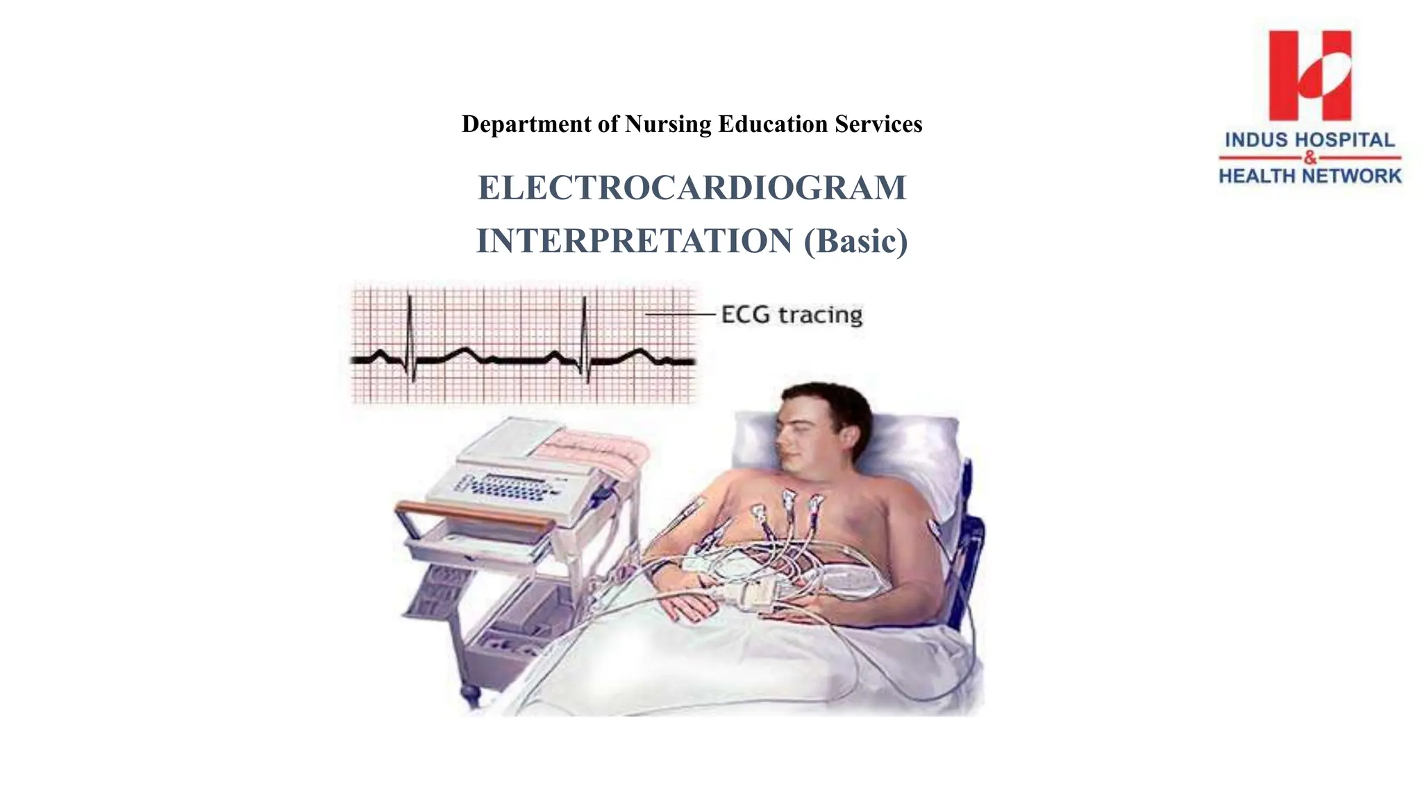
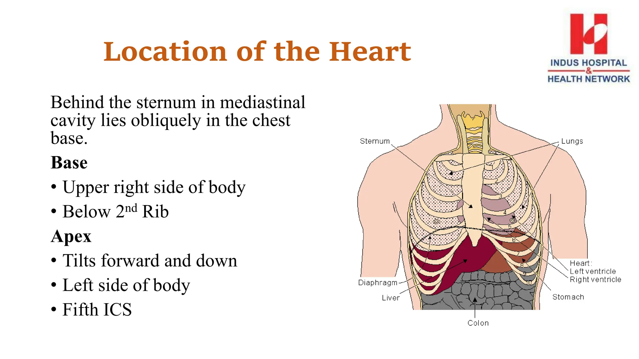
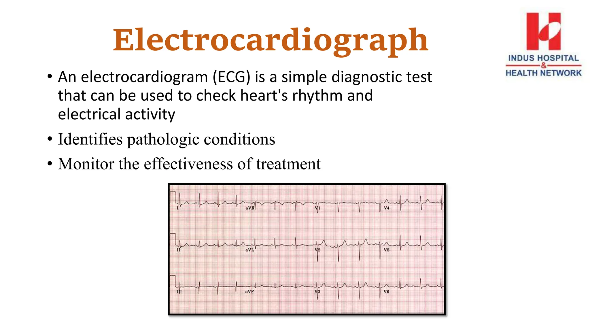
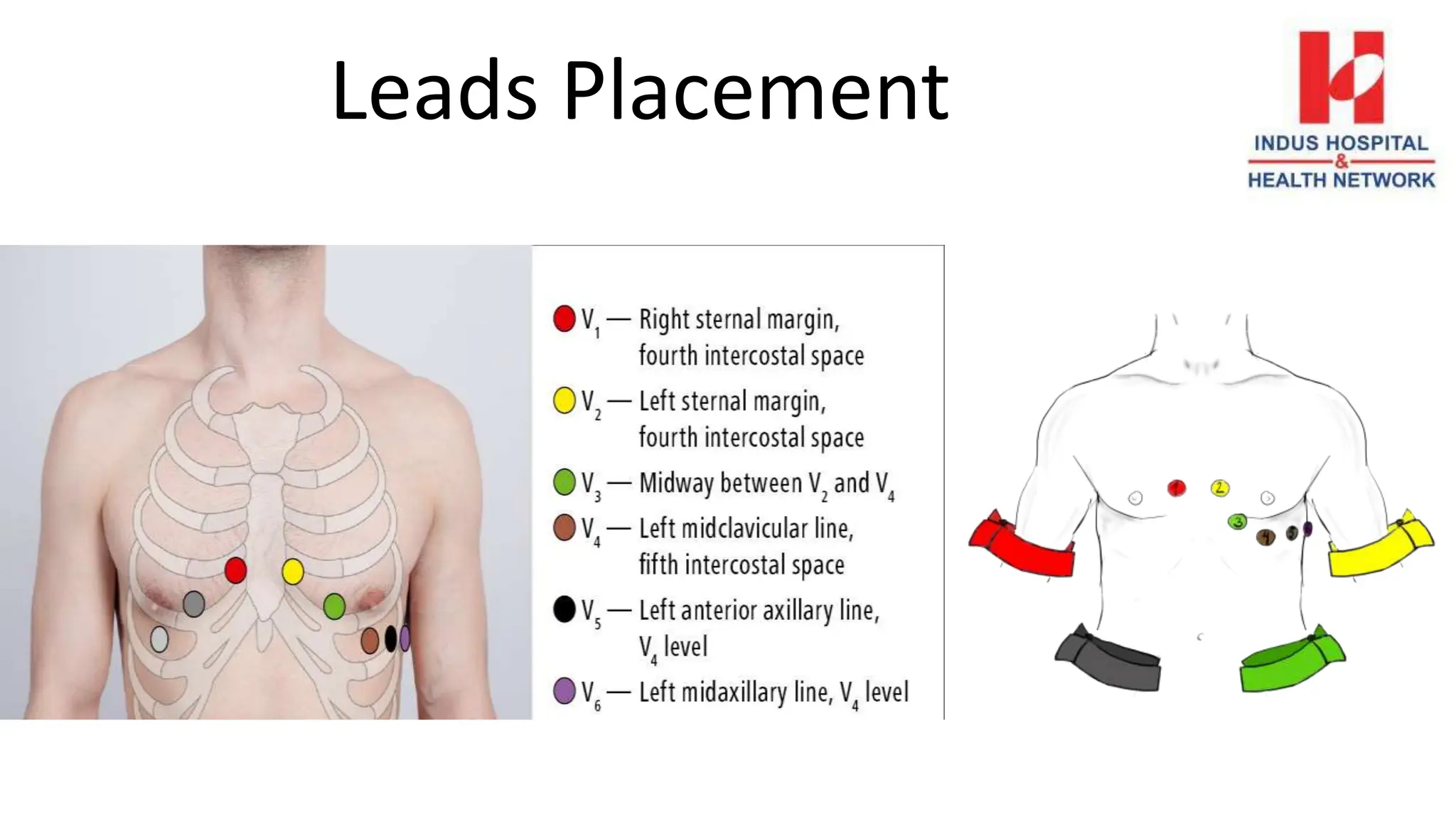
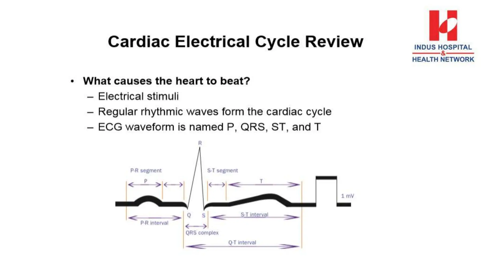
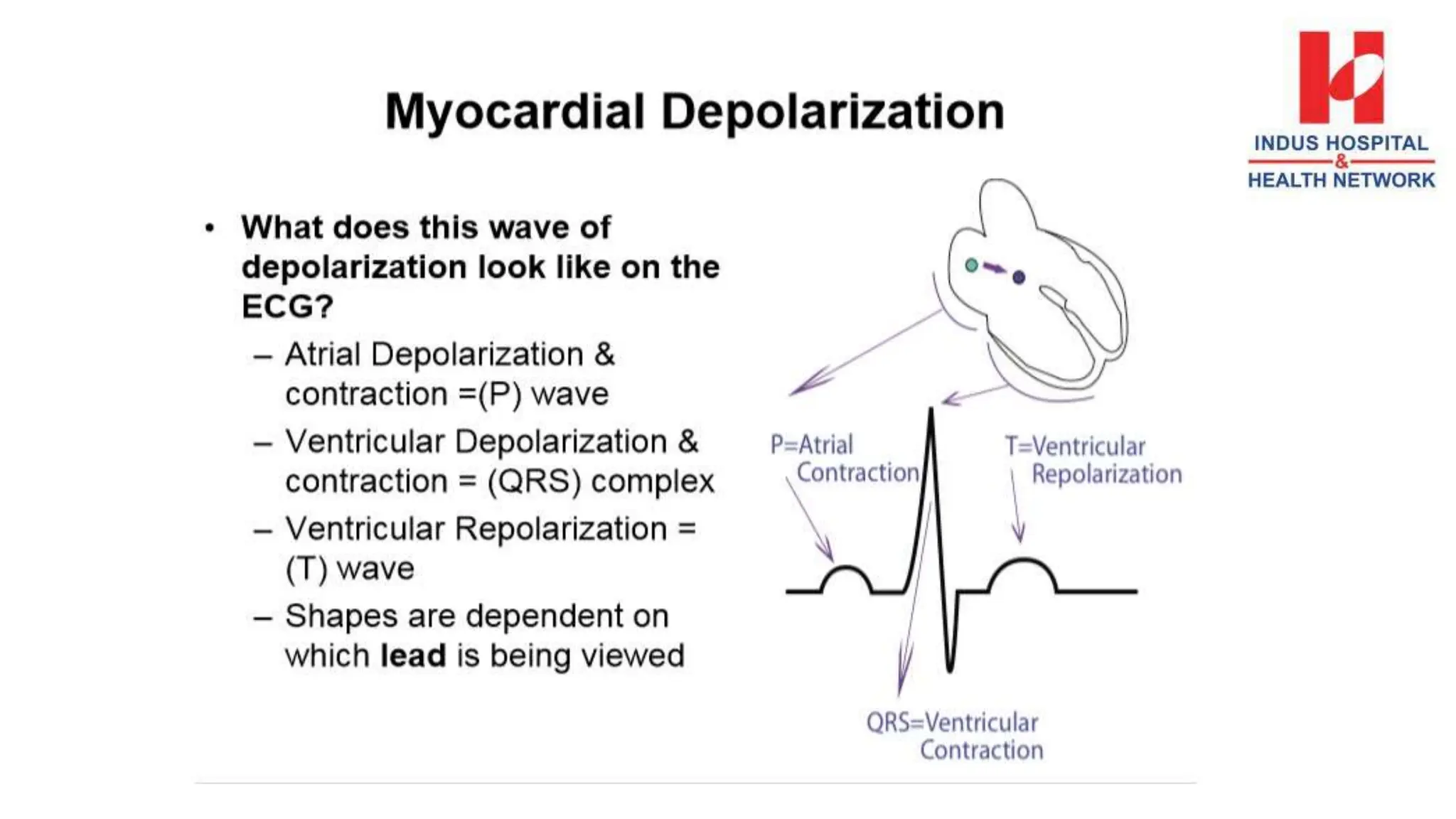
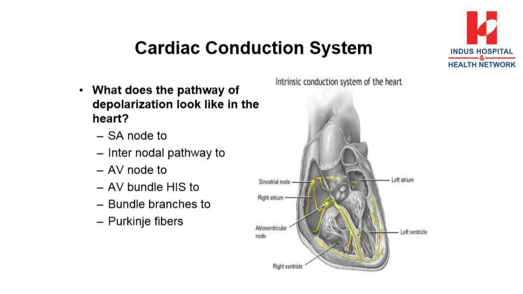
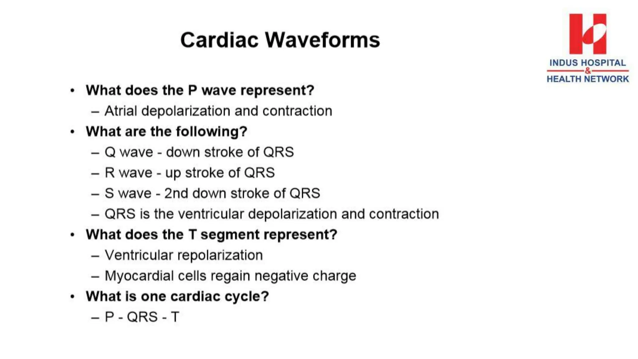
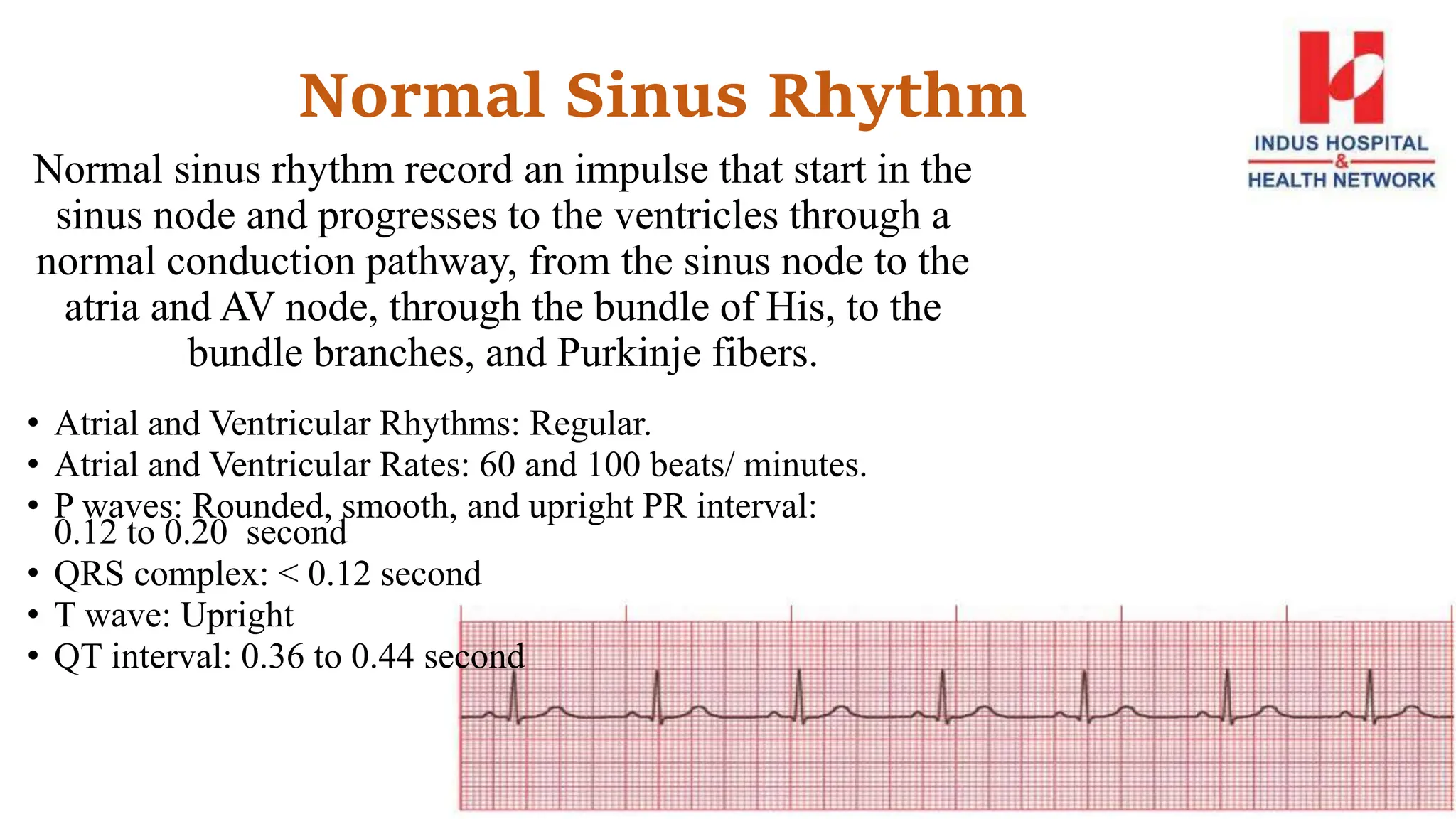
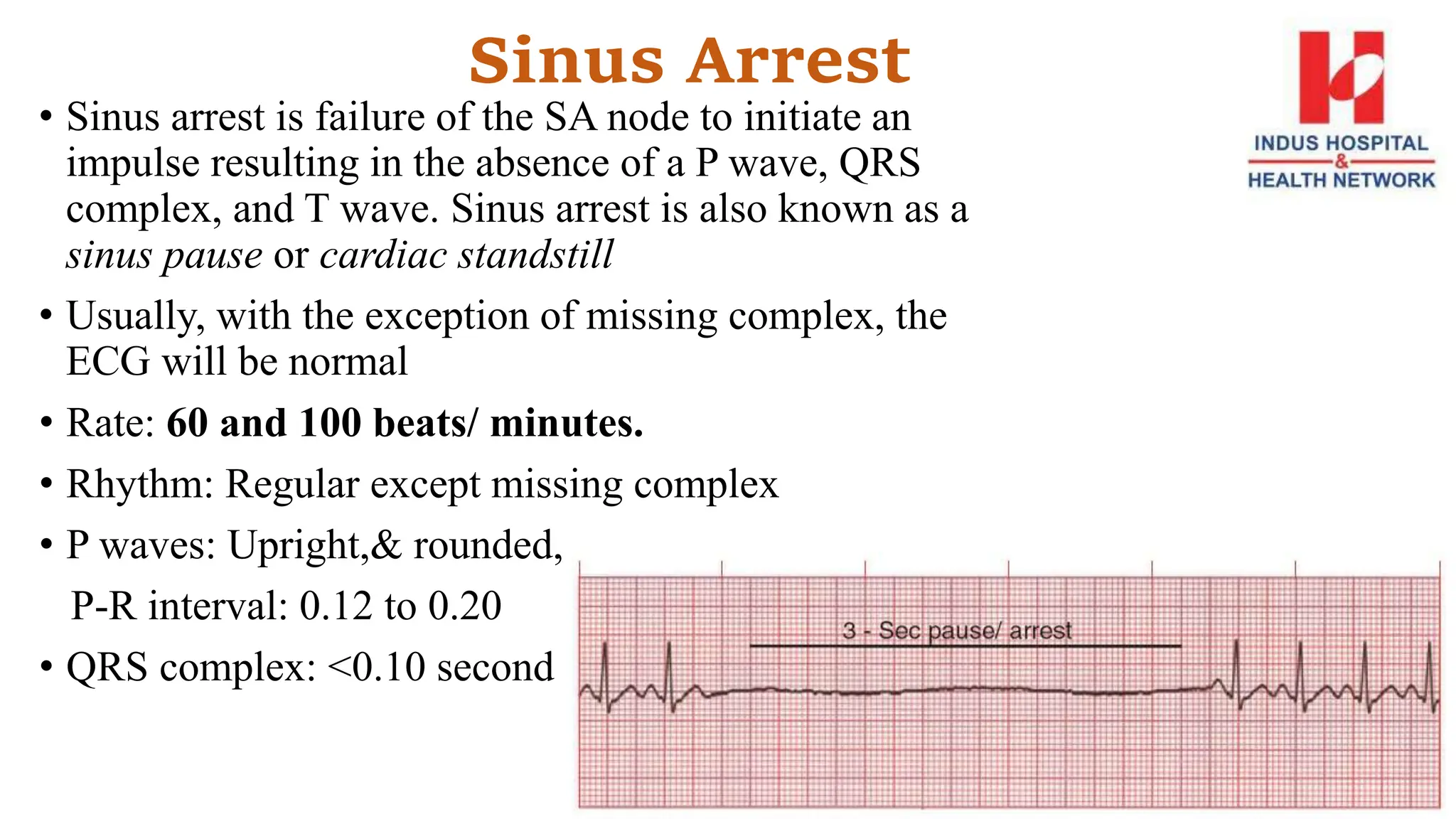
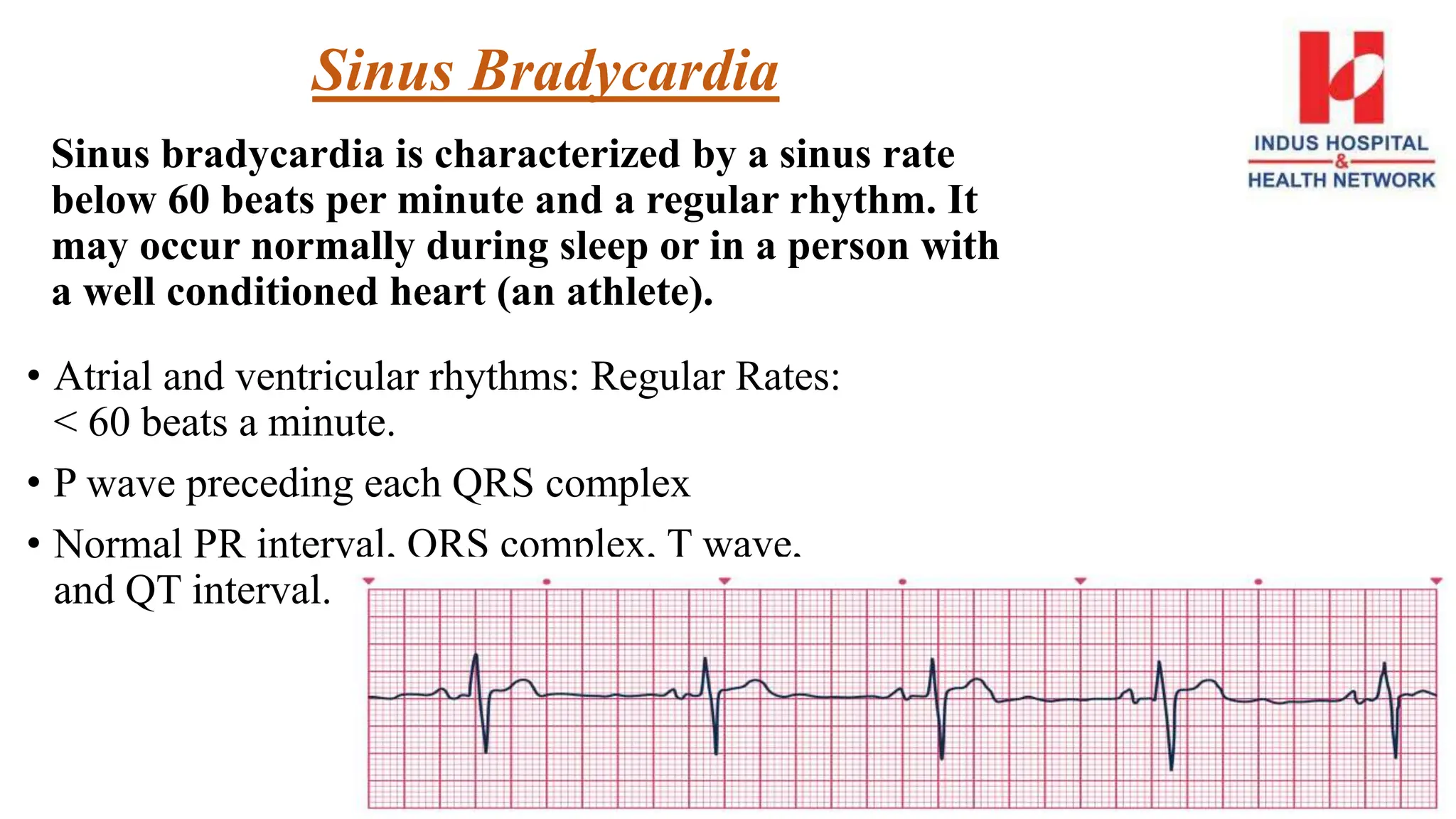
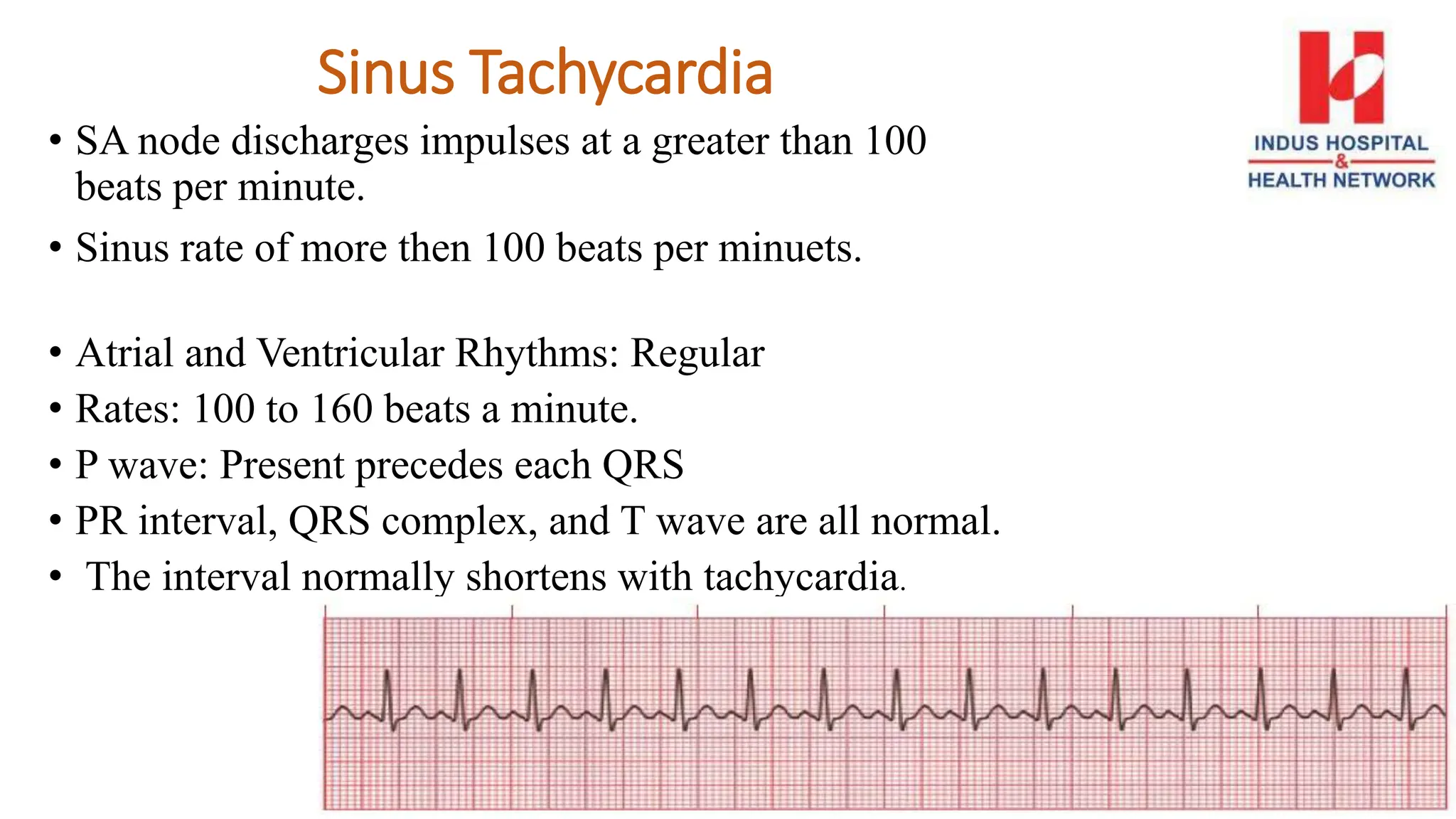
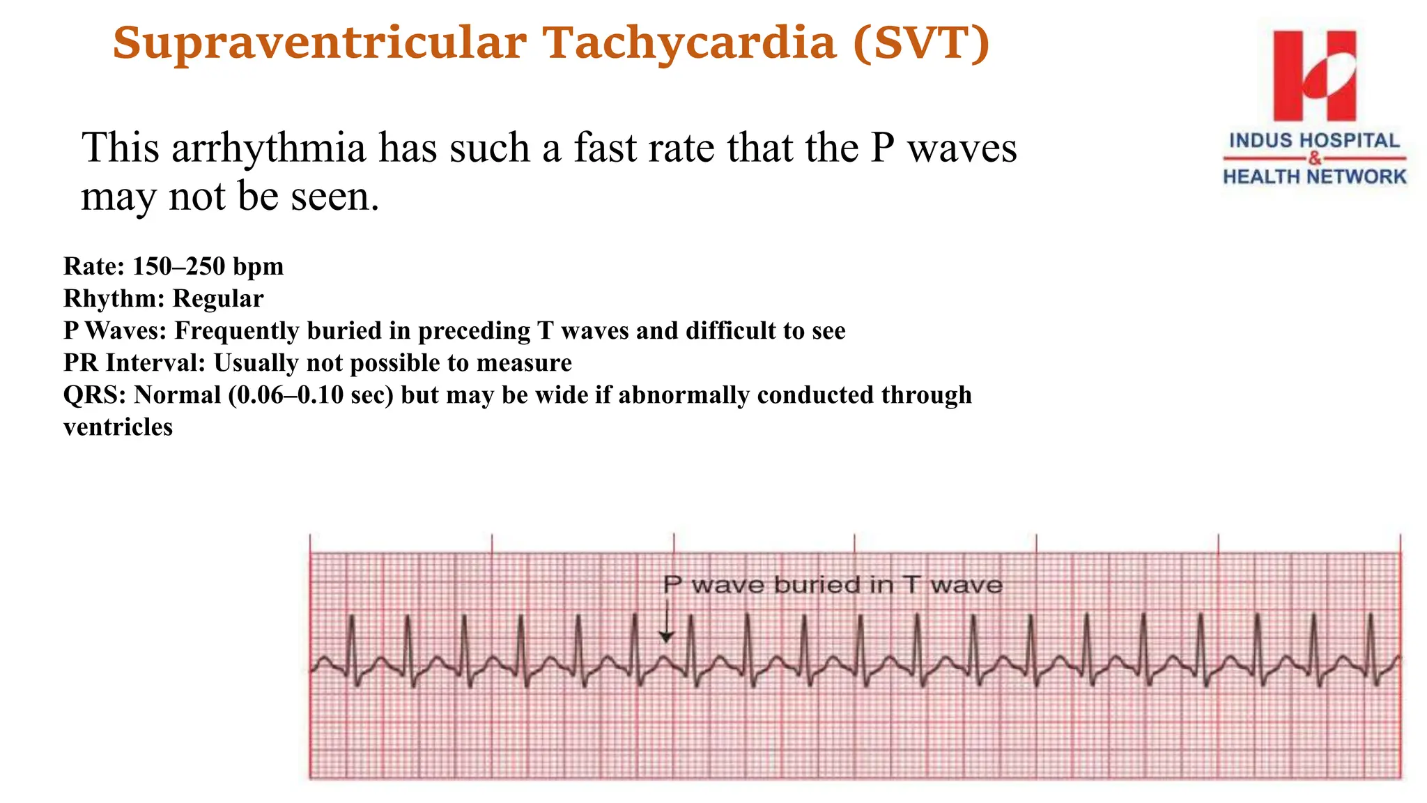
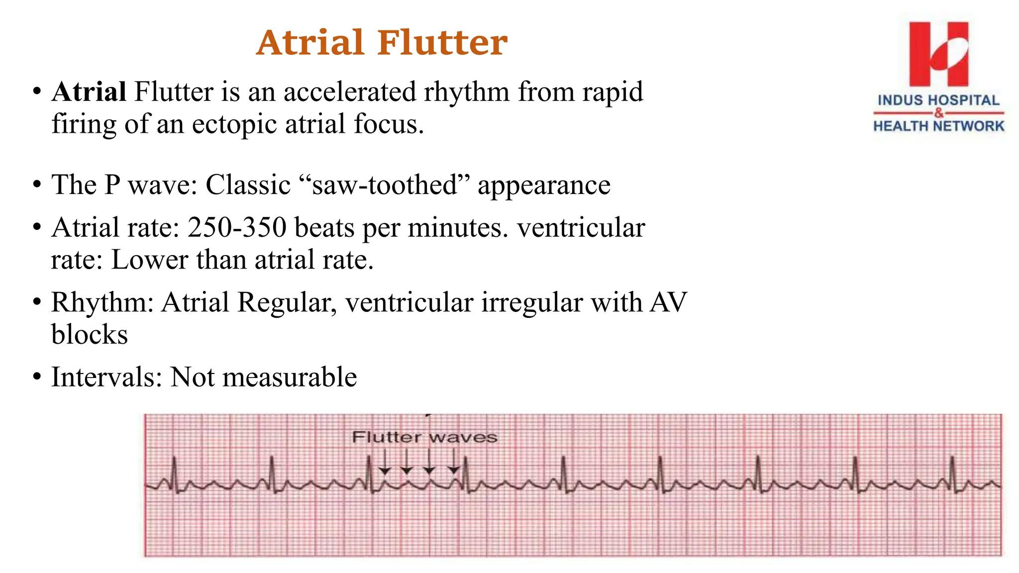
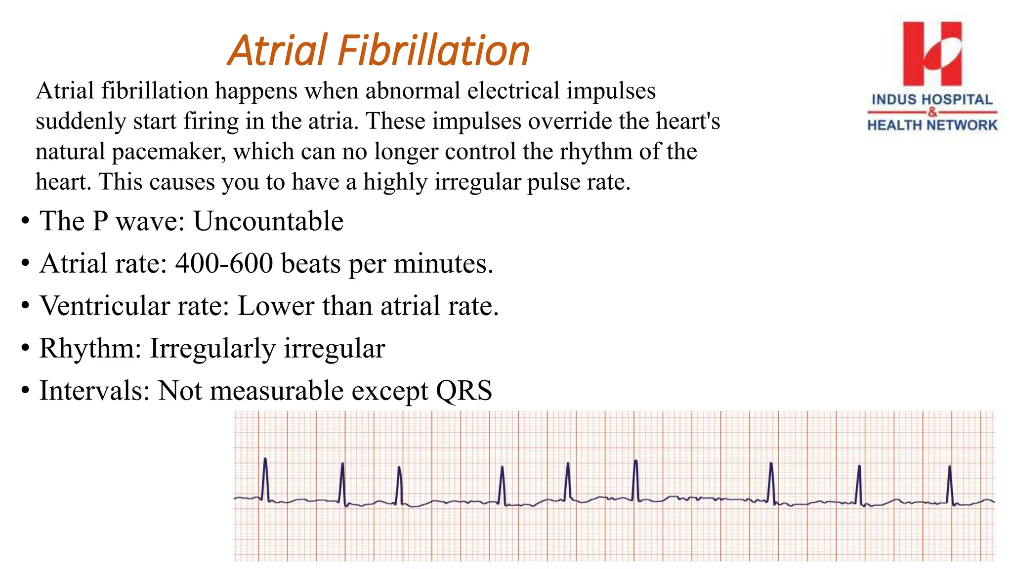
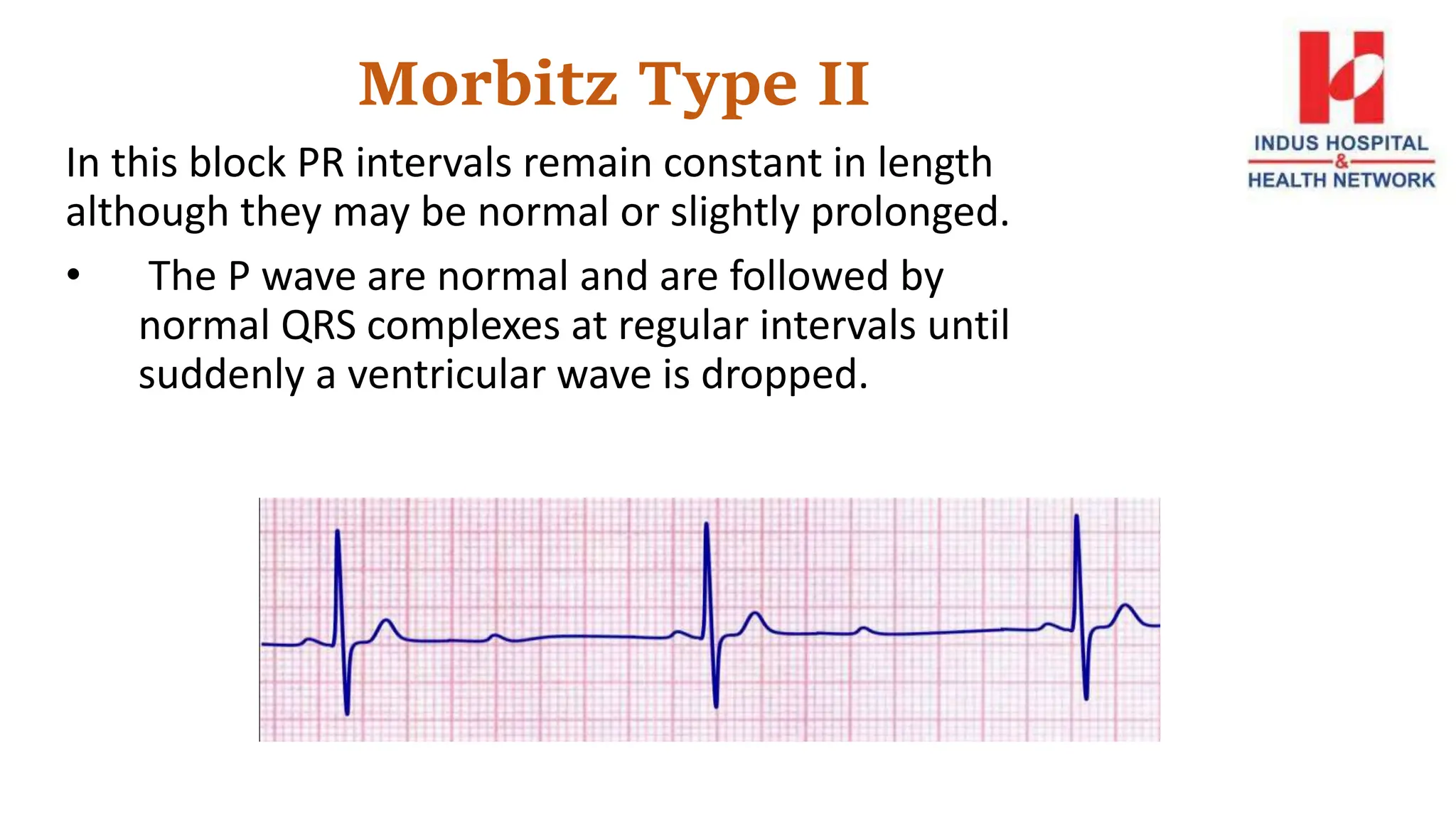
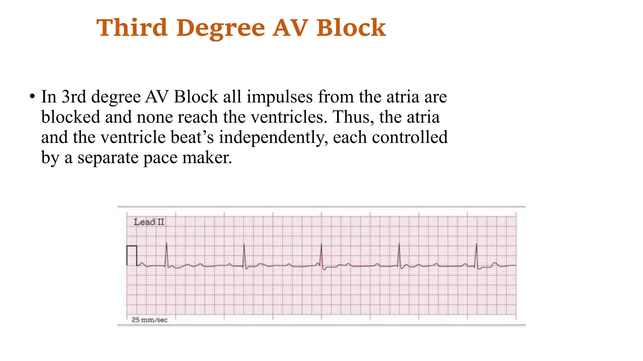
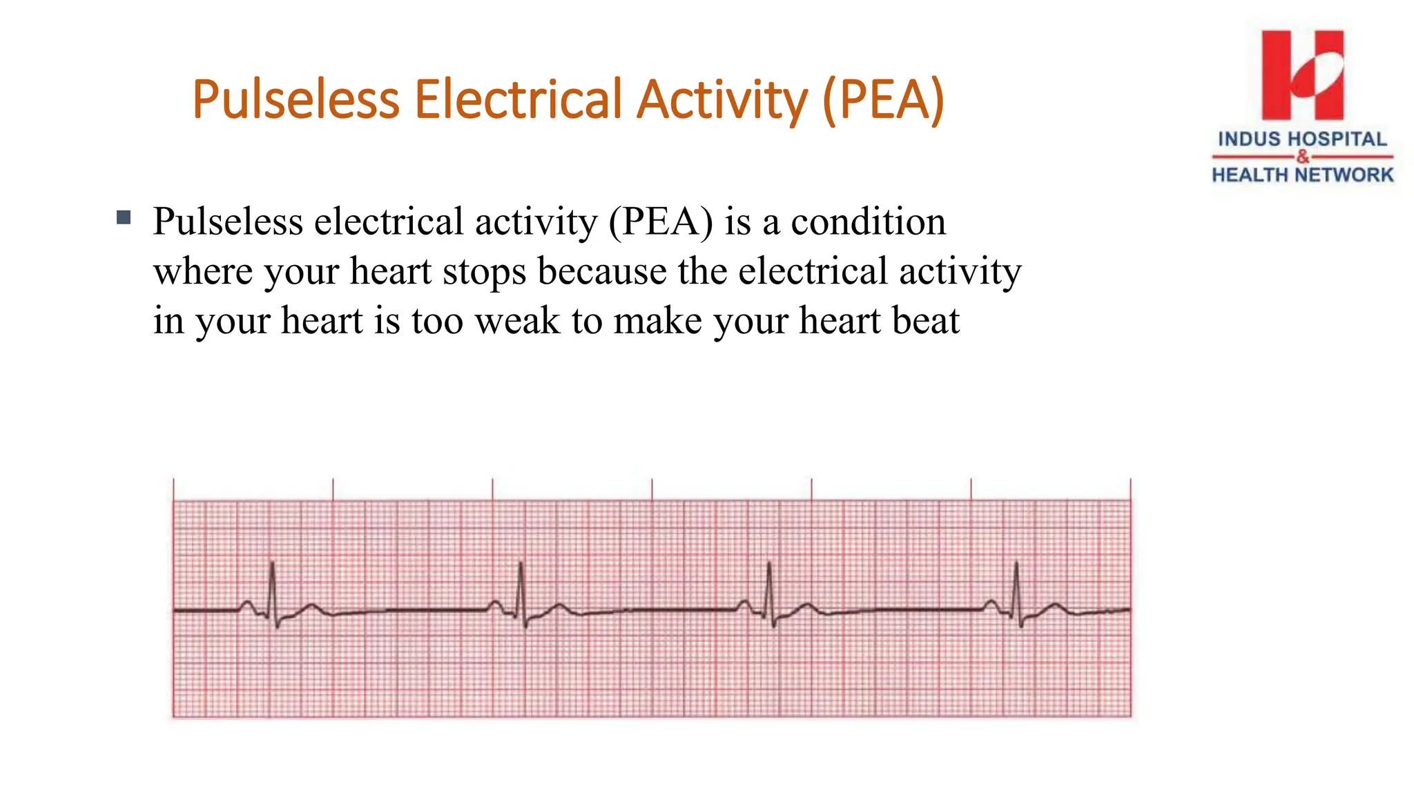
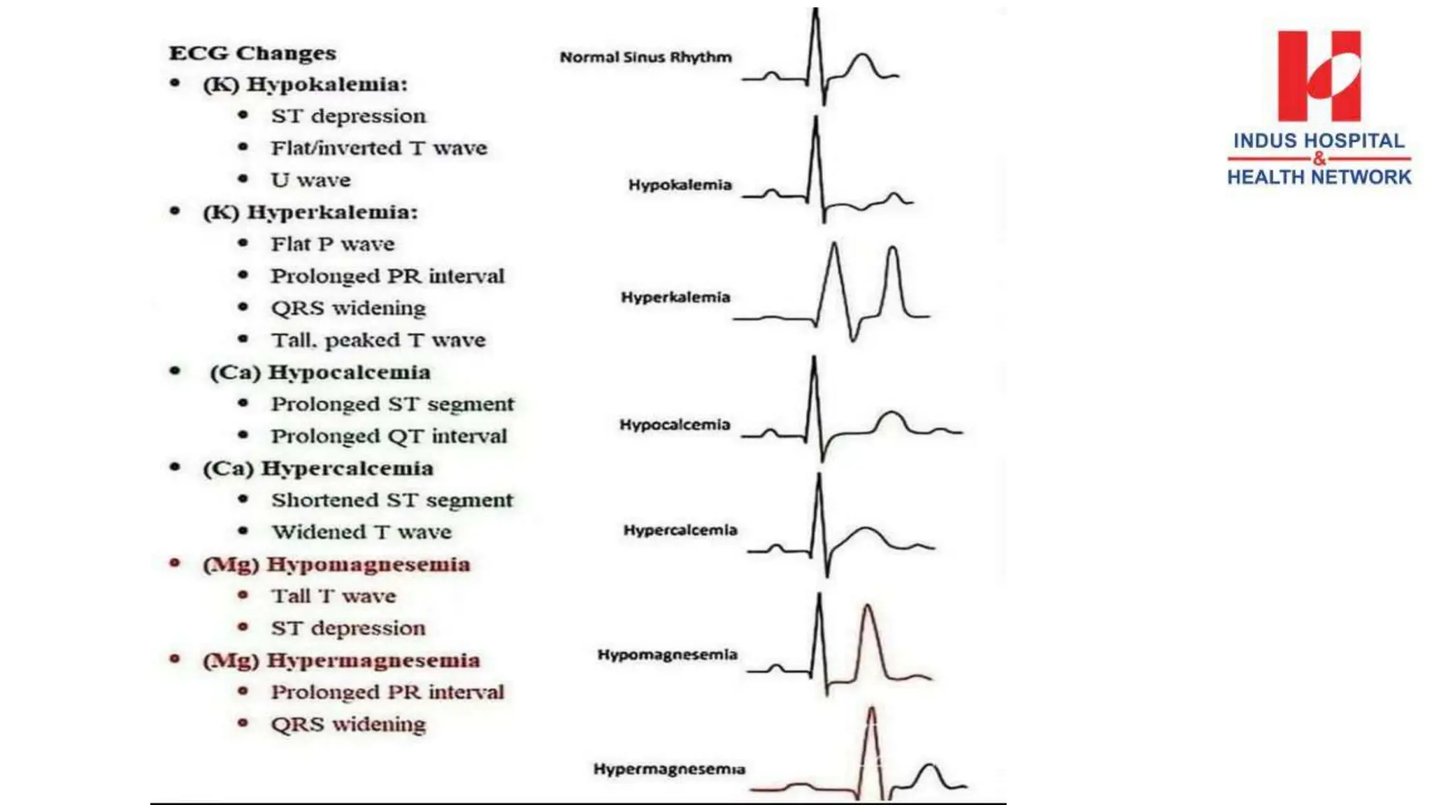
Department of Nursing Education Services
ELECTROCARDIOGRAM
INTERPRETATION (Basic)
Objectives
At the end of the session, participants will be able to:
• Discuss location of heart
• Describe normal pattern of ECG.
• List or trace different types of atrial & ventricular
arrhythmias.
• Discuss atrial & ventricular arrhythmias
characteristics.
Location of the Heart
Behind the sternum in mediastinal
cavity lies obliquely in the chest
base.
Base
• Upper right side of body
• Below 2nd Rib
Apex
• Tilts forward and down
• Left side of body
• Fifth ICS
Electrocardiograph
• An electrocardiogram (ECG) is a simple diagnostic test
that can be used to check heart's rhythm and
electrical activity
• Identifies pathologic conditions
• Monitor the effectiveness of treatment
Leads Placement
ECG Paper
Normal Sinus Rhythm
Normal sinus rhythm record an impulse that start in the
sinus node and progresses to the ventricles through a
normal conduction pathway, from the sinus node to the
atria and AV node, through the bundle of His, to the
bundle branches, and Purkinje fibers.
• Atrial and Ventricular Rhythms: Regular.
• Atrial and Ventricular Rates: 60 and 100 beats/ minutes.
• P waves: Rounded, smooth, and upright PR interval:
0.12 to 0.20 second
• QRS complex: < 0.12 second
• T wave: Upright
• QT interval: 0.36 to 0.44 second
Sinus Arrest
• Sinus arrest is failure of the SA node to initiate an
impulse resulting in the absence of a P wave, QRS
complex, and T wave. Sinus arrest is also known as a
sinus pause or cardiac standstill
• Usually, with the exception of missing complex, the
ECG will be normal
• Rate: 60 and 100 beats/ minutes.
• Rhythm: Regular except missing complex
• P waves: Upright,& rounded,
P-R interval: 0.12 to 0.20
• QRS complex: <0.10 second
Sinus Bradycardia
Sinus bradycardia is characterized by a sinus rate
below 60 beats per minute and a regular rhythm. It
may occur normally during sleep or in a person with
a well conditioned heart (an athlete).
• Atrial and ventricular rhythms: Regular Rates:
< 60 beats a minute.
• P wave preceding each QRS complex
• Normal PR interval, QRS complex, T wave,
and QT interval.
Sinus Tachycardia
• SA node discharges impulses at a greater than 100
beats per minute.
• Sinus rate of more then 100 beats per minuets.
• Atrial and Ventricular Rhythms: Regular
• Rates: 100 to 160 beats a minute.
• P wave: Present precedes each QRS
• PR interval, QRS complex, and T wave are all normal.
• The interval normally shortens with tachycardia.
Supraventricular Tachycardia (SVT)
This arrhythmia has such a fast rate that the P waves
may not be seen.
Rate: 150–250 bpm
Rhythm: Regular
P Waves: Frequently buried in preceding T waves and difficult to see
PR Interval: Usually not possible to measure
QRS: Normal (0.06–0.10 sec) but may be wide if abnormally conducted through
ventricles
Atrial Flutter
• Atrial Flutter is an accelerated rhythm from rapid
firing of an ectopic atrial focus.
• The P wave: Classic “saw-toothed” appearance
• Atrial rate: 250-350 beats per minutes. ventricular
rate: Lower than atrial rate.
• Rhythm: Atrial Regular, ventricular irregular with AV
blocks
• Intervals: Not measurable
Atrial fibrillation happens when abnormal electrical impulses
suddenly start firing in the atria. These impulses override the heart's
natural pacemaker, which can no longer control the rhythm of the
heart. This causes you to have a highly irregular pulse rate.
Atrial Fibrillation
• The P wave: Uncountable
• Atrial rate: 400-600 beats per minutes.
• Ventricular rate: Lower than atrial rate.
• Rhythm: Irregularly irregular
• Intervals: Not measurable except QRS
Ventricular Tachycardia (V-Tach)
• Ventricular tachycardia (VT or V-tach) is a type of abnormal heart
rhythm, or arrhythmia. It occurs when the lower chamber of the
heart beats too fast to pump well and the body doesn't receive
enough oxygenated blood.
• P waves: Absent
• QRS complex: Wide and bizarre.
• Ventricular Rate: 100-250 beats per minutes
• Rhythm: Regular
• PR Intervals: Not measurable
• QRS complex: Wide & bizarre
Ventricular Fibrillation
• This arrhythmia is characterized by rapid erratic
impulse formation and conduction.
• The ECG tracing display bizarre fibrillatory wave
pattern and its’ impossible to identify P wave, QRS
complex and T wave.
• P waves: Can not determine
• QRS complex: Duration can not determine
• Rate: Can not determine
• Rhythm: Can not determine
• Intervals: Not measurable
First Degree AV Block
This disturbance occurs when conduction in the AV
node slows so that the PR interval is longer than .20
seconds.
Second Degree AV Block
Morbitz Type I
This is composed of recurrent cycle in which the PR
interval become progressive prolong until eventually no
QRS complex follows the P wave.
Morbitz Type II
In this block PR intervals remain constant in length
although they may be normal or slightly prolonged.
• The P wave are normal and are followed by
normal QRS complexes at regular intervals until
suddenly a ventricular wave is dropped.
Third Degree AV Block
• In 3rd degree AV Block all impulses from the atria are
blocked and none reach the ventricles. Thus, the atria
and the ventricle beat’s independently, each controlled
by a separate pace maker.
Pulseless Electrical Activity (PEA)
 Pulseless electrical activity (PEA) is a condition
where your heart stops because the electrical activity
in your heart is too weak to make your heart beat
Asystole
Asystole is when your heart's electrical system fails, causing
your heart to stop pumping.
Basics of ECG.pptx
Basics of ECG.pptx

Basics of ECG.pptx

  • 1.
    Department of NursingEducation Services ELECTROCARDIOGRAM INTERPRETATION (Basic)
  • 2.
    Objectives At the endof the session, participants will be able to: • Discuss location of heart • Describe normal pattern of ECG. • List or trace different types of atrial & ventricular arrhythmias. • Discuss atrial & ventricular arrhythmias characteristics.
  • 3.
    Location of theHeart Behind the sternum in mediastinal cavity lies obliquely in the chest base. Base • Upper right side of body • Below 2nd Rib Apex • Tilts forward and down • Left side of body • Fifth ICS
  • 4.
    Electrocardiograph • An electrocardiogram(ECG) is a simple diagnostic test that can be used to check heart's rhythm and electrical activity • Identifies pathologic conditions • Monitor the effectiveness of treatment
  • 5.
  • 10.
  • 11.
    Normal Sinus Rhythm Normalsinus rhythm record an impulse that start in the sinus node and progresses to the ventricles through a normal conduction pathway, from the sinus node to the atria and AV node, through the bundle of His, to the bundle branches, and Purkinje fibers. • Atrial and Ventricular Rhythms: Regular. • Atrial and Ventricular Rates: 60 and 100 beats/ minutes. • P waves: Rounded, smooth, and upright PR interval: 0.12 to 0.20 second • QRS complex: < 0.12 second • T wave: Upright • QT interval: 0.36 to 0.44 second
  • 12.
    Sinus Arrest • Sinusarrest is failure of the SA node to initiate an impulse resulting in the absence of a P wave, QRS complex, and T wave. Sinus arrest is also known as a sinus pause or cardiac standstill • Usually, with the exception of missing complex, the ECG will be normal • Rate: 60 and 100 beats/ minutes. • Rhythm: Regular except missing complex • P waves: Upright,& rounded, P-R interval: 0.12 to 0.20 • QRS complex: <0.10 second
  • 13.
    Sinus Bradycardia Sinus bradycardiais characterized by a sinus rate below 60 beats per minute and a regular rhythm. It may occur normally during sleep or in a person with a well conditioned heart (an athlete). • Atrial and ventricular rhythms: Regular Rates: < 60 beats a minute. • P wave preceding each QRS complex • Normal PR interval, QRS complex, T wave, and QT interval.
  • 14.
    Sinus Tachycardia • SAnode discharges impulses at a greater than 100 beats per minute. • Sinus rate of more then 100 beats per minuets. • Atrial and Ventricular Rhythms: Regular • Rates: 100 to 160 beats a minute. • P wave: Present precedes each QRS • PR interval, QRS complex, and T wave are all normal. • The interval normally shortens with tachycardia.
  • 15.
    Supraventricular Tachycardia (SVT) Thisarrhythmia has such a fast rate that the P waves may not be seen. Rate: 150–250 bpm Rhythm: Regular P Waves: Frequently buried in preceding T waves and difficult to see PR Interval: Usually not possible to measure QRS: Normal (0.06–0.10 sec) but may be wide if abnormally conducted through ventricles
  • 16.
    Atrial Flutter • AtrialFlutter is an accelerated rhythm from rapid firing of an ectopic atrial focus. • The P wave: Classic “saw-toothed” appearance • Atrial rate: 250-350 beats per minutes. ventricular rate: Lower than atrial rate. • Rhythm: Atrial Regular, ventricular irregular with AV blocks • Intervals: Not measurable
  • 17.
    Atrial fibrillation happenswhen abnormal electrical impulses suddenly start firing in the atria. These impulses override the heart's natural pacemaker, which can no longer control the rhythm of the heart. This causes you to have a highly irregular pulse rate. Atrial Fibrillation • The P wave: Uncountable • Atrial rate: 400-600 beats per minutes. • Ventricular rate: Lower than atrial rate. • Rhythm: Irregularly irregular • Intervals: Not measurable except QRS
  • 18.
    Ventricular Tachycardia (V-Tach) •Ventricular tachycardia (VT or V-tach) is a type of abnormal heart rhythm, or arrhythmia. It occurs when the lower chamber of the heart beats too fast to pump well and the body doesn't receive enough oxygenated blood. • P waves: Absent • QRS complex: Wide and bizarre. • Ventricular Rate: 100-250 beats per minutes • Rhythm: Regular • PR Intervals: Not measurable • QRS complex: Wide & bizarre
  • 19.
    Ventricular Fibrillation • Thisarrhythmia is characterized by rapid erratic impulse formation and conduction. • The ECG tracing display bizarre fibrillatory wave pattern and its’ impossible to identify P wave, QRS complex and T wave. • P waves: Can not determine • QRS complex: Duration can not determine • Rate: Can not determine • Rhythm: Can not determine • Intervals: Not measurable
  • 20.
    First Degree AVBlock This disturbance occurs when conduction in the AV node slows so that the PR interval is longer than .20 seconds.
  • 21.
    Second Degree AVBlock Morbitz Type I This is composed of recurrent cycle in which the PR interval become progressive prolong until eventually no QRS complex follows the P wave.
  • 22.
    Morbitz Type II Inthis block PR intervals remain constant in length although they may be normal or slightly prolonged. • The P wave are normal and are followed by normal QRS complexes at regular intervals until suddenly a ventricular wave is dropped.
  • 23.
    Third Degree AVBlock • In 3rd degree AV Block all impulses from the atria are blocked and none reach the ventricles. Thus, the atria and the ventricle beat’s independently, each controlled by a separate pace maker.
  • 24.
    Pulseless Electrical Activity(PEA)  Pulseless electrical activity (PEA) is a condition where your heart stops because the electrical activity in your heart is too weak to make your heart beat
  • 25.
    Asystole Asystole is whenyour heart's electrical system fails, causing your heart to stop pumping.

Editor's Notes