Downloaded 490 times

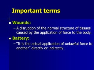
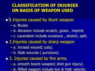
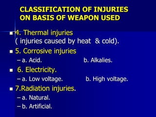
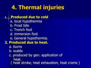
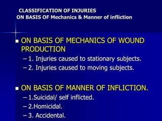
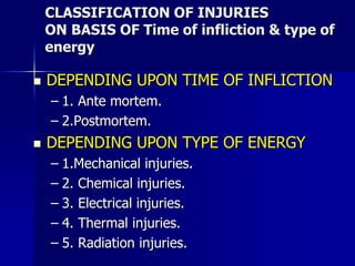

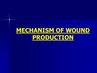

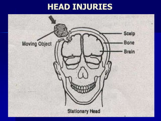
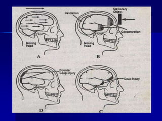
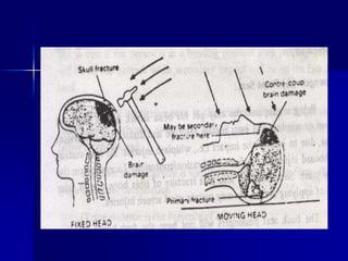
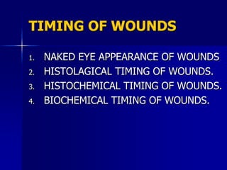


The document discusses mechanical injuries and traumatology. It defines important terms like trauma, injury, wounds, and classifications of injuries based on the weapon used, mechanics of infliction, and time of infliction. It also covers the mechanism of wound production, factors affecting wound appearance, and methods of determining the timing of wounds including naked eye appearance, histological timing by examining wound healing stages, histochemical timing by studying enzyme activity, and biochemical timing by measuring substances like histamine and serotonin.


![wound healing [Autosaved].pptx](https://cdn.slidesharecdn.com/ss_thumbnails/woundhealingautosaved-231005112651-37261d70-thumbnail.jpg?width=640&height=640&fit=bounds)


