This document describes several techniques used to study the brain:
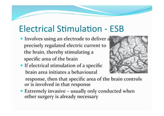
1. Electrical stimulation involves using electrodes to deliver electric currents to specific brain areas to stimulate them and observe behavioral responses, helping identify brain regions' functions. However, it is highly invasive.
2. Transcranial magnetic stimulation uses magnetic pulses through the skull to stimulate neurons near the surface, allowing non-invasive study of brain region functions. Long term effects are unclear.
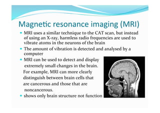
3. Imaging techniques like CT scans, MRI, fMRI, PET scans provide information about brain structure and activity with varying levels of detail and invasiveness.