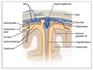
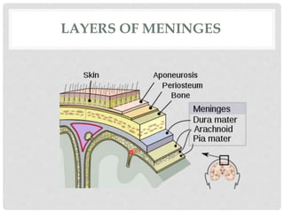
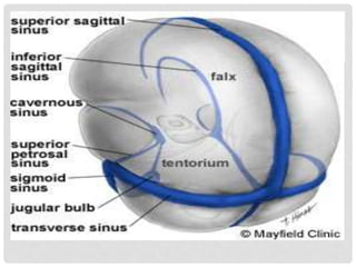
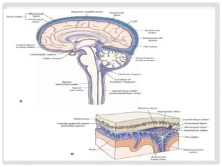
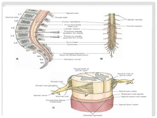
P R E S E N T E D B Y - S H R U T I R I C H A
A S S I S T A N T P R O F E S S O R
A . I . P . R .
MENINGES
CNS
• The Central Nervous System (CNS) is the part of the nervous
system consisting of the center most region of nervous system the
body.
• The central nervous system is so named because it integrates
information it receives from, and coordinates and influences the
activity of, all parts of the bodies.
• The central nervous system consists of the two major structures: the
brain and spinal cord.
STRUCTURE
• The brain is encased in the skull, and protected by the cranium & the
spinal cord is in continuous with the brain and lies caudally to the
brain, and is protected by the vertebrae.
• The spinal cord reaches from the base of the skull, continues
through or starting below the foramen magnum, and terminates
roughly level with the first or second lumbar vertebra, occupying the
upper sections of the vertebral canal.
MENINGES
• It is a Greek word, meaning ‘membrane’.
• It is the plural form of meninx.
• These meninges consist of fibroblasts and collagen fibrils.
• Basically, meninges are the three membranes that envelop the brain
and spinal cord.
• Thus, the brain and spinal cord are covered and protected by three
layers of tissue called meninges.
Dura mater
Meninges Arachanoid mater
Pia mater
• But there are several differences in the meninges covering the brain
and spinal cord.
• Like- the amount of collagen varies in different meningeal layers.
For example, the dura mater contains copious amounts of collagen
fibrils, whereas the arachnoid mater has no collagen.
• From the outermost layer of meninges, from inward they are: the
dura mater, arachnoid mater, and pia mater.
Function- Its function is to protect the central nervous system.
LAYERS OF MENINGES
1. DURA MATER
• The dura mater is a Latin word meaning ‘tough mother’ or hard & and
inflexible.
• It is also called as ‘meninx fibrosa’ or ‘pachymeninx’.
• It is the outermost part, thick & durable membrane, closest to the skull
and vertebrae.
• And is made up of loosely arranged, fibro-elastic layer of cells. But its
middle region is a mostly fibrous portion.
• It is characterized by- multiple interdigitating (interlock like the
fingers) cell processes,
- no extracellular collagen, and
- significant extracellular spaces.
• It consists of two layers:
• Its two layers, the periosteal dura and meningeal dura, are fused and
separate to form venous sinuses.
Periosteal dura
Layers of dura
Meningeal dura
• The dura creates two little folds or compartments.
Falx (The falx separates the right and
left hemispheres of the brain).
Two special dural folds
Tentorium (The tentorium separates the
cerebrum from the cerebellum).
FUNCTION
• Dura mater forms several structures that separate the cranial cavity
into compartments and protect the brain from displacement.
• The falx cerebri (separates the hemispheres of the cerebrum).
• The falx cerebelli (separates the lobes of the cerebellum).
• The tentorium cerebelli (separates the cerebrum from the
cerebellum).
• The dura mater also forms several vein-like sinuses that carry blood
(which has already given its supply of oxygen and nutrients to the
brain) back to the heart.
• The epidural space is a potential space between the dura
mater and the skull. (If there is hemorrhaging in the brain,
blood may collect here. Adults are more likely than children to
bleed here as a result of closed head injury.)
• The subdural space is another potential space. It is between
the dura mater and the middle layer of the meninges, the
arachnoid mater. (When bleeding occurs in the cranium, blood
may collect here and push down on the lower layers of the
meninges. If bleeding continues, brain damage will result from
this pressure. Children are especially likely to have bleeding in
the subdural space in cases of head injury.)
2. ARACHNOID MATER
• The arachnoid mater is a middle thin, spider web-like membrane
that covers the entire brain.
• The term arachnoid refers to the spider web like appearance of the
blood vessels within the space.
• The arachnoid is made of flat elastic tissue which is impermeable to
fluid.
• It looks like a loosely fitting sac.
• In some areas, it projects into the sinuses (cavity) formed by the
dura mater.
• These projections are the arachnoid granulation/ arachnoid villi.
• The subarachanoid space lies between the arachnoid and pia
mater. It is filled with cerebrospinal fluid. All blood vessels entering
the brain, as well as cranial nerves pass through this space.
Function- They transfer cerebrospinal fluid from the ventricles
back into the bloodstream.
3. PIA MATER
• The pia mater is a Latin word meaning tender mother or soft mother.
• It is a very delicate & the innermost membrane.
• It is the meningeal envelope that firmly adheres to the surface of the
brain and spinal cord, following all of the brain's outlines (the gyri
and sulci).
• It is a very thin membrane composed of fibrous tissue covered on its
outer surface by a sheet of flat cells thought to be impermeable to
fluid.
• The pia mater is pierced by blood vessels to the brain and spinal
cord, and its capillaries nourish the brain.
• Unlike the other layers, this tissue adheres closely to the brain,
running down into the sulci and fissures of the cortex.
• It fuses with the ependyma, the membranous lining of the
ventricles to form structures called the choroid plexes which
produce cerebrospinal fluid.
LEPTOMENINGES
• The arachnoid and pia mater together are sometimes called the
leptomeninges, which means "thin meninges" (Greek: leptos - thin).
.
SPACES
• The subarachnoid space is the space between the arachnoid and the
pia mater, which is filled with cerebrospinal fluid.
• The epidural space is a potential space between the dura mater and
the skull. The subdural space is between the dura mater and the
middle layer of the meninges, the arachnoid mater. The dura mater is
attached to the skull, whereas in the spinal cord, the dura mater is
separated from the bone (vertebrae) by a space called the epidural
space, which contains fat and blood vessels. The arachnoid is
attached to the dura mater.
• There is a subpial space underneath the pia mater that separates it
from the glia limitans.
SPINAL DURA MATER
• The spinal dura mater consists of only the meningeal layer and
lacks the periosteal layer of the cranial dura.
• The spinal dura joins the meningeal layer of the cranial dura at the
margins of the foramen magnum.
• The spinal epidural space separates the spinal dura from the
periosteum of the vertebra and is filled with fatty connective tissue
and plexuses of veins.
• Caudally, the spinal dura ends at the level of the second sacral
vertebra.
• As mentioned earlier, at this level, it becomes a thin extension (the
coccygeal ligament or filum termi-nale externum) and serves to
anchor the fluid-filled spinal dural sac to the base of the vertebral
canal.
SPINAL ARACHNOID MATER
• The spinal arachnoid mater invests the spinal cord and is connected
to the dura via connective tissue trabeculae (line) (Fig. C).
• It passes through the foramen magnum to join the cranial arachnoid,
and caudally it surrounds the cauda equina.
• The cauda equina consists of a bundle of nerve roots of all the
spinal nerves caudal to the second lumbar vertebra (Fig. B).
SPINAL PIA MATER
• The spinal pia mater (Fig. C) is thicker than the cranial pia mater.
• It is a vascular membrane and projects into the ventral fissure of the
spinal cord.
• At intervals, toothed ligaments of pia tissues, called dentate
ligaments, extend from the lateral surfaces of the spinal cord; these
ligaments serve to anchor the spinal cord to the arachnoid and the
inner surface of the dura.
ANY QUERY???????????
Meninges

Meninges

  • 1.
    P R ES E N T E D B Y - S H R U T I R I C H A A S S I S T A N T P R O F E S S O R A . I . P . R . MENINGES
  • 2.
    CNS • The CentralNervous System (CNS) is the part of the nervous system consisting of the center most region of nervous system the body. • The central nervous system is so named because it integrates information it receives from, and coordinates and influences the activity of, all parts of the bodies. • The central nervous system consists of the two major structures: the brain and spinal cord.
  • 3.
    STRUCTURE • The brainis encased in the skull, and protected by the cranium & the spinal cord is in continuous with the brain and lies caudally to the brain, and is protected by the vertebrae. • The spinal cord reaches from the base of the skull, continues through or starting below the foramen magnum, and terminates roughly level with the first or second lumbar vertebra, occupying the upper sections of the vertebral canal.
  • 4.
    MENINGES • It isa Greek word, meaning ‘membrane’. • It is the plural form of meninx. • These meninges consist of fibroblasts and collagen fibrils. • Basically, meninges are the three membranes that envelop the brain and spinal cord. • Thus, the brain and spinal cord are covered and protected by three layers of tissue called meninges. Dura mater Meninges Arachanoid mater Pia mater
  • 6.
    • But thereare several differences in the meninges covering the brain and spinal cord. • Like- the amount of collagen varies in different meningeal layers. For example, the dura mater contains copious amounts of collagen fibrils, whereas the arachnoid mater has no collagen. • From the outermost layer of meninges, from inward they are: the dura mater, arachnoid mater, and pia mater. Function- Its function is to protect the central nervous system.
  • 7.
  • 8.
    1. DURA MATER •The dura mater is a Latin word meaning ‘tough mother’ or hard & and inflexible. • It is also called as ‘meninx fibrosa’ or ‘pachymeninx’. • It is the outermost part, thick & durable membrane, closest to the skull and vertebrae. • And is made up of loosely arranged, fibro-elastic layer of cells. But its middle region is a mostly fibrous portion. • It is characterized by- multiple interdigitating (interlock like the fingers) cell processes, - no extracellular collagen, and - significant extracellular spaces. • It consists of two layers:
  • 9.
    • Its twolayers, the periosteal dura and meningeal dura, are fused and separate to form venous sinuses. Periosteal dura Layers of dura Meningeal dura • The dura creates two little folds or compartments. Falx (The falx separates the right and left hemispheres of the brain). Two special dural folds Tentorium (The tentorium separates the cerebrum from the cerebellum).
  • 10.
    FUNCTION • Dura materforms several structures that separate the cranial cavity into compartments and protect the brain from displacement. • The falx cerebri (separates the hemispheres of the cerebrum). • The falx cerebelli (separates the lobes of the cerebellum). • The tentorium cerebelli (separates the cerebrum from the cerebellum). • The dura mater also forms several vein-like sinuses that carry blood (which has already given its supply of oxygen and nutrients to the brain) back to the heart.
  • 13.
    • The epiduralspace is a potential space between the dura mater and the skull. (If there is hemorrhaging in the brain, blood may collect here. Adults are more likely than children to bleed here as a result of closed head injury.) • The subdural space is another potential space. It is between the dura mater and the middle layer of the meninges, the arachnoid mater. (When bleeding occurs in the cranium, blood may collect here and push down on the lower layers of the meninges. If bleeding continues, brain damage will result from this pressure. Children are especially likely to have bleeding in the subdural space in cases of head injury.)
  • 14.
    2. ARACHNOID MATER •The arachnoid mater is a middle thin, spider web-like membrane that covers the entire brain. • The term arachnoid refers to the spider web like appearance of the blood vessels within the space. • The arachnoid is made of flat elastic tissue which is impermeable to fluid. • It looks like a loosely fitting sac. • In some areas, it projects into the sinuses (cavity) formed by the dura mater. • These projections are the arachnoid granulation/ arachnoid villi.
  • 15.
    • The subarachanoidspace lies between the arachnoid and pia mater. It is filled with cerebrospinal fluid. All blood vessels entering the brain, as well as cranial nerves pass through this space. Function- They transfer cerebrospinal fluid from the ventricles back into the bloodstream.
  • 16.
    3. PIA MATER •The pia mater is a Latin word meaning tender mother or soft mother. • It is a very delicate & the innermost membrane. • It is the meningeal envelope that firmly adheres to the surface of the brain and spinal cord, following all of the brain's outlines (the gyri and sulci). • It is a very thin membrane composed of fibrous tissue covered on its outer surface by a sheet of flat cells thought to be impermeable to fluid. • The pia mater is pierced by blood vessels to the brain and spinal cord, and its capillaries nourish the brain.
  • 17.
    • Unlike theother layers, this tissue adheres closely to the brain, running down into the sulci and fissures of the cortex. • It fuses with the ependyma, the membranous lining of the ventricles to form structures called the choroid plexes which produce cerebrospinal fluid.
  • 18.
    LEPTOMENINGES • The arachnoidand pia mater together are sometimes called the leptomeninges, which means "thin meninges" (Greek: leptos - thin). .
  • 19.
    SPACES • The subarachnoidspace is the space between the arachnoid and the pia mater, which is filled with cerebrospinal fluid. • The epidural space is a potential space between the dura mater and the skull. The subdural space is between the dura mater and the middle layer of the meninges, the arachnoid mater. The dura mater is attached to the skull, whereas in the spinal cord, the dura mater is separated from the bone (vertebrae) by a space called the epidural space, which contains fat and blood vessels. The arachnoid is attached to the dura mater. • There is a subpial space underneath the pia mater that separates it from the glia limitans.
  • 20.
    SPINAL DURA MATER •The spinal dura mater consists of only the meningeal layer and lacks the periosteal layer of the cranial dura. • The spinal dura joins the meningeal layer of the cranial dura at the margins of the foramen magnum. • The spinal epidural space separates the spinal dura from the periosteum of the vertebra and is filled with fatty connective tissue and plexuses of veins. • Caudally, the spinal dura ends at the level of the second sacral vertebra. • As mentioned earlier, at this level, it becomes a thin extension (the coccygeal ligament or filum termi-nale externum) and serves to anchor the fluid-filled spinal dural sac to the base of the vertebral canal.
  • 21.
    SPINAL ARACHNOID MATER •The spinal arachnoid mater invests the spinal cord and is connected to the dura via connective tissue trabeculae (line) (Fig. C). • It passes through the foramen magnum to join the cranial arachnoid, and caudally it surrounds the cauda equina. • The cauda equina consists of a bundle of nerve roots of all the spinal nerves caudal to the second lumbar vertebra (Fig. B).
  • 23.
    SPINAL PIA MATER •The spinal pia mater (Fig. C) is thicker than the cranial pia mater. • It is a vascular membrane and projects into the ventral fissure of the spinal cord. • At intervals, toothed ligaments of pia tissues, called dentate ligaments, extend from the lateral surfaces of the spinal cord; these ligaments serve to anchor the spinal cord to the arachnoid and the inner surface of the dura.
  • 24.