Downloaded 455 times

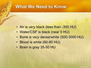
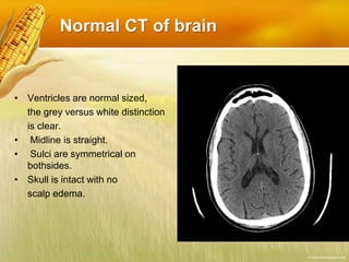
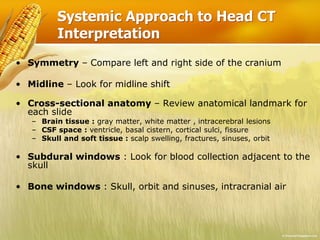
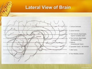
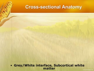
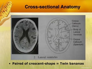
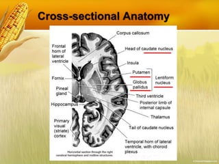
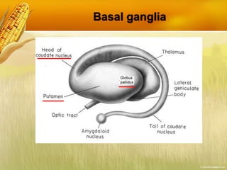
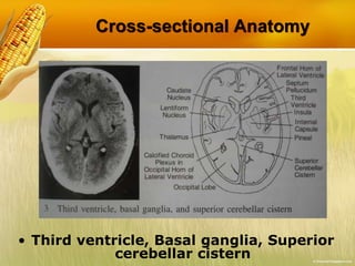
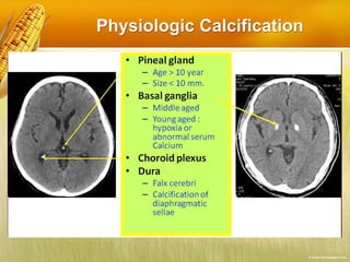
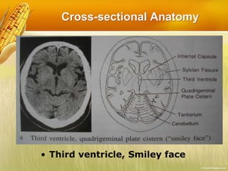
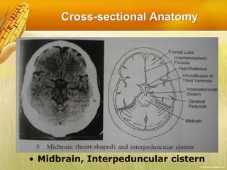

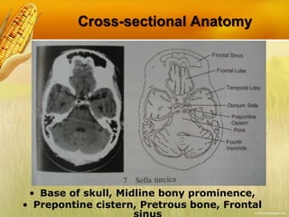
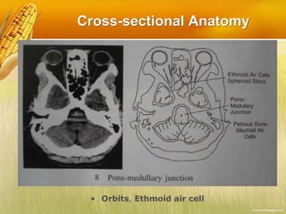
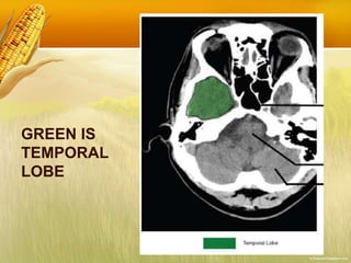
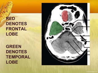
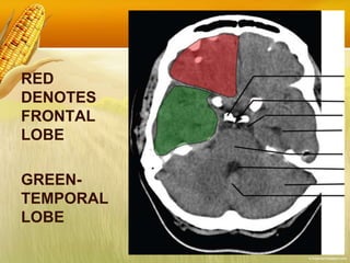
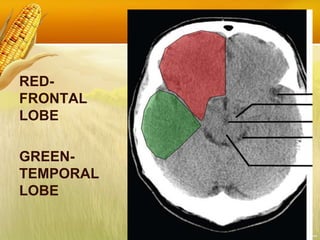
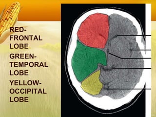
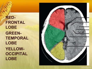
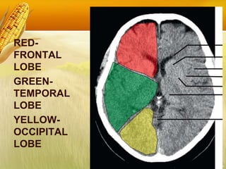
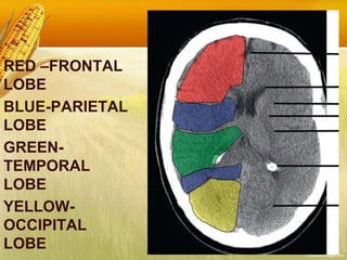
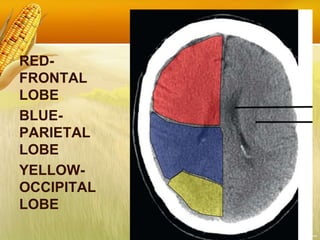
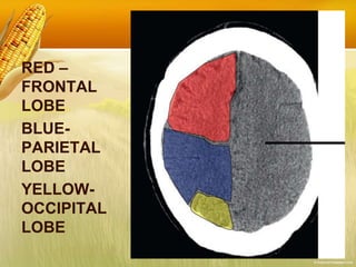
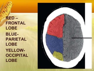


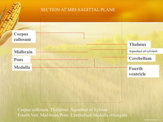


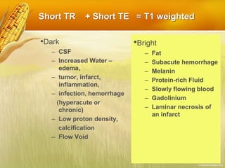
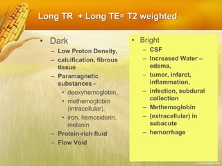


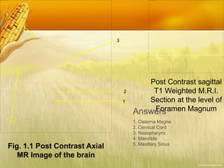


1. The document discusses the basics of neuroimaging using CT and MRI. It explains how different tissues appear on CT and MRI scans and provides examples of normal anatomy. 2. It then covers the systematic approach to interpreting head CT scans and analyzing different areas of the brain. Examples of cross-sectional anatomy at different brain levels are shown on CT scans. 3. The document also discusses the physics behind MRI and how tissues appear differently on T1-weighted, T2-weighted, and FLAIR sequences. Multiple images demonstrate normal brain anatomy on post-contrast MRI scans.