Neuroimaging involves using various imaging techniques to assess brain structure and function in order to diagnose diseases. The main techniques discussed are computed tomography (CT), which combines multiple X-rays to create 3D brain images; positron emission tomography (PET), which images brain activity by detecting radioactive tracers; and magnetic resonance imaging (MRI), which produces highly detailed structural images using magnetic fields and radio waves. Functional MRI (fMRI) is a specialized MRI technique that maps brain activity by detecting changes in oxygenated blood flow.
What is a Pet Scan : Nuclear 3-D imaging test that uses a radioactive substance called a tracer to look for disease in the body.
Shows how organs and tissues are working at a molecular and cellular level. Scan is non-invasive, but does involve exposure to ionizing radiation.
Best known for its role in detecting cancer imaging.
A small amount of a radioactive sugar molecule, 18 fluoro-2-deoxyglucose (FDG), is injected into the bloodstream (can also be inhaled as gas or swallowed in pill form).
A PET Scan is used to detect and generate images that indicate areas of high FDG uptake.
Many cancers require more energy than normal cells, and the FDG tracer accumulates in these cells.
This allows cancers to be seen on the Pet images as hot spots.
Positron emission tomography pet scan and its applicationsYashawant Yadav
Slides contains physic about the PET scan that is positron emission tomography , its principle , detector configuration types , clinical application of PET Scan and advancement with CT and MRI
This presentation discusees a brief history of the MRI, it's mechanism of action, applications in dentistry and recent advancements in its technology. Also it's advantages and disadvantages in comparison with the CT scan
Multimodality Molecular Imaging – An Overview With Special Focus on PET/CTApollo Hospitals
Imaging capabilities have evolved from those that provide anatomical pictures to those that capture functional information and, more recently, molecular information (nuclear medicine, PET, SPECT, PET/CT, SPECT/CT, MRS, contrast-enhanced ultrasound, fluorescence and bioluminescence imaging). Multimodality imaging has emerged as a technology that utilizes the strengths of different modalities and yields a hybrid imaging platform with benefits superior to those of any of its individual components, considered alone. Leading edge hybrid imaging (combining multiple, complementary imaging technologies such as PET and CT) offer unique opportunities to “view” the molecular biology of disease, and the use of this equipment is on the rise.
Brief explanation of what is PET, the main components for a PET system along with their basic functions. The principle behind PET inclusive of positron emission and emission detection. Acquisition and reconstruction of the collected data to produce the final image. Finally the pros and cons of Positron emission tomography.
https://www.snmclub.com/presentation
PET/MRI Current & Future Status
DALE BAILEY PhD , Principal Physicist
Departement of Nuclear Medicine, Royal North Shore Hospital
Professor in Medical Radiation Sciences, University of Sydney
Sydney, Australia
icrm2018
It includes history, components, principle, it's benefits and risk in very concise way and point to point information. Points are in bullet and bold form, so you can easy grab it.
International Journal of Engineering and Science Invention (IJESI)inventionjournals
International Journal of Engineering and Science Invention (IJESI) is an international journal intended for professionals and researchers in all fields of computer science and electronics. IJESI publishes research articles and reviews within the whole field Engineering Science and Technology, new teaching methods, assessment, validation and the impact of new technologies and it will continue to provide information on the latest trends and developments in this ever-expanding subject. The publications of papers are selected through double peer reviewed to ensure originality, relevance, and readability. The articles published in our journal can be accessed online.
Image Processing Technique for Brain Abnormality DetectionCSCJournals
Medical imaging is expensive and very much sophisticated because of proprietary software and expert personalities. This paper introduces an inexpensive, user friendly general-purpose image processing tool and visualization program specifically designed in MATLAB to detect much of the brain disorders as early as possible. The application provides clinical and quantitative analysis of medical images. Minute structural difference of brain gradually results in major disorders such as schizophrenia, Epilepsy, inherited speech and language disorder, Alzheimer's dementia etc. Here the main focusing is given to diagnose the disease related to the brain and its psychic nature (Alzheimer’s disease).
Classification of EEG Signals for Brain-Computer InterfaceAzoft
This e-book gives you a sneak peak into how the classification of right hand movements via EEG could contribute to the development of a brain-computer interface. The Azoft R&D department, along with Sergey Alyamkin and Expasoft provide detailed data from research done for the "Grasp-and-Lift EEG Detection" competition organized by Kaggle. You’ll learn why the deep learning algorithms can be effective in various types of signal classifications and how to apply convolutional neural networks for a specific task such as identifying hand motions from EEG recordings.
See more details on our website: http://rnd.azoft.com/classification-eeg-signals-brain-computer-interface/
Brain mapping can capture a window of brain activity. The brain is a multi-billion neuron organ. Neurons communicate with every cell in your body. It is carried by electrical impulses that form brain waves. This application helps us analyze your brainwaves and find ways to improve communication across different brain regions.
Significance of Brain imaging in Psychiatry. Most of the major Psychiatric disorders are associated with statistically significant differences on various Neuroimaging measures, when comparing groups of patients and controls.
Francesca Gottschalk - How can education support child empowerment.pptxEduSkills OECD
Francesca Gottschalk from the OECD’s Centre for Educational Research and Innovation presents at the Ask an Expert Webinar: How can education support child empowerment?
Introduction to AI for Nonprofits with Tapp NetworkTechSoup
Dive into the world of AI! Experts Jon Hill and Tareq Monaur will guide you through AI's role in enhancing nonprofit websites and basic marketing strategies, making it easy to understand and apply.
Model Attribute Check Company Auto PropertyCeline George
In Odoo, the multi-company feature allows you to manage multiple companies within a single Odoo database instance. Each company can have its own configurations while still sharing common resources such as products, customers, and suppliers.
Welcome to TechSoup New Member Orientation and Q&A (May 2024).pdfTechSoup
In this webinar you will learn how your organization can access TechSoup's wide variety of product discount and donation programs. From hardware to software, we'll give you a tour of the tools available to help your nonprofit with productivity, collaboration, financial management, donor tracking, security, and more.
Embracing GenAI - A Strategic ImperativePeter Windle
Artificial Intelligence (AI) technologies such as Generative AI, Image Generators and Large Language Models have had a dramatic impact on teaching, learning and assessment over the past 18 months. The most immediate threat AI posed was to Academic Integrity with Higher Education Institutes (HEIs) focusing their efforts on combating the use of GenAI in assessment. Guidelines were developed for staff and students, policies put in place too. Innovative educators have forged paths in the use of Generative AI for teaching, learning and assessments leading to pockets of transformation springing up across HEIs, often with little or no top-down guidance, support or direction.
This Gasta posits a strategic approach to integrating AI into HEIs to prepare staff, students and the curriculum for an evolving world and workplace. We will highlight the advantages of working with these technologies beyond the realm of teaching, learning and assessment by considering prompt engineering skills, industry impact, curriculum changes, and the need for staff upskilling. In contrast, not engaging strategically with Generative AI poses risks, including falling behind peers, missed opportunities and failing to ensure our graduates remain employable. The rapid evolution of AI technologies necessitates a proactive and strategic approach if we are to remain relevant.
How to Make a Field invisible in Odoo 17Celine George
It is possible to hide or invisible some fields in odoo. Commonly using “invisible” attribute in the field definition to invisible the fields. This slide will show how to make a field invisible in odoo 17.
Macroeconomics- Movie Location
This will be used as part of your Personal Professional Portfolio once graded.
Objective:
Prepare a presentation or a paper using research, basic comparative analysis, data organization and application of economic information. You will make an informed assessment of an economic climate outside of the United States to accomplish an entertainment industry objective.
2. NEUROIMAGING
It is the process of imaging the brains structure to
assess brain functions and diagnosis.
The study of brain is incomplete without imaging the
structure of the brain, imaging of the brain is
necessary not only for research but also for
diagnosing diseases like tumors and lesions.
Since conventional X-rays are useless in
neuroimaging as the brain consists of more or less
similar tissues performing different functions. So
better techniques are used in brain imaging.
3. TECHNIQUES USED IN BRAIN IMAGING
Following methods are useful in brain imaging.
1. CONTRAST X-RAY
2. COMPUTED TOMOGRAPHY
3. POSITRON EMISSION TOMOGRAPHY
4. M.R.I
5. fM.R.I
4. CONTRAST X-RAY
It involves injecting a substance that absorbs x-rays
more or less than the surrounding brain tissues. The
injected substance then highlights the contrast
between the compartment and the surrounding
tissue during x-ray photography. Making the injected
area appear different from the surrounding tissue.
A good example of contrast x-ray technique is the
cerebral angiography.
5. CEREBRAL ANGIOGRAPHY
This technique involves injecting a radio-opaque dye
into a cerebral artery to visualize the cerebral
circulatory system during x-ray photography.
Cerebral angiograms are most useful to study the
brain circulation and localizing vascular damage.
Also, the displacement of blood vessels from their
normal position also can indicate the location of a
tumor.
7. COMPUTED TOMOGRAPHY
It is revolutionary process founded in 1970s. It combines
multiple x-rays to create a combined image of the brain.
The machine consists of a cylindrical x-ray tube that
projects an x-ray beam through the head on an x-ray
detector mounted on the other side. The projector and
the detector automatically rotate around the head of the
patient at one level of the brain and collect several x-rays
which are combined by a computer to generate a CT
SCAN. This process is repeated for 5-7 different levels of
the patient’s brain to obtain 3-D representations of the
brain.
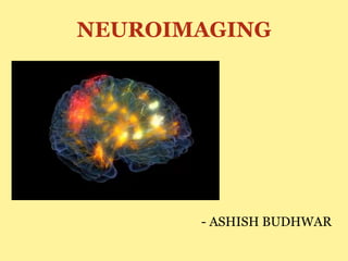
9. POSITRON EMISSION TOMOGRAPHY
It was the first brain imaging technique to provide
images of brain activity rather than brain structure.
These functional brain images are very much helpful
in the study of the brain structure and its functions.
In a commonly used method of P.E.T, radioactive
Fluorodeoxyglucose (FDG) is injected into the
patient’s carotid artery (artery of the neck that feeds
the ipsilateral cerebral hemisphere).
10. POSITRON EMISSION TOMOGRAPHY
Because of the similarity of FDG to GLUCOSE, the
primary source of energy to the brain, FDG is rapidly
taken up by active cells. But unlike Glucose, FDG
cannot be metabolized, so it accumulates in active
neurons or in associated astrocytes until it is
gradually broken down.
The scan therefore images the levels of
radioactivity(indicated by color coding) in various
parts of the brain.
11. POSITRON IMAGING TOMOGRAPHY
Therefore, if a PET scan is taken of a patient
engaging in some activity, such as reading, after FDG
is administered, the PET scan will indicate the areas
of the brain that were most active during the activity.
PET scan are not the images of the brain, it is merely
a colored map of the amount of radioactivity present
in the particular area.
PET scan is useful in identifying the distribution of
the neurotransmitters, receptors and transporters in
the brain.
13. MAGNETIC RESONANCE IMAGING
An MRI is a structural brain imaging procedure in
which high resolution images are constructed from
the measurement of radio frequency waves that
hydrogen atoms emit as they align with a powerful
magnetic field.
An MRI provide better brain structure images than
CT. Despite this, the MRI is capable of producing
images in 3 dimensions.
15. FUNCTIONAL MRI
Functional MRI is the use of MRI technology to
produce functional images of the brain. fMRI has
become the most influential tool of cognitive
neuroscience and is widely used for medical
diagnosis.
Functional MRI produces images representing the
increase in oxygen flow in the blood to active areas of
the brain. This is possible because of the two
attributes of blood in brain which are;
16. FUNCTIONAL MRI
First, the active areas of the brain take up more
oxygenated blood than they need for their energy
requirements and thus oxygenated blood
accumulates in active areas of the brain.
Second, oxygenated blood has magnetic properties
that influence the radio-frequency waves emitted by
hydrogen atoms in an MRI.
The signal recorded by the fMRI is called the BOLD
signal which is Blood Oxygen Level Dependent
signal.
18. ADVANTAGES OF fMRI
fMRI has 4 advantages over PET;
1. Nothing is injected in the volunteer/patient
2. It provides both structural and functional images of
the brain.
3. It’s spatial resolution is better than PET and CT.
4. It is capable of producing 3-D images of activity
over the entire brain.
19. DISADVANTAGES OF fMRI
The images provides by fMRI are only the images of
the BOLD signal which is very complex when related
to neural activity.
Also the fMRI has poor temporal resolution. It takes
2-3 seconds to measure the BOLD signal and many
neural responses such as action potentials, occurs in
the millisecond range.
Editor's Notes
A growing tumor can be seen colored red in the second image.