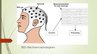
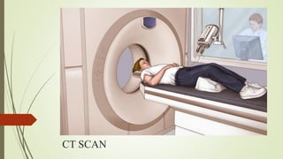
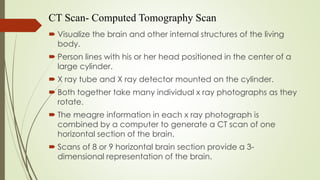
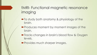
RECORDING BRAIN
ACTIVITY
- NITHIN NARAYANAN P B (21037)
RECORDING BRAIN ACTIVITY
The recording of changes in electric
potentials in various areas of the brain by
means of electrodes placed on the scalp
Function can be defined as the ability to
perform a given cognitive or physiological
task
EEG- Electroencephalogram
 An EEG is a test that detects abnormalities in your brain
waves
 Records electrical activity of the brain through electrodes
attached to the scalp.
 Electrodes measure the average activity of cells under the
electrode at any moment.
 The output is amplified and recorded.
 Records spontaneous brain activity as well as the evoked
potential or responses (activity in response to stimulus.)
CT SCAN
CT Scan- Computed Tomography Scan
 Visualize the brain and other internal structures of the living
body.
 Person lines with his or her head positioned in the center of a
large cylinder.
 X ray tube and X ray detector mounted on the cylinder.
 Both together take many individual x ray photographs as they
rotate.
 The meagre information in each x ray photograph is
combined by a computer to generate a CT scan of one
horizontal section of the brain.
 Scans of 8 or 9 horizontal brain section provide a 3-
dimensional representation of the brain.
MEG-Magnetoencephalography
MEG- Magnetoencephalography
• Measures the faint magnetic fields generated by
brain activity
• Change in magnetic fields on the surface of the
scalp that are produced by change in underlying
patterns of neural activity
MEG- Magnetoencephalography
MRI - Magnetic resonance imaging
MRIS doesn’t use x-rays instead it passes strong
magnetic field.
Magnetic field makes nuclei or some molecules to
in the body to spin.
In return to the magnetic field, the nuclei also emit
radio wares their own.
Waves packed up by the scanner and constructs a
three-dimensional image.
MRI can be used in frontal planes.
FMRI- Functional magnetic resonance imaging
fMRI- Functional magnetic resonance
imaging
To study both anatomy & physiology of the
brain.
Produces moment by moment images of the
brain.
Tracks changes in brain's blood flow & Oxygen
levels.
Provides much sharper images.
PET - Positron Emission Tomography
 Studies the functioning of brain
 Assess the metabolic activity of the brain
 Records the emission of radioactivity from injected chemical
 Injection of radioactive tracer which are taken up by active brain
areas
 Amount of tracer taken is proportional to metabolic activity of the
brain.
 Radioactive tracer emits positron (positively charged electrons)
 Positrons collide with nearby electrons.
 Collision- gamma rays are released detector pick up these rays &
construct the image
 Most radioactive area - most blood flow
THANK YOU

RECORDING BRAIN ACTIVITY.pdf

  • 1.
    RECORDING BRAIN ACTIVITY - NITHINNARAYANAN P B (21037)
  • 2.
    RECORDING BRAIN ACTIVITY Therecording of changes in electric potentials in various areas of the brain by means of electrodes placed on the scalp Function can be defined as the ability to perform a given cognitive or physiological task
  • 3.
  • 4.
    EEG- Electroencephalogram  AnEEG is a test that detects abnormalities in your brain waves  Records electrical activity of the brain through electrodes attached to the scalp.  Electrodes measure the average activity of cells under the electrode at any moment.  The output is amplified and recorded.  Records spontaneous brain activity as well as the evoked potential or responses (activity in response to stimulus.)
  • 5.
  • 6.
    CT Scan- ComputedTomography Scan  Visualize the brain and other internal structures of the living body.  Person lines with his or her head positioned in the center of a large cylinder.  X ray tube and X ray detector mounted on the cylinder.  Both together take many individual x ray photographs as they rotate.  The meagre information in each x ray photograph is combined by a computer to generate a CT scan of one horizontal section of the brain.  Scans of 8 or 9 horizontal brain section provide a 3- dimensional representation of the brain.
  • 7.
  • 8.
    MEG- Magnetoencephalography • Measuresthe faint magnetic fields generated by brain activity • Change in magnetic fields on the surface of the scalp that are produced by change in underlying patterns of neural activity
  • 9.
  • 10.
    MRI - Magneticresonance imaging
  • 11.
    MRI - Magneticresonance imaging MRIS doesn’t use x-rays instead it passes strong magnetic field. Magnetic field makes nuclei or some molecules to in the body to spin. In return to the magnetic field, the nuclei also emit radio wares their own. Waves packed up by the scanner and constructs a three-dimensional image. MRI can be used in frontal planes.
  • 12.
    FMRI- Functional magneticresonance imaging
  • 13.
    fMRI- Functional magneticresonance imaging To study both anatomy & physiology of the brain. Produces moment by moment images of the brain. Tracks changes in brain's blood flow & Oxygen levels. Provides much sharper images.
  • 14.
    PET - PositronEmission Tomography
  • 15.
    PET - PositronEmission Tomography  Studies the functioning of brain  Assess the metabolic activity of the brain  Records the emission of radioactivity from injected chemical  Injection of radioactive tracer which are taken up by active brain areas  Amount of tracer taken is proportional to metabolic activity of the brain.  Radioactive tracer emits positron (positively charged electrons)  Positrons collide with nearby electrons.  Collision- gamma rays are released detector pick up these rays & construct the image  Most radioactive area - most blood flow
  • 16.