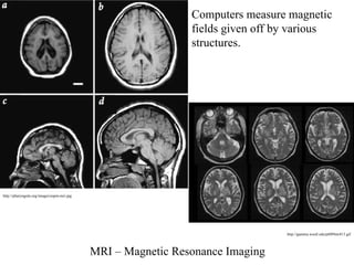
MRI – MagneticResonance Imaging
Computers measure magnetic
fields given off by various
structures.
http://pharyngula.org/images/aspm-mri.jpg
http://gamma.wustl.edu/pt089mr413.gif
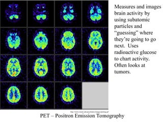
PET – PositronEmission Tomography
Measures and images
brain activity by
using subatomic
particles and
“guessing” where
they’re going to go
next. Uses
radioactive glucose
to chart activity.
Often looks at
tumors.
http://www.mssm.edu/psychiatry/images/petimg.gif
8.
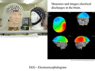
EEG - Electroencephalogram
Measuresand images electrical
discharges in the brain.
http://www.bsp.brain.riken.jp/Photos/EEG%20Acquisition%20-%20%2064-QuickCap%
http://www.ant-software.nl/images/eeg-pain_small.gif
9.
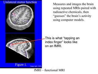
fMRI – functionalMRI
Measures and images the brain
using repeated MRIs paired with
radioactive chemicals, then
“guesses” the brain’s activity
using computer models.
This is what “tapping an
index finger” looks like
on an fMRI.
10.
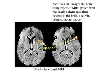
fMRI – functionalMRI
Measures and images the brain
using repeated MRIs paired with
radioactive chemicals, then
“guesses” the brain’s activity
using computer models.