Download as PDF, PPTX


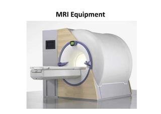


MRI uses magnetic fields and radio waves to produce detailed images of organs and tissues in the body. It is commonly used to evaluate the chest, abdomen, pelvis, and breasts to diagnose conditions like tumors, heart problems, and liver or kidney diseases. During an MRI exam, the patient lies still inside the machine while images are taken. MRI has benefits over other tests as it does not use radiation and can clearly depict soft tissues, though movement can cause blurred images and certain implants are not compatible.


![CASE_PRESENTATION_ON_subdural_hematoma(SDH)[1 FINAL PPT]-1.pptx](https://cdn.slidesharecdn.com/ss_thumbnails/casepresentationonsubduralhematomasdh1finalppt-1-260129172522-d405d375-thumbnail.jpg?width=640&height=640&fit=bounds)

