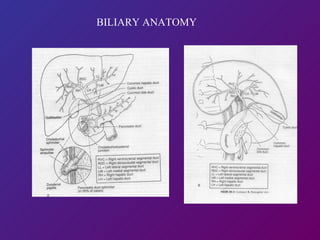
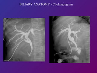
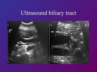
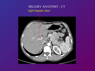
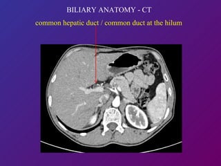
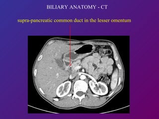
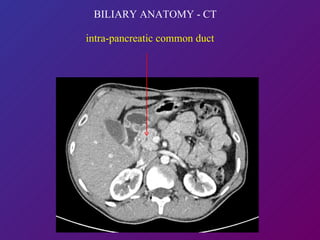
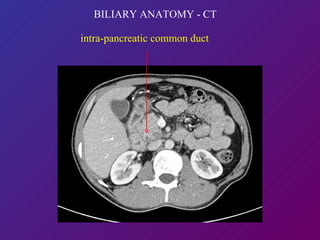
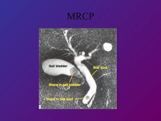
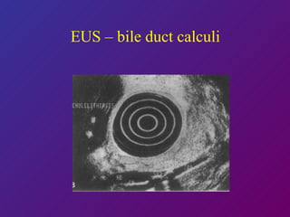
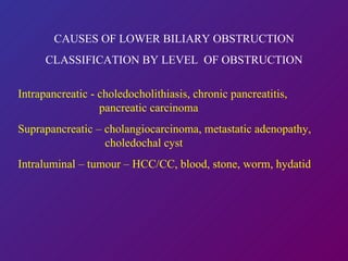
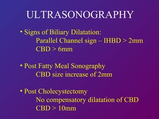
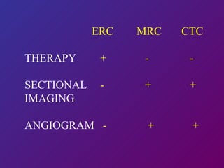
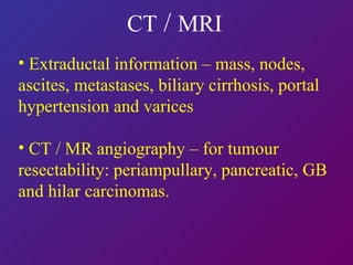
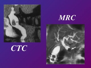
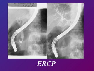
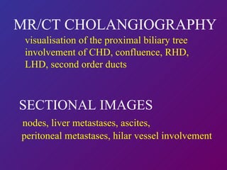
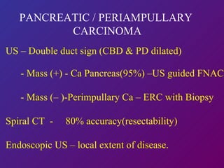
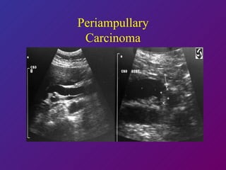
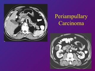
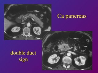
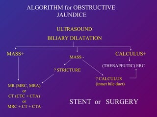
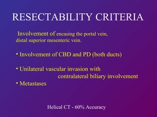
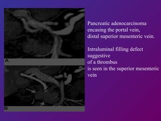
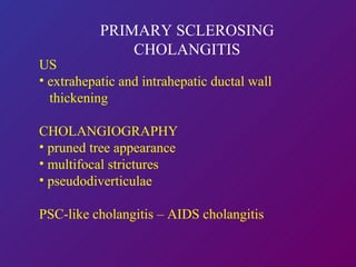
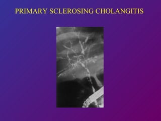
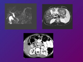
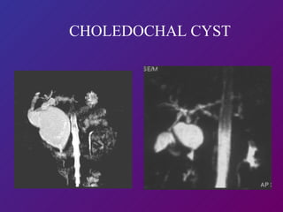
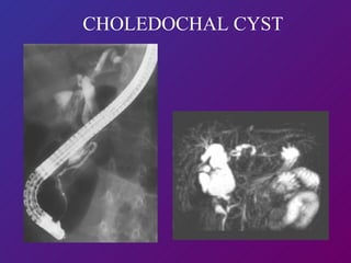
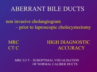
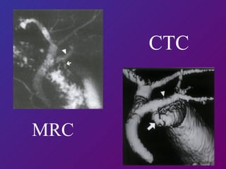
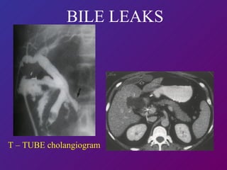
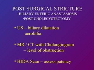
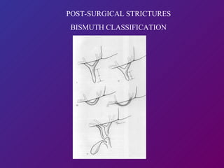
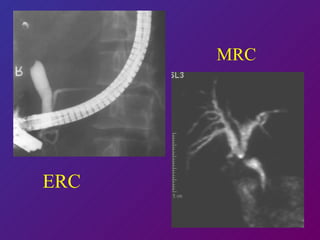
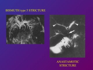
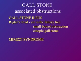
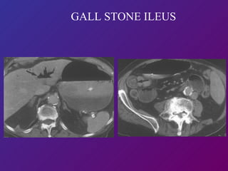
IMAGING OF BILE DUCT DR.SUDHEER HEGDE CONSULTANT RADIOLOGIST DEPARTMENT OF RADIOLOGY COLUMBIA ASIA HOSPITALS Courtesy : Dr.Shalini Govil
NORMAL ANATOMY  cross-sectional and cholangiographic CAUSES OF LOWER BILIARY OBSTRUCTION APPEARANCES ON DIFFERENT  IMAGING MODALITIES ALGORITHM FOR  OBSTRUCTIVE JAUNDICE
  BILIARY ANATOMY
IMAGING MODALITIES Ultrasound - transabdominal, EUS, intraductal Cholangiography - invasive : ERCP / PTC - non-invasive : MR Cholangiography   CT Cholangiography - minIP and maxIP Cross Sectional - spiral CT / MRI as part of MRC/CTC Non-invasive biliary package – MRC with spiral CT DSA  Biliary Scintigraphy
BILIARY ANATOMY - Cholangiogram
Ultrasound biliary tract
  BILIARY ANATOMY - CT right hepatic duct
common hepatic duct / common duct at the hilum   BILIARY ANATOMY - CT
supra-pancreatic common duct in the lesser omentum BILIARY ANATOMY - CT
BILIARY ANATOMY - CT intra-pancreatic common duct
MRCP
EUS – bile duct calculi
CAUSES OF LOWER BILIARY OBSTRUCTION CLASSIFICATION BY LEVEL  OF OBSTRUCTION Intrapancreatic -   choledocholithiasis, chronic pancreatitis,    pancreatic carcinoma Suprapancreatic – cholangiocarcinoma, metastatic adenopathy,    choledochal cyst Intraluminal – tumour – HCC/CC, blood, stone, worm, hydatid
ULTRASONOGRAPHY Signs of Biliary Dilatation: Parallel Channel sign – IHBD > 2mm CBD > 6mm Post Fatty Meal Sonography CBD size increase of 2mm Post Cholecystectomy No compensatory dilatation of CBD CBD > 10mm
CHOLANGIOGRAPHY Invasive  (ERCP / PTC) - High spatial resolution   Possible therapeutic options   Complication rate (2-3%) Non-invasive  CT Cholangiogram - with IV contrast (maxIP)   bilirubin > 2mg% - ineffective - without IV contrast (minIP) MR Cholangiogram
MR CHOLANGIOGRAPHY   Breath-hold (HASTE, RARE) Non-breath-hold (IRTSE) Bile appears bright on heavily T2W images Mapping of biliary tree proximal to obstruction Contraindicated in presence of aneurysm clips, cardiac pacemakers.
MR CHOLANGIOGRAPHY     SENSITIVITY SPECIFICITY   Biliary Obstruction  91 – 100%   100% Level of Obstruction  91 – 100%   100% Choledocholithiasis  81 – 100%   85 – 100% (2mm)
MR CHOLANGIOGRAPHY
ERC MRC CTC THERAPY   +   -   - SECTIONAL   -   +   + IMAGING ANGIOGRAM  -   +   +
CT  /  MRI   Extraductal information – mass, nodes, ascites, metastases, biliary cirrhosis, portal hypertension and varices CT / MR angiography – for tumour resectability: periampullary, pancreatic, GB and hilar carcinomas.
CTC  MRC
ERCP
MR/CT CHOLANGIOGRAPHY visualisation of the proximal biliary tree  involvement of CHD, confluence, RHD, LHD, second order ducts SECTIONAL IMAGES nodes, liver metastases, ascites,  peritoneal metastases, hilar vessel involvement
PANCREATIC / PERIAMPULLARY  CARCINOMA US – Double duct sign (CBD & PD dilated) - Mass (+) - Ca Pancreas(95%) –US guided FNAC - Mass (– )-Perimpullary Ca – ERC with Biopsy Spiral CT  -  80% accuracy(resectability) Endoscopic US – local extent of disease.
Periampullary Carcinoma
Ca pancreas double duct sign
ALGORITHM for OBSTRUCTIVE  JAUNDICE ULTRASOUND BILIARY DILATATION MASS+   MR (MRC, MRA) or CT (CTC + CTA) or MRC + CT + CTA STENT  or  SURGERY ? STRICTURE ? CALCULUS (intact bile duct) (THERAPEUTIC) ERC CALCULUS+ MASS -
RESECTABILITY CRITERIA Involvement of  encasing the portal vein, distal superior mesenteric vein.  Involvement of CBD and PD (both ducts) Unilateral vascular invasion with    contralateral biliary involvement Metastases Helical CT - 60% Accuracy
Pancreatic adenocarcinoma encasing the portal vein, distal superior mesenteric vein.  Intraluminal filling defect suggestive  of a thrombus  is seen in the superior mesenteric vein
THANK YOU
CHOLANGIOCARCINOMA Intraductal ultrasound bile duct wall thickening -  carcinoma vs inflammation semicircular, eccentric, asymmetric wall thickening notched outer margin rigid, papillary inner margin heterogeneous echoes
NON SURGICAL  THERAPEUTIC DRAINAGE Low Obstruction  –  ERCP with Stent Placement Cholangitis – Drainage  (Nasobiliary/PTBD)
PTBD with STENT PLACEMENT   ERC with STENT
DISTAL CHOLANGIOCARCINOMA
GB CARCINOMA
PRIMARY SCLEROSING CHOLANGITIS US  extrahepatic and intrahepatic ductal wall    thickening  CHOLANGIOGRAPHY pruned tree appearance multifocal strictures pseudodiverticulae PSC-like cholangitis – AIDS cholangitis NON INVASIVE CHOLANGIOGRAM PREFERABLE
PRIMARY SCLEROSING CHOLANGITIS
 
HYDATID CYSTS
CHOLEDOCHAL CYST US / NON INVASIVE CHOLANGIOGRAPHY - Todani type  - abnormal pancreatico biliary junction
CHOLEDOCHAL CYST
CHOLEDOCHAL CYST
ABERRANT BILE DUCTS non invasive cholangiogram  –  prior to laproscopic cholecystectomy MRC  HIGH DIAGNOSTIC CT C  ACCURACY  MRC 0.5 T – SUBOPTIMAL VISUALISATION    OF NORMAL CALIBER DUCTS
MRC   CTC
POST SURGICAL  COMPLICATIONS Retained calculi –  T tube Cholangiogram / ERCP Biliary leak  Biliary stenosis/stricture
 
 
BILE LEAKS Site of Leak T-Tube Cholangiogram ERCP with sphincterotomy / Stent Scintigram Infected Biloma US / CT – pigtail drainage
T – TUBE cholangiogram BILE LEAKS
POST SURGICAL STRICTURE BILIARY ENTERIC ANASTAMOSIS POST CHOLECYSTECTOMY   US – biliary dilatation  aerobilia MR / CT with Cholangiogram  –  level of obstruction HIDA Scan – assess patency
POST-SURGICAL STRICTURES BISMUTH CLASSIFICATION
ERC   MRC
BISMUTH type 5 STRICTURE ANASTAMOTIC STRICTURE
GALL STONE  associated obstructions GALL STONE ILEUS Rigler’s triad - air in the biliary tree small bowel obstruction ectopic gall stone MIRIZZI SYNDROME
GALL STONE ILEUS

Imaging of Bile Duct - Columbia Asia Workshop

  • 1.
    IMAGING OF BILEDUCT DR.SUDHEER HEGDE CONSULTANT RADIOLOGIST DEPARTMENT OF RADIOLOGY COLUMBIA ASIA HOSPITALS Courtesy : Dr.Shalini Govil
  • 2.
    NORMAL ANATOMY cross-sectional and cholangiographic CAUSES OF LOWER BILIARY OBSTRUCTION APPEARANCES ON DIFFERENT IMAGING MODALITIES ALGORITHM FOR OBSTRUCTIVE JAUNDICE
  • 3.
    BILIARYANATOMY
  • 4.
    IMAGING MODALITIES Ultrasound- transabdominal, EUS, intraductal Cholangiography - invasive : ERCP / PTC - non-invasive : MR Cholangiography CT Cholangiography - minIP and maxIP Cross Sectional - spiral CT / MRI as part of MRC/CTC Non-invasive biliary package – MRC with spiral CT DSA Biliary Scintigraphy
  • 5.
    BILIARY ANATOMY -Cholangiogram
  • 6.
  • 7.
    BILIARYANATOMY - CT right hepatic duct
  • 8.
    common hepatic duct/ common duct at the hilum BILIARY ANATOMY - CT
  • 9.
    supra-pancreatic common ductin the lesser omentum BILIARY ANATOMY - CT
  • 10.
    BILIARY ANATOMY -CT intra-pancreatic common duct
  • 11.
    BILIARY ANATOMY -CT intra-pancreatic common duct
  • 12.
  • 13.
    EUS – bileduct calculi
  • 14.
    CAUSES OF LOWERBILIARY OBSTRUCTION CLASSIFICATION BY LEVEL OF OBSTRUCTION Intrapancreatic - choledocholithiasis, chronic pancreatitis, pancreatic carcinoma Suprapancreatic – cholangiocarcinoma, metastatic adenopathy, choledochal cyst Intraluminal – tumour – HCC/CC, blood, stone, worm, hydatid
  • 15.
    ULTRASONOGRAPHY Signs ofBiliary Dilatation: Parallel Channel sign – IHBD > 2mm CBD > 6mm Post Fatty Meal Sonography CBD size increase of 2mm Post Cholecystectomy No compensatory dilatation of CBD CBD > 10mm
  • 16.
    CHOLANGIOGRAPHY Invasive (ERCP / PTC) - High spatial resolution Possible therapeutic options Complication rate (2-3%) Non-invasive CT Cholangiogram - with IV contrast (maxIP) bilirubin > 2mg% - ineffective - without IV contrast (minIP) MR Cholangiogram
  • 17.
    MR CHOLANGIOGRAPHY Breath-hold (HASTE, RARE) Non-breath-hold (IRTSE) Bile appears bright on heavily T2W images Mapping of biliary tree proximal to obstruction Contraindicated in presence of aneurysm clips, cardiac pacemakers.
  • 18.
    MR CHOLANGIOGRAPHY SENSITIVITY SPECIFICITY Biliary Obstruction 91 – 100% 100% Level of Obstruction 91 – 100% 100% Choledocholithiasis 81 – 100% 85 – 100% (2mm)
  • 19.
  • 20.
    ERC MRC CTCTHERAPY + - - SECTIONAL - + + IMAGING ANGIOGRAM - + +
  • 21.
    CT / MRI Extraductal information – mass, nodes, ascites, metastases, biliary cirrhosis, portal hypertension and varices CT / MR angiography – for tumour resectability: periampullary, pancreatic, GB and hilar carcinomas.
  • 22.
  • 23.
  • 24.
    MR/CT CHOLANGIOGRAPHY visualisationof the proximal biliary tree involvement of CHD, confluence, RHD, LHD, second order ducts SECTIONAL IMAGES nodes, liver metastases, ascites, peritoneal metastases, hilar vessel involvement
  • 25.
    PANCREATIC / PERIAMPULLARY CARCINOMA US – Double duct sign (CBD & PD dilated) - Mass (+) - Ca Pancreas(95%) –US guided FNAC - Mass (– )-Perimpullary Ca – ERC with Biopsy Spiral CT - 80% accuracy(resectability) Endoscopic US – local extent of disease.
  • 26.
  • 27.
  • 28.
  • 29.
    ALGORITHM for OBSTRUCTIVE JAUNDICE ULTRASOUND BILIARY DILATATION MASS+ MR (MRC, MRA) or CT (CTC + CTA) or MRC + CT + CTA STENT or SURGERY ? STRICTURE ? CALCULUS (intact bile duct) (THERAPEUTIC) ERC CALCULUS+ MASS -
  • 30.
    RESECTABILITY CRITERIA Involvementof encasing the portal vein, distal superior mesenteric vein. Involvement of CBD and PD (both ducts) Unilateral vascular invasion with contralateral biliary involvement Metastases Helical CT - 60% Accuracy
  • 31.
    Pancreatic adenocarcinoma encasingthe portal vein, distal superior mesenteric vein. Intraluminal filling defect suggestive of a thrombus is seen in the superior mesenteric vein
  • 32.
  • 33.
    CHOLANGIOCARCINOMA Intraductal ultrasoundbile duct wall thickening - carcinoma vs inflammation semicircular, eccentric, asymmetric wall thickening notched outer margin rigid, papillary inner margin heterogeneous echoes
  • 34.
    NON SURGICAL THERAPEUTIC DRAINAGE Low Obstruction – ERCP with Stent Placement Cholangitis – Drainage (Nasobiliary/PTBD)
  • 35.
    PTBD with STENTPLACEMENT ERC with STENT
  • 36.
  • 37.
  • 38.
    PRIMARY SCLEROSING CHOLANGITISUS extrahepatic and intrahepatic ductal wall thickening CHOLANGIOGRAPHY pruned tree appearance multifocal strictures pseudodiverticulae PSC-like cholangitis – AIDS cholangitis NON INVASIVE CHOLANGIOGRAM PREFERABLE
  • 39.
  • 40.
  • 41.
  • 42.
    CHOLEDOCHAL CYST US/ NON INVASIVE CHOLANGIOGRAPHY - Todani type - abnormal pancreatico biliary junction
  • 43.
  • 44.
  • 45.
    ABERRANT BILE DUCTSnon invasive cholangiogram – prior to laproscopic cholecystectomy MRC HIGH DIAGNOSTIC CT C ACCURACY MRC 0.5 T – SUBOPTIMAL VISUALISATION OF NORMAL CALIBER DUCTS
  • 46.
    MRC CTC
  • 47.
    POST SURGICAL COMPLICATIONS Retained calculi – T tube Cholangiogram / ERCP Biliary leak Biliary stenosis/stricture
  • 48.
  • 49.
  • 50.
    BILE LEAKS Siteof Leak T-Tube Cholangiogram ERCP with sphincterotomy / Stent Scintigram Infected Biloma US / CT – pigtail drainage
  • 51.
    T – TUBEcholangiogram BILE LEAKS
  • 52.
    POST SURGICAL STRICTUREBILIARY ENTERIC ANASTAMOSIS POST CHOLECYSTECTOMY US – biliary dilatation aerobilia MR / CT with Cholangiogram – level of obstruction HIDA Scan – assess patency
  • 53.
  • 54.
    ERC MRC
  • 55.
    BISMUTH type 5STRICTURE ANASTAMOTIC STRICTURE
  • 56.
    GALL STONE associated obstructions GALL STONE ILEUS Rigler’s triad - air in the biliary tree small bowel obstruction ectopic gall stone MIRIZZI SYNDROME
  • 57.