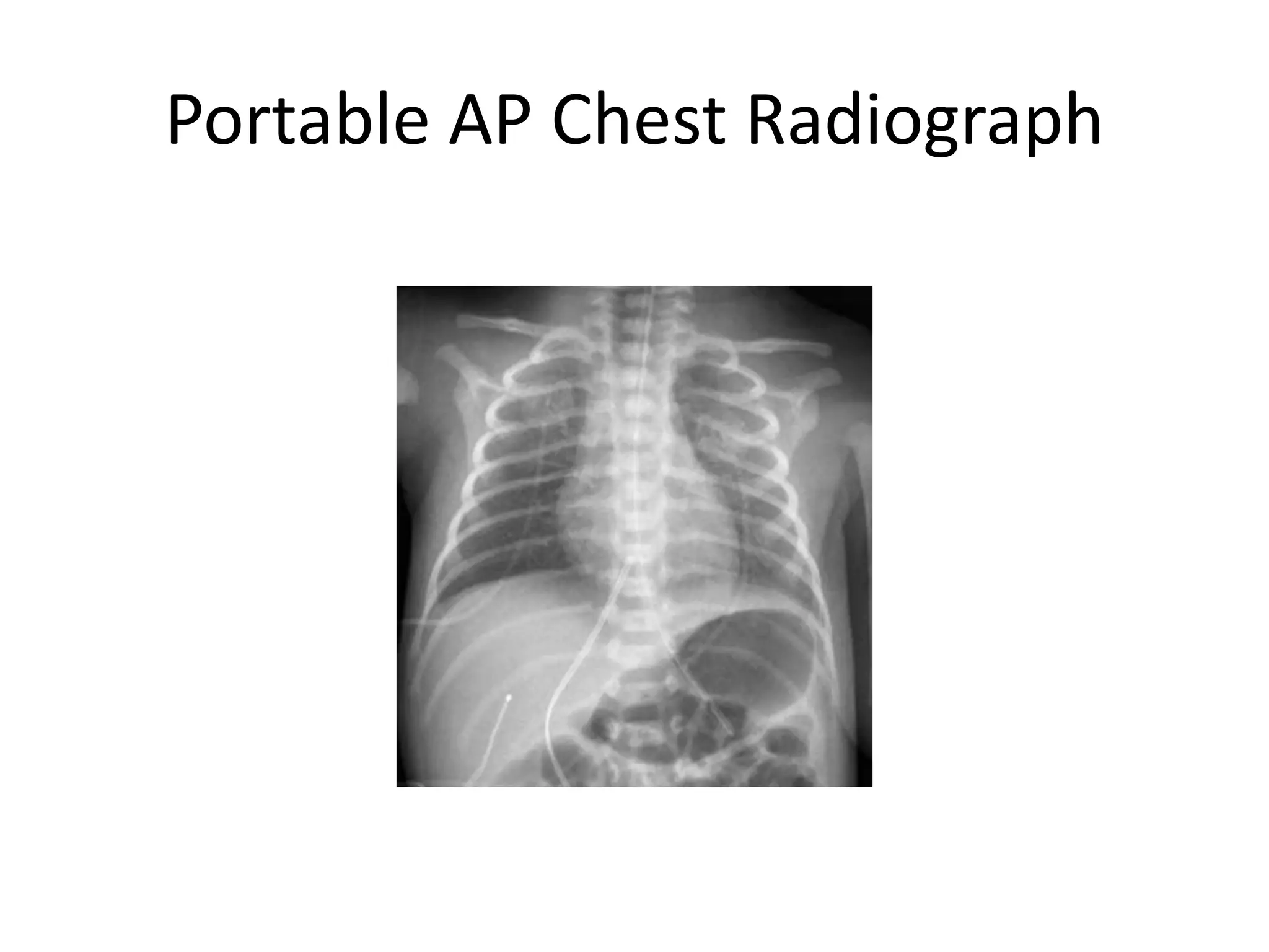
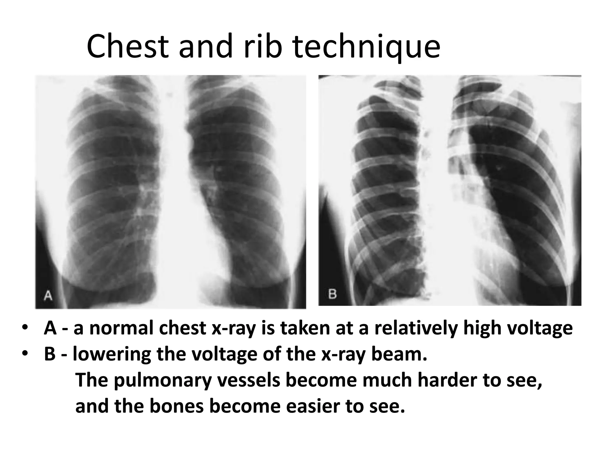
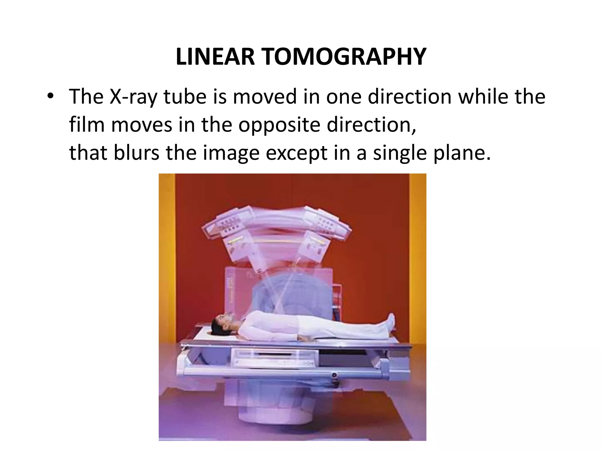
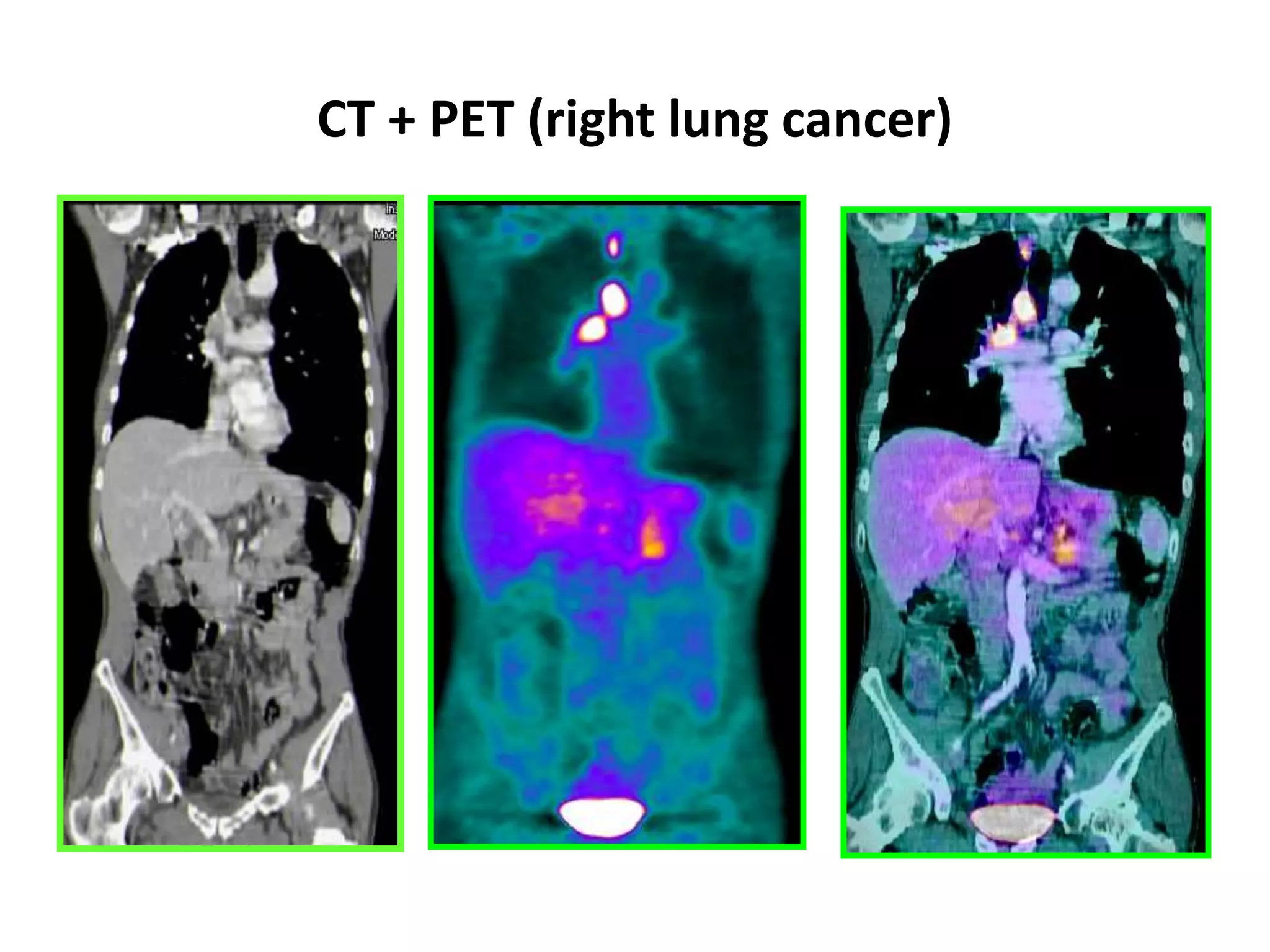
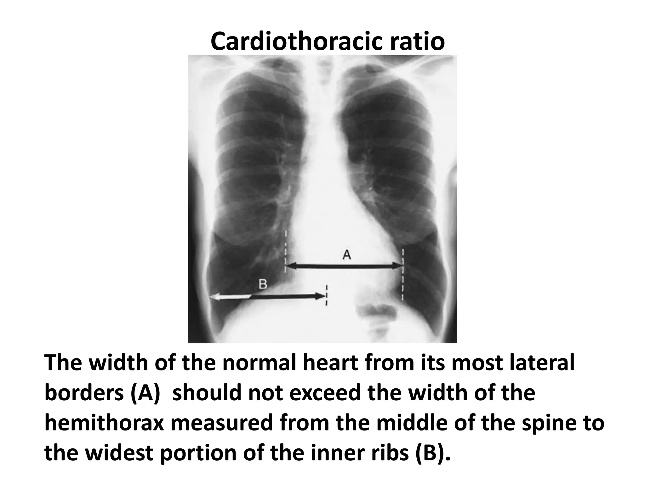
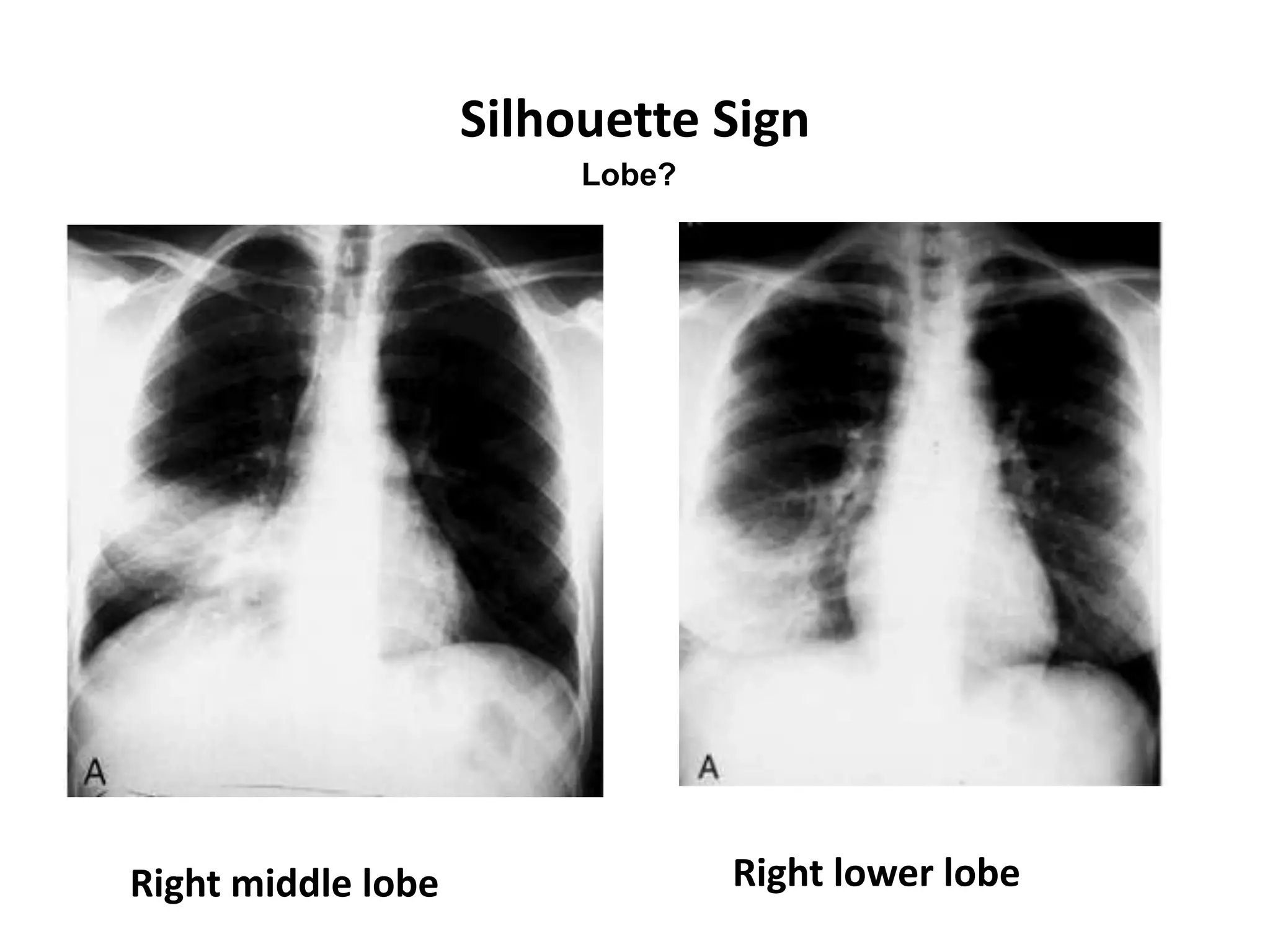
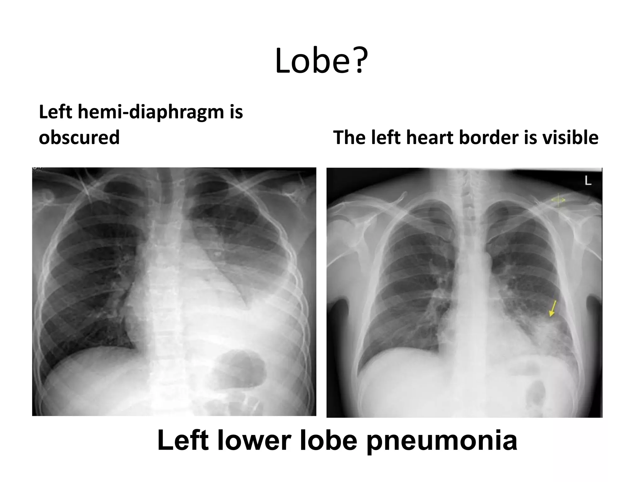
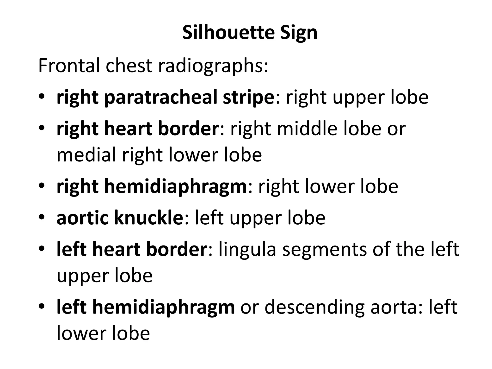
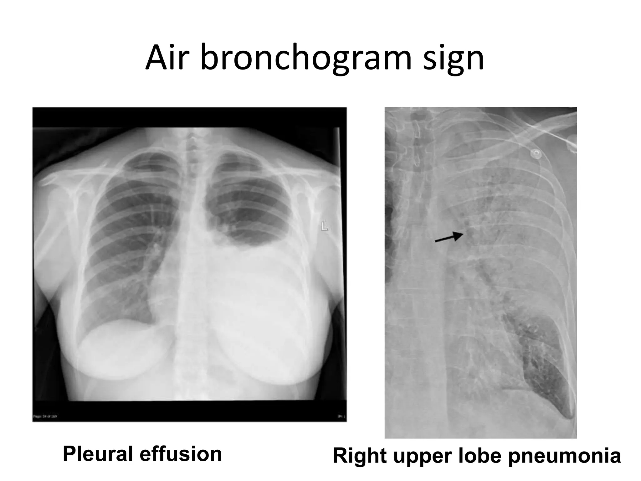
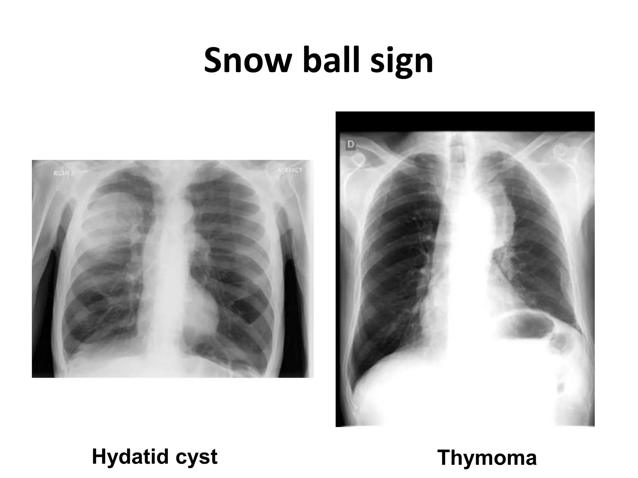
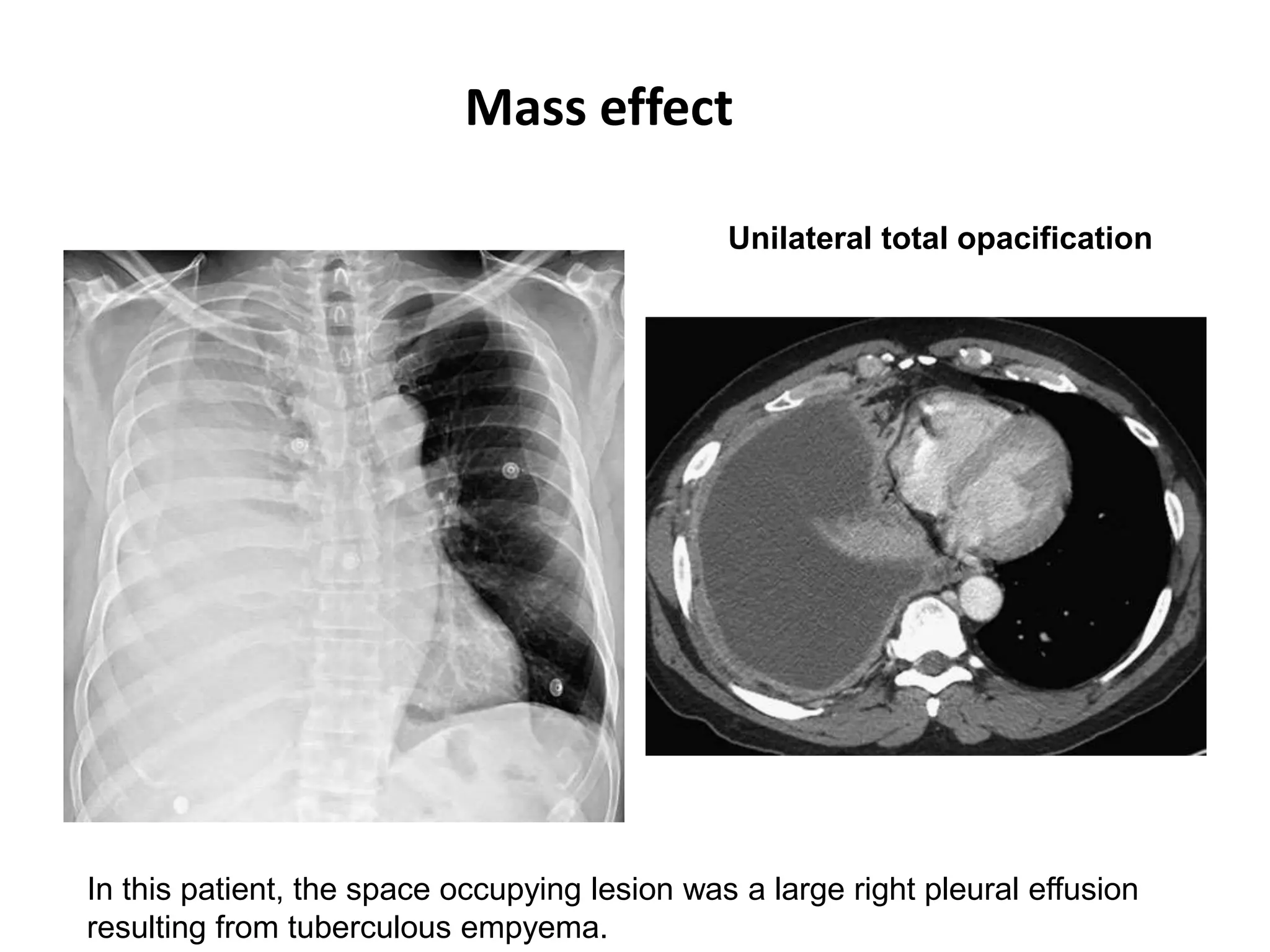
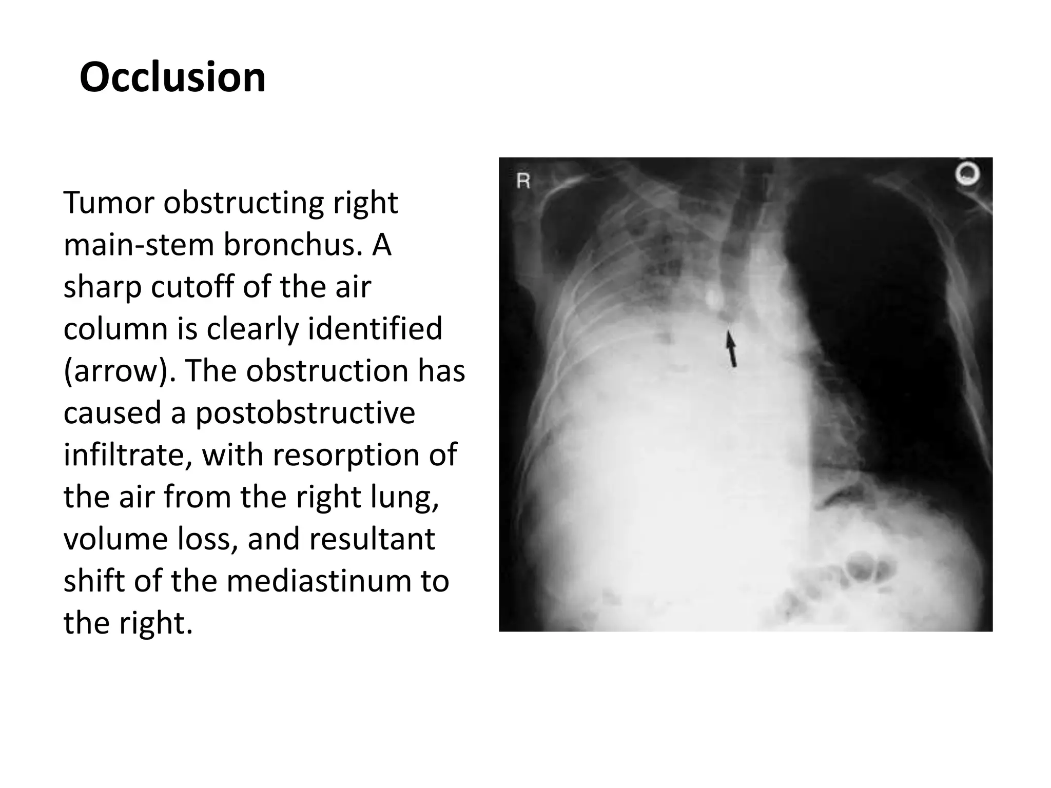
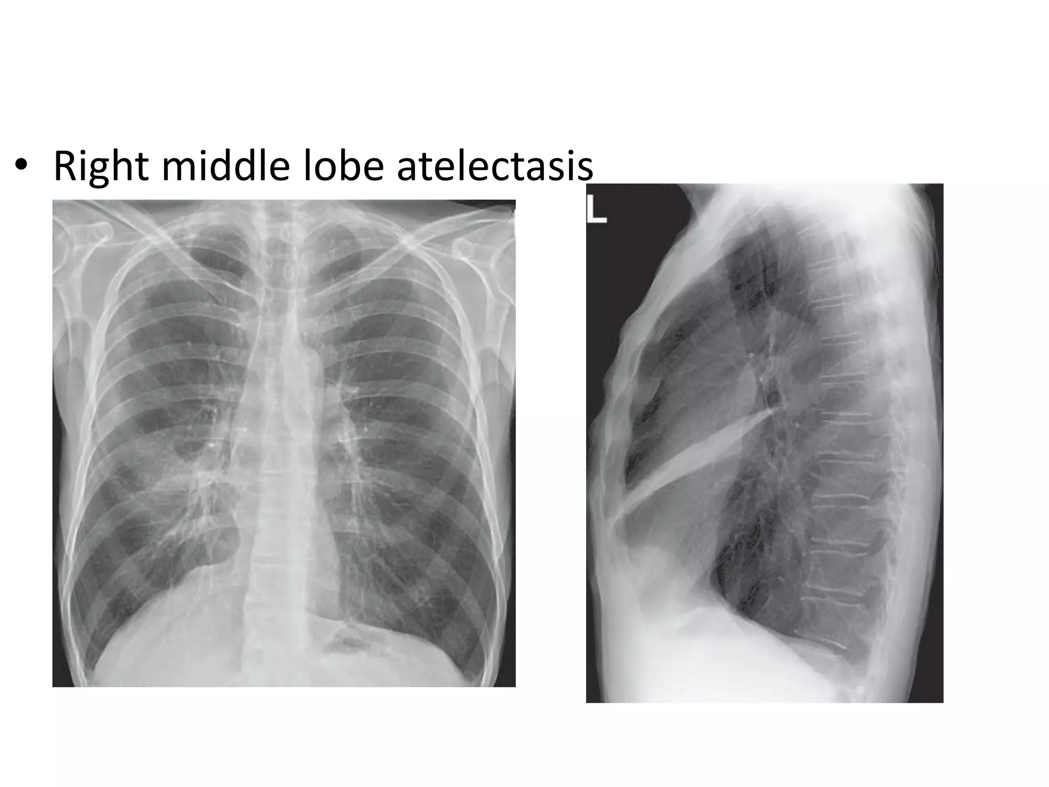
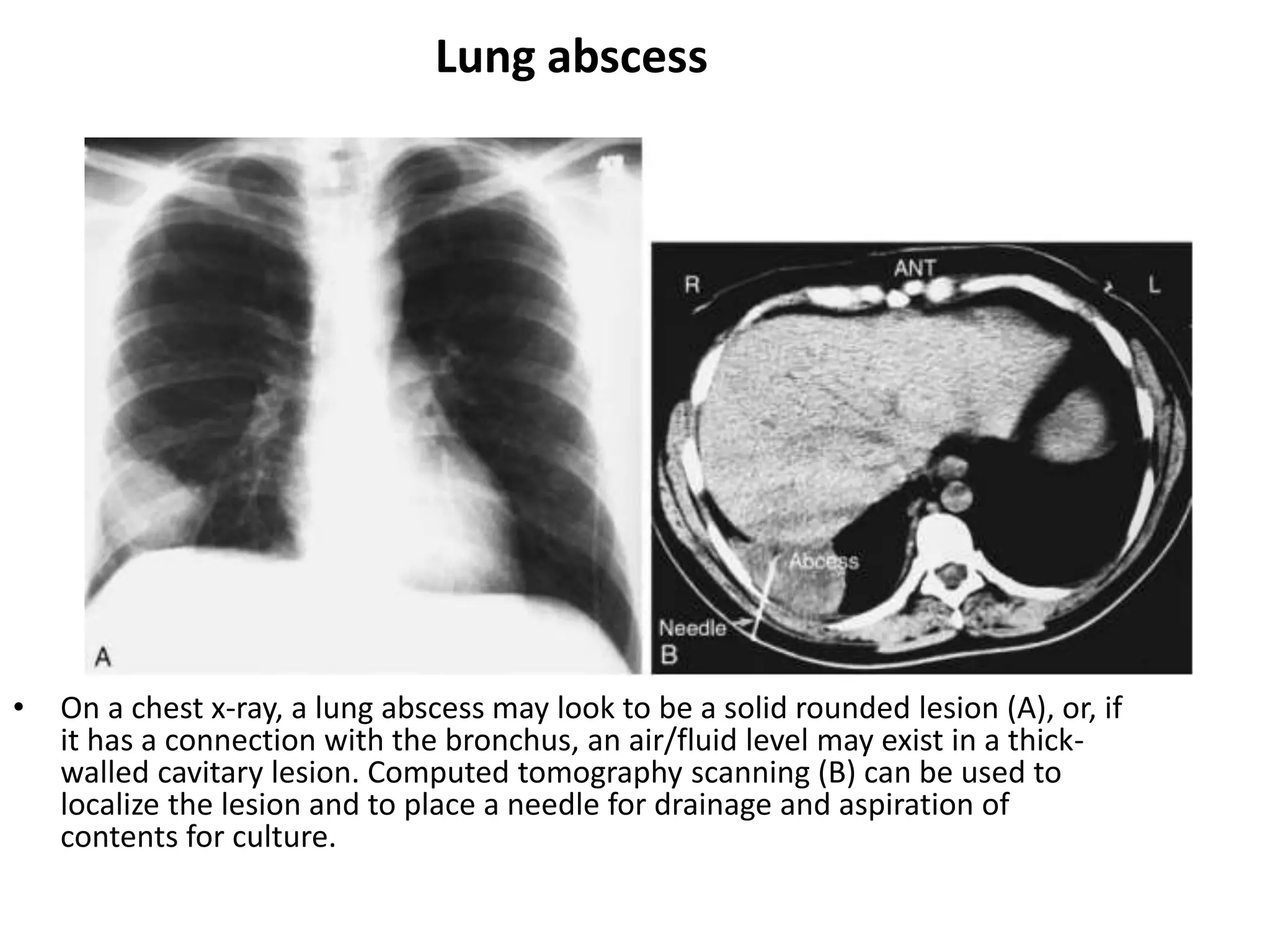
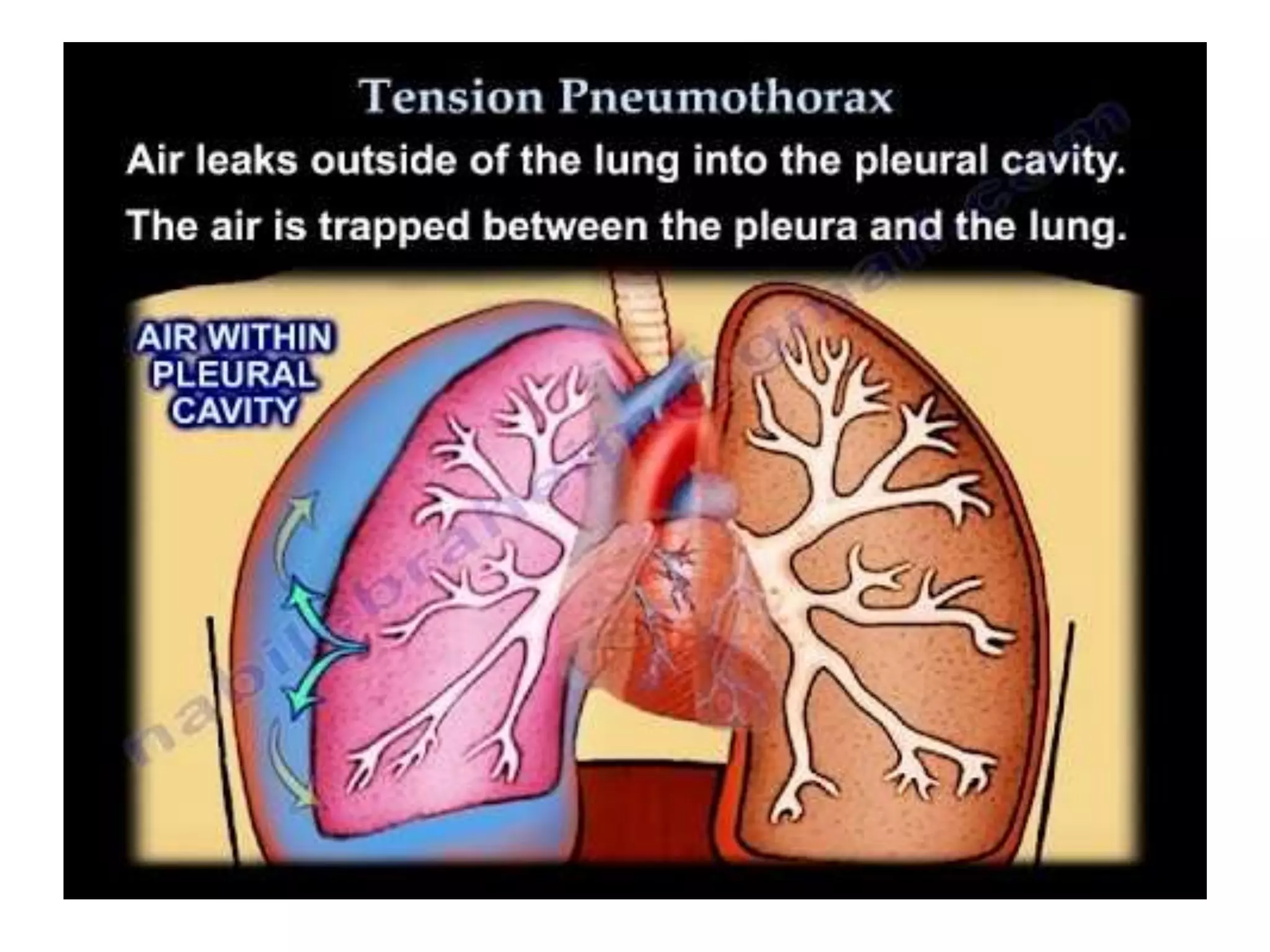
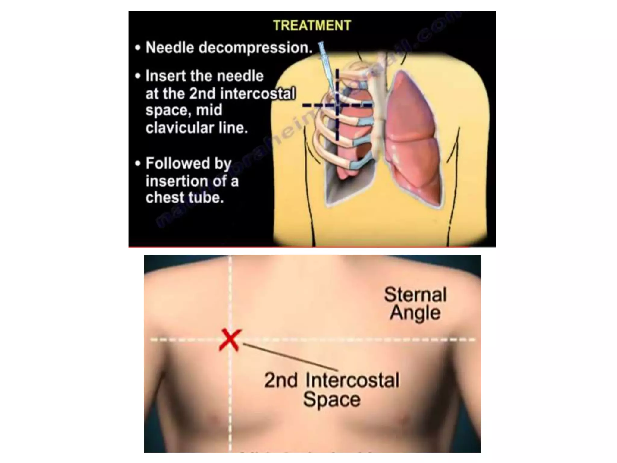
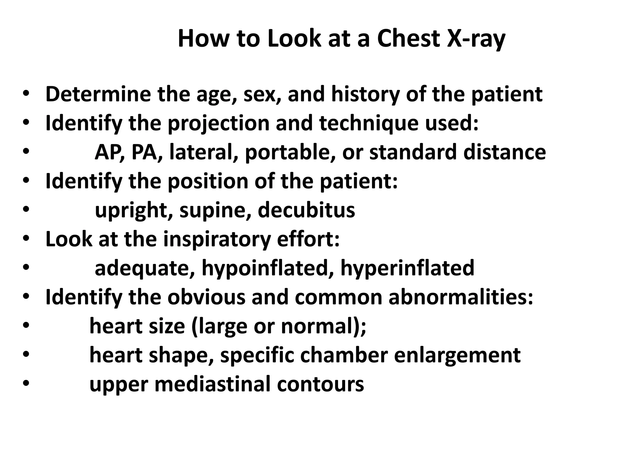
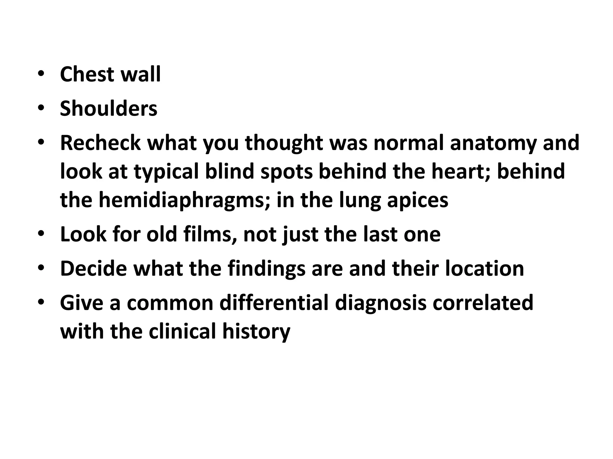
This document discusses various radiology methods used to examine the respiratory system including x-rays, CT scans, fluoroscopy, tomography, angiography, ultrasound, MRI, PET scans, and endoscopy. It provides details on positioning and normal anatomy seen on chest x-rays. It also describes signs seen on chest imaging such as the silhouette sign, air bronchogram sign, snowball sign, and signs of mass effect or volume loss that can help determine the location and characteristics of abnormalities.