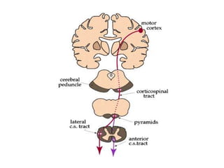
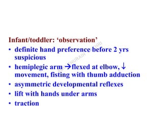
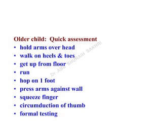
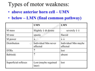
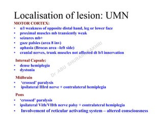
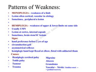
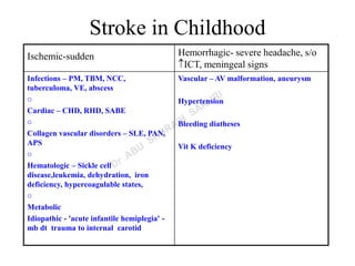
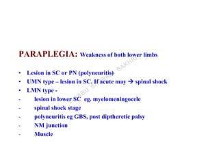
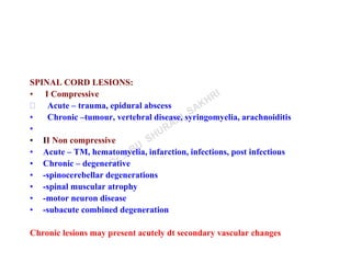
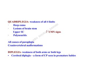
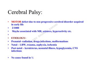
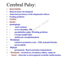
Motor Weakness- APPROACH
Pathways
Types of weakness
Upper motor neuron
Lower motor neuron
Localization of lesion
Patterns of weakness
monoplegia
hemiplegia
paraplegia
quadriplegia
diplegia
Cerebral palsy
MOTOR WEAKNESS:
• Types & pattern
• Anatomical localization
• Etiology
History
• Onset
• Course
• H/o fever
• H/o seizures
• Developmental milestones
Examination
• Young child - ‘observation’
• Older child - ‘play’
 Gait & Posture
 Muscle mass
 Tone
 Power
 Coordination
 Abnormal movements
 Reflexes –superficial & deep
Examination:
Posture
– Frog leg
– Scissoring
– Decerebrate/decorticate/ophisthotonus
– Others
Gait
– Foot drop
– Circumduction
– Limp
– Waddling
Examination:
Muscle mass:
• Compare 2 sides
• Measure in relation to fixed points
•  in lower motor neuron
• Slightly  in upper motor neuron
Muscle Tone
•  in LMN, cerebellar lesions
•  in UMN
• physiological  in newborn
• frog leg position/scissoring
Muscle power
• maximum strength maybe impossible to test in uncooperative
children
• normal strength in pure cerebellar/basal ganglia lesions
Infant/toddler: ‘observation’
• definite hand preference before 2 yrs
suspicious
• hemiplegic arm flexed at elbow, 
movement, fisting with thumb adduction
• asymmetric developmental reflexes
• lift with hands under arms
• traction
Older child: Quick assessment
• hold arms over head
• walk on heels & toes
• get up from floor
• run
• hop on 1 foot
• press arms against wall
• squeeze finger
• circumduction of thumb
• formal testing
Types of motor weakness:
• above anterior horn cell – UMN
• below – LMN (final common pathway)
UMN LMN
M mass Slightly  dt disuse · severely  
M tone spastic flaccid
M power   
Distribution Individual Mm never
affected
Individual Mm maybe
affected
DTRs  lost
Babinski  absent
Superficial reflexes Lost (maybe regained
later)
lost
Localisation of lesion: UMN
MOTOR CORTEX:
• u/l weakness of opposite distal hand, leg or lower face
• proximal muscles mb transiently weak
• seizures mb+
• gaze palsies (area 8 inv)
• aphasia (Brocas area –left side)
• cranial nerves, trunk muscles not affected dt b/l innervation
Internal Capsule:
• dense hemiplegia
• dystonia
Midbrain
• ‘crossed’ paralysis
• ipsilateral IIIrd nerve + contralateral hemiplegia
Pons
• ‘crossed’ paralysis
• ipsilateral Vith/VIIth nerve palsy + contralateral hemiplegia
• Involvement of reticular activating system – altered consciousness
Localisation of lesion : UMN
Medulla
• ‘crossed’ paralysis
• ipsilateral XII th nerve palsy + contralateral hemiplegia
• Involvement of reticular activating system – altered consciousness
Spinal cord
• LMN signs at level of lesion
• UMN signs below
Acute destructive lesions of UMN  hypotonia
All cranial nerves have b/l representation except
part of VII & XII
The Final Common Pathway
Localisation of lesion: LMN
Spinal Cord lesion:
• LMN signs at level of lesion + UMN signs below
• Acute lesions  spinal shock  recovery in few weeks
• Bladder & bowel involvement
Anterior horn cell/ventral root/plexus lesions:
• Weakness in specific myotomes
• Slow degeneration of anterior horn cells fasciculations
Peripheral Nerves:
• Single nerve lesion  mononeuritis –weakness in distribution
• Polyneuritis:
– Distal weakness
– Early loss of reflexes – may not correlate with degree of weakness
Neuromuscular junction:
• Prediliction for ocular/pharyngeal or proximal muscles
• Reflexes lost late in affected muscles
Muscle:
• Proximal weakness
• Deep reflexes maybe  but elicitable
• Myotonia in some
LEVEL
SC PN NMJ M
Weakness ++ +/- +/- +
DTRs - - - 
(early) (late)
Distribution upper level distal ocular+ proximal
or patchy pharyngeal
fasciculations + - - -
(in chronic degenerative
disorder)
tone    
NCV n  n n
EMG fibrillations ,, fatigue pattern BSAPP
 amplitude of MUP
Patterns of Weakness:
• MONOPLEGIA – weakness of a limb
• Lesion often cortical, vascular in etiology
• Sometimes, peripheral n lesion
•
• HEMIPLEGIA – weakness of upper & lower limbs on same side
• Usually UMN
• Lesion at cortex, internal capsule
• Sometimes, brain stem/SC lesions
Signs:
• hand preference before 2 yrs of age
• circumduction gait
• asymmetrical reflexes
• hemiplegic hand kept flexed at elbow, fisted with adducted thum
Causes:
• Hemiplegic cerebral palsy
• Todds palsy
 Tumour
• Trauma
Migraine
Abscess
Granuloma
Vascular - Stroke :Sudden onset --
> gradual recovery
Stroke in Childhood
Ischemic-sudden Hemorrhagic- severe headache, s/o
ICT, meningeal signs
Infections – PM, TBM, NCC,
tuberculoma, VE, abscess
o
Cardiac – CHD, RHD, SABE
o
Collagen vascular disorders – SLE, PAN,
APS
o
Hematologic – Sickle cell
disease,leukemia, dehydration, iron
deficiency, hypercoagulable states,
o
Metabolic
Idiopathic - 'acute infantile hemiplegia' -
mb dt trauma to internal carotid
Vascular – AV malformation, aneurysm
Hypertension
Bleeding diatheses
Vit K deficiency
PARAPLEGIA: Weakness of both lower limbs
• Lesion in SC or PN (polyneuritis)
• UMN type – lesion in SC. If acute may  spinal shock
• LMN type -
- lesion in lower SC eg. myelomeningocele
- spinal shock stage
- polyneuritis eg GBS, post diptheretic palsy
- NM junction
- Muscle
SPINAL CORD LESIONS:
• I Compressive
 Acute – trauma, epidural abscess
• Chronic –tumour, vertebral disease, syringomyelia, arachnoiditis
•
• II Non compressive
• Acute – TM, hematomyelia, infarction, infections, post infectious
• Chronic – degenerative
• -spinocerebellar degenerations
• -spinal muscular atrophy
• -motor neuron disease
• -subacute combined degeneration
Chronic lesions may present acutely dt secondary vascular changes
QUADRIPLEGIA- weakness of all 4 limbs
· Deep coma
· Lesions of brain stem
Upper SC  UMN signs
· Polyneuritis
All causes of paraplegia
Craniovertebral malformations
DIPLEGIA- weakness of both arms or both legs
• Cerebral diplegia – a form of CP seen in premature babies
Cerebral Palsy:
• MOTOR defect due to non progressive cerebral disorder acquired
in early life
• 2/1000
• Maybe associated with MR, seizures, hyperactivity etc.
•
• ETIOLOGY:
• Prenatal- radiation, drugs,infections, malformations
• Natal – LBW, trauma, asphyxia, ischemia
• Post natal – kernicterus, neonatal illness, hypoglycemia, CNS
infections
• No cause found in ¼
Cerebral Palsy:
• DIAGNOSIS:
• Delayed motor development
• Abnormal persistence of developmental reflexes
• Feeding problems
• TYPES:
• Spastic
• quadriplegia
• - most common
• - severe disability, MR
• - pseudobulbar palsy feeding problems
• - extrapyramidal signs
• - multicystic encephalomalacia
• Hemiplegia
• - related to perinatal events – PIH, ischemiastroke
• - porencephaly
• Diplegia
• - prematurity periventricular leukomalacia
• ·Dyskinetic – kernicterus, circulatory failure, asphyxia
 · Ataxic – often due to unrecognized cerebellar malformation
MANAGEMENT
Multidisciplinary approach with involvement of
neurologist, physiotherapist, speech therapist,
occupational therapist
Drugs to reduce tone , appliances
THANK YOU

Approach to motor weakness

  • 1.
  • 2.
    Pathways Types of weakness Uppermotor neuron Lower motor neuron Localization of lesion Patterns of weakness monoplegia hemiplegia paraplegia quadriplegia diplegia Cerebral palsy
  • 4.
    MOTOR WEAKNESS: • Types& pattern • Anatomical localization • Etiology
  • 5.
    History • Onset • Course •H/o fever • H/o seizures • Developmental milestones
  • 6.
    Examination • Young child- ‘observation’ • Older child - ‘play’  Gait & Posture  Muscle mass  Tone  Power  Coordination  Abnormal movements  Reflexes –superficial & deep
  • 7.
    Examination: Posture – Frog leg –Scissoring – Decerebrate/decorticate/ophisthotonus – Others Gait – Foot drop – Circumduction – Limp – Waddling
  • 8.
    Examination: Muscle mass: • Compare2 sides • Measure in relation to fixed points •  in lower motor neuron • Slightly  in upper motor neuron Muscle Tone •  in LMN, cerebellar lesions •  in UMN • physiological  in newborn • frog leg position/scissoring Muscle power • maximum strength maybe impossible to test in uncooperative children • normal strength in pure cerebellar/basal ganglia lesions
  • 9.
    Infant/toddler: ‘observation’ • definitehand preference before 2 yrs suspicious • hemiplegic arm flexed at elbow,  movement, fisting with thumb adduction • asymmetric developmental reflexes • lift with hands under arms • traction
  • 10.
    Older child: Quickassessment • hold arms over head • walk on heels & toes • get up from floor • run • hop on 1 foot • press arms against wall • squeeze finger • circumduction of thumb • formal testing
  • 11.
    Types of motorweakness: • above anterior horn cell – UMN • below – LMN (final common pathway) UMN LMN M mass Slightly  dt disuse · severely   M tone spastic flaccid M power    Distribution Individual Mm never affected Individual Mm maybe affected DTRs  lost Babinski  absent Superficial reflexes Lost (maybe regained later) lost
  • 12.
    Localisation of lesion:UMN MOTOR CORTEX: • u/l weakness of opposite distal hand, leg or lower face • proximal muscles mb transiently weak • seizures mb+ • gaze palsies (area 8 inv) • aphasia (Brocas area –left side) • cranial nerves, trunk muscles not affected dt b/l innervation Internal Capsule: • dense hemiplegia • dystonia Midbrain • ‘crossed’ paralysis • ipsilateral IIIrd nerve + contralateral hemiplegia Pons • ‘crossed’ paralysis • ipsilateral Vith/VIIth nerve palsy + contralateral hemiplegia • Involvement of reticular activating system – altered consciousness
  • 13.
    Localisation of lesion: UMN Medulla • ‘crossed’ paralysis • ipsilateral XII th nerve palsy + contralateral hemiplegia • Involvement of reticular activating system – altered consciousness Spinal cord • LMN signs at level of lesion • UMN signs below Acute destructive lesions of UMN  hypotonia All cranial nerves have b/l representation except part of VII & XII
  • 14.
  • 15.
    Localisation of lesion:LMN Spinal Cord lesion: • LMN signs at level of lesion + UMN signs below • Acute lesions  spinal shock  recovery in few weeks • Bladder & bowel involvement Anterior horn cell/ventral root/plexus lesions: • Weakness in specific myotomes • Slow degeneration of anterior horn cells fasciculations Peripheral Nerves: • Single nerve lesion  mononeuritis –weakness in distribution • Polyneuritis: – Distal weakness – Early loss of reflexes – may not correlate with degree of weakness Neuromuscular junction: • Prediliction for ocular/pharyngeal or proximal muscles • Reflexes lost late in affected muscles Muscle: • Proximal weakness • Deep reflexes maybe  but elicitable • Myotonia in some
  • 16.
    LEVEL SC PN NMJM Weakness ++ +/- +/- + DTRs - - -  (early) (late) Distribution upper level distal ocular+ proximal or patchy pharyngeal fasciculations + - - - (in chronic degenerative disorder) tone     NCV n  n n EMG fibrillations ,, fatigue pattern BSAPP  amplitude of MUP
  • 17.
    Patterns of Weakness: •MONOPLEGIA – weakness of a limb • Lesion often cortical, vascular in etiology • Sometimes, peripheral n lesion • • HEMIPLEGIA – weakness of upper & lower limbs on same side • Usually UMN • Lesion at cortex, internal capsule • Sometimes, brain stem/SC lesions Signs: • hand preference before 2 yrs of age • circumduction gait • asymmetrical reflexes • hemiplegic hand kept flexed at elbow, fisted with adducted thum Causes: • Hemiplegic cerebral palsy • Todds palsy  Tumour • Trauma Migraine Abscess Granuloma Vascular - Stroke :Sudden onset -- > gradual recovery
  • 18.
    Stroke in Childhood Ischemic-suddenHemorrhagic- severe headache, s/o ICT, meningeal signs Infections – PM, TBM, NCC, tuberculoma, VE, abscess o Cardiac – CHD, RHD, SABE o Collagen vascular disorders – SLE, PAN, APS o Hematologic – Sickle cell disease,leukemia, dehydration, iron deficiency, hypercoagulable states, o Metabolic Idiopathic - 'acute infantile hemiplegia' - mb dt trauma to internal carotid Vascular – AV malformation, aneurysm Hypertension Bleeding diatheses Vit K deficiency
  • 19.
    PARAPLEGIA: Weakness ofboth lower limbs • Lesion in SC or PN (polyneuritis) • UMN type – lesion in SC. If acute may  spinal shock • LMN type - - lesion in lower SC eg. myelomeningocele - spinal shock stage - polyneuritis eg GBS, post diptheretic palsy - NM junction - Muscle
  • 20.
    SPINAL CORD LESIONS: •I Compressive  Acute – trauma, epidural abscess • Chronic –tumour, vertebral disease, syringomyelia, arachnoiditis • • II Non compressive • Acute – TM, hematomyelia, infarction, infections, post infectious • Chronic – degenerative • -spinocerebellar degenerations • -spinal muscular atrophy • -motor neuron disease • -subacute combined degeneration Chronic lesions may present acutely dt secondary vascular changes
  • 21.
    QUADRIPLEGIA- weakness ofall 4 limbs · Deep coma · Lesions of brain stem Upper SC  UMN signs · Polyneuritis All causes of paraplegia Craniovertebral malformations DIPLEGIA- weakness of both arms or both legs • Cerebral diplegia – a form of CP seen in premature babies
  • 22.
    Cerebral Palsy: • MOTORdefect due to non progressive cerebral disorder acquired in early life • 2/1000 • Maybe associated with MR, seizures, hyperactivity etc. • • ETIOLOGY: • Prenatal- radiation, drugs,infections, malformations • Natal – LBW, trauma, asphyxia, ischemia • Post natal – kernicterus, neonatal illness, hypoglycemia, CNS infections • No cause found in ¼
  • 23.
    Cerebral Palsy: • DIAGNOSIS: •Delayed motor development • Abnormal persistence of developmental reflexes • Feeding problems • TYPES: • Spastic • quadriplegia • - most common • - severe disability, MR • - pseudobulbar palsy feeding problems • - extrapyramidal signs • - multicystic encephalomalacia • Hemiplegia • - related to perinatal events – PIH, ischemiastroke • - porencephaly • Diplegia • - prematurity periventricular leukomalacia • ·Dyskinetic – kernicterus, circulatory failure, asphyxia  · Ataxic – often due to unrecognized cerebellar malformation
  • 24.
    MANAGEMENT Multidisciplinary approach withinvolvement of neurologist, physiotherapist, speech therapist, occupational therapist Drugs to reduce tone , appliances
  • 25.