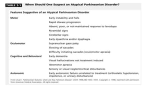
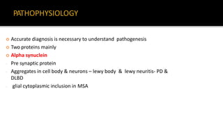
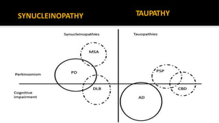
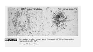
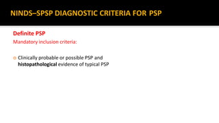
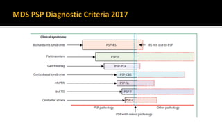
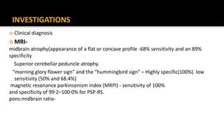
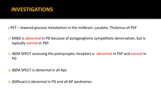
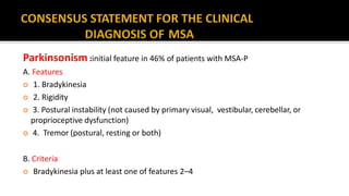
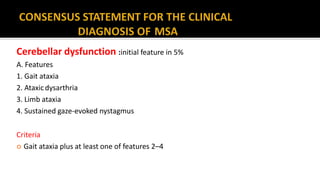
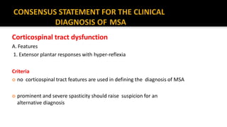
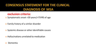
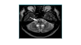
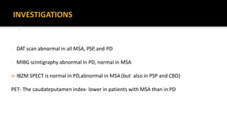
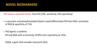
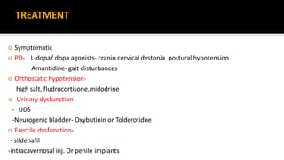
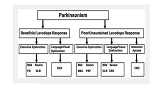
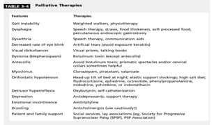
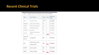
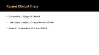
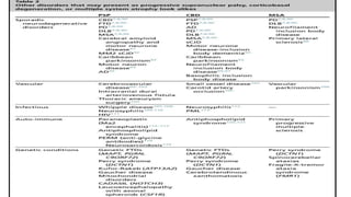
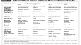
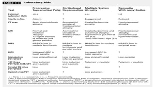
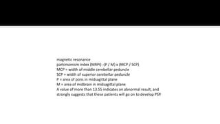
Dr. RAHI KIRAN.B
SR NEUROLOGY
GOVT MEDICAL COLLEGE, KOTA
 Classification
 Red flag signs
 Diagnostic criteria
 Phenotypic spectrum
 Investigations
 Novel biomarkers
 Treatment
 Future trends
 Common problem in neurology OPD
 Wide variety of sporadic / heredodegenerative syndromes
 80-85% -IPD
 Differentiation from other syndromes
 important in prognostication and management
 Progressive Supranuclear Palsy
 Multiple System Atrophy [(Shy-Dragger syn.) SND (MSAP) OPCA (MSAC)]
 Corticobasal Degeneration
 Dementia with Lewy Body Disease
 Accurate diagnosis is necessary to understand pathogenesis
 Two proteins mainly
 Alpha synuclein
- Pre synaptic protein
- Aggregates in cell body & neurons – lewy body & lewy neuritis- PD &
DLBD
- glial cytoplasmic inclusion in MSA
SYNUCLEINOPATHY TAUPATHY
 Steele et al—1964
 5% of parkinsonian pts
 male-to-female ratio is 1.5:1
 Commonly misdiagnosed as PD
 diagnosis is purely clinical
 always sporadic, few familial cases-
 The usual interval from initial symptom occurrence to the need for a cane or a walker is
3.1 years,
 confinement to a chair or bed is 8.2 years.
 median disease duration of 9.7 years
Postural Instability & EP Features :
 Falls—backward
 Rigidity –axial
 Hypophonic
 Widely based ataxic
 Frontal release signs
 Pseudobulbar palsy
 L-DOPA UNRESPONSIVENESS
 early signs- Slow vertical saccades and square wave jerks
 Reduced blink rate and apraxia of eyelid opening
 on doll’s eye maneuver, there is improved range
 Subcortical-type dementia
 Typical facies- “surprised look”
 advanced PSP - Complete ophthalmoparesis
Possible PSP(highly sensitive)
Mandatory inclusion criteria:
 Gradually progressive disorder
 Onset age 40 or later
 Either vertical supranuclear palsy or both slowing of vertical saccades
 Postural instability with falls within a year of disease onset
 No evidence of other diseases that could explain the foregoing features, as
indicated by exclusion criteria
Mandatory exclusion criteria:
 Recent history of encephalitis
 Alien limb syndrome
 Cortical sensory deficits
 Focal frontal or temporoparietal atrophy
 Hallucinations or delusions unrelated to dopaminergic
therapy
 Cortical dementia of Alzheimer type
 Prominent, early cerebellar symptoms
 Unexplained dysautonomia
NINDS–SPSP DIAGNOSTIC CRITERIA FOR PSP
Supportive features:
 symmetrical akinesia or rigidity
 proximal more than distal
 abnormal neck posture especially retrocollis
 poor or absent response of parkinsonism to levodopa
 early dysphagia and dysarthria
 early onset of cognitive impairment including two or more of: apathy,
impairment in abstract thought, decreased verbal fluency, utilisation or
imitation behaviour, or frontal release signs
NINDS–SPSP DIAGNOSTIC CRITERIA FOR PSP
Probable PSP(highly specific)
Mandatory inclusion criteria
 gradually progressive disorder
 onset age 40 or later
 vertical supranuclear palsy
 prominent postural instability with falls within a year of disease onset
 no evidence of other diseases that could explain the foregoing features,
as indicated by exclusion criteria
NINDS–SPSP DIAGNOSTIC CRITERIA FOR PSP
Definite PSP
Mandatory inclusion criteria:
 Clinically probable or possible PSP and
histopathological evidence of typical PSP
NINDS–SPSP DIAGNOSTIC CRITERIA FOR PSP
INVESTIGATIONS
 Clinical diagnosis
 MRI-
midbrain atrophy(appearance of a flat or concave profile -68% sensitivity and an 89%
specificity
Superior cerebellar peduncle atrophy.
“morning glory flower sign” and the “hummingbird sign” – Highly specific(100%) low
sensitivity (50% and 68.4%)
magnetic resonance parkinsonism index (MRPI) - sensitivity of 100%
and specificity of 99·2–100·0% for PSP-RS.
pons:midbrain ratio-
oPET – lowered glucose metabolism in the midbrain ,caudate, Thalamus of PSP
 MIBG is abnormal in PD because of postganglionic sympathetic denervation, but is
typically normal in PSP.
 IBZM SPECT assessing the postsynaptic receptors is abnormal in PSP and normal in
PD
 IBZM SPECT is abnormal in all Aps
 (DATscan) is abnormal in PD and all AP syndromes
INVESTIGATIONS
PATHOLOGY
 Neurofibrillary tangles are present in reticular formation and ocular motor nuclei.
 Tufted astrocytes (Gallyas-positive) -feature of PSP that differentiates other
tauopathies such as CBD (astrocytic plaques)
-CSF tau protein- CSF phospho-tau and total tau concentrations lower than AD
- 2–5 times increased neurofilament light chain concentrations in PSP
Novel diagnostic approaches and biomarkers
Treatment
 No effective symptomatic or neuroprotective treatments
 A trial of levodopa (up to 1 g/d) and amantadine (up to 450mg/d)
 Botulinum toxin injections can be used to treat levator inhibition,rigiditY, dystonia
 Serotonin reuptake inhibitors (SSRIs) may be used for apathy with no clear benefit.
 Supportive measures such as physiotherapy, walking aids, speech therapy and PEG
 A small study with Coenzyme Q10- no RCTstudy
 Recent large, double-blind studies with GSK-3b inhibitors (Tideglusib,
Davunetide) - failed.
 Tideglusib reduced the rate of brain atrophy in onestudy.
 Sporadic neurodegenerative disorder clinically any combination of parkinsonian,
autonomic, cerebellar, or pyramidal signs
 MSA is an a-synucleinopathy
 usually a sporadic disease; however, rarely, familial cases - mutations in COQ2
gene
 Prevalence of MSA - ranged from 1·9 to 4·9 cases per 100 000 people.
Clinical presentation :
 Affects both men and women
 sixth decade of life
 mean survival of 6–9 years.(upto 15yrs)
Main features –
 Autonomic failure
 Parkinsonism
 Cerebellar ataxia
 Pyramidal signs in any combination.
 Distinguished clinically–
1. Parkinsonian features predominate in 80% of patients (MSA-P
subtype),
2. Cerebellar ataxia is the main motor feature in 20% of patients (MSA-C
subtype).
 Both similar survival times.
 MSA-P - more rapid functional deterioration
 progressive akinesia and rigidity
 jerky postural tremor and tremor at rest.
 orofacial or craniocervical dystonia
 recurrent falls at disease onset are unusual .
 90% of the MSA-P pts- unresponsive to levodopa in the long term.
 gait ataxia
 scanning dysarthria
 cerebellar oculomotor disturbances.
 may be indistinguishable from other patients with idiopathic late onset cerebellar
ataxia
Dysautonomia :
 urogenital and orthostatic dysfunction.
 Early erectile dysfunction is nearly universal in men with MSA
 Female- genital insensitivity
 urinary incontinence or retention are common
Autonomic and urinary dysfunction :
 Features
 1. Orthostatic hypotension(68% of patients)
 2. Urinary incontinence or incomplete bladder emptying
Criteria
 Reduction of least 30mmHg or in diastolic blood pressure by at least 15 mm Hg
after 3 min of standing
 urinary incontinence (persistent, involuntary partial or total bladder emptying,
accompanied by erectile dysfunction in men) or both
Parkinsonism :initial feature in 46% of patients with MSA-P
A. Features
 1. Bradykinesia
 2. Rigidity
 3. Postural instability (not caused by primary visual, vestibular, cerebellar, or
proprioceptive dysfunction)
 4. Tremor (postural, resting or both)
B. Criteria
 Bradykinesia plus at least one of features 2–4
Cerebellar dysfunction :initial feature in 5%
A. Features
1. Gait ataxia
2. Ataxicdysarthria
3. Limb ataxia
4. Sustained gaze-evoked nystagmus
Criteria
 Gait ataxia plus at least one of features 2–4
Corticospinal tract dysfunction
A. Features
1. Extensor plantar responses with hyper-reflexia
Criteria
 no corticospinal tract features are used in defining the diagnosis of MSA
 prominent and severe spasticity should raise suspicion for an
alternative diagnosis
exclusion criteria:
 Symptomatic onset <30 years/>75YRS of age
 Family history of a similar disorder
 Systemic disease or other identifiable causes
 Hallucinations unrelated to medication
 Dementia
exclusion criteria:
 Prominent slowing of vertical saccades or vertical supranuclear gaze palsy
 Evidence of focal cortical dysfunction
 Laboratory investigation- Metabolic, molecular genetic and imaging evidence of an
alternative cause of features
 Possible MSA
 A sporadic, progressive, adult (>30y) with onset disease* characterized by the following:
 Parkinsonism or cerebellar syndrome
 At least 1 feature of autonomic or urogenital dysfunction
 At least 1 additional feature
 Probable MSA
 A sporadic, progressive, adult (>30y) with onset disease* characterized by the following:
 Autonomic failure involving urinary dysfunction
 Poorly levodopa-responsive parkinsonism or
 cerebellar dysfunction
 Definitive MSA
 A sporadic, progressive, adult (>30y) with onset disease pathologically confirmed by
presence of high density GCIs in association with degenerative changes in
striatonigral and olivopontocerebellar pathways
 Pyramidal signs
 Orofacial dystonia or dyskinesias
 dyskinesia mainly affecting orofacial muscles
 Axial dystonia -Pisa syndrome (subacute axial dystonia with a severe
tonic lateral flexion of the trunk, head, and neck)
 early severe camptocormia
MSA - Additional features
 Jerky tremor
 Dysarthria- Atypical, irregular and severely hypophonic
 Dysphagia within 5 years of motor onset
 Neuropsychiatric features - • Depression (41%) • Hallucinations (5·5%)
• Dementia (4·5%) • Insomnia (19%)
• Daytime sleepiness (17%) • Restless legs (10%)
MSA - Additional features
 Atrophy on MRI of putamen, middle cerebellar peduncle, pons, or cerebellum
 Hypometabolism on FDG-PET in putamen, brainstem, or cerebellum
Investigations
 Autonomic function tests
 Cardiovascular function test-within in 2-3 ys
 Standard urine analysis will exclude infection.
 The residual volume –usg,cystometry ,UDS
MSA
IMAGING
 MRI
- Hot cross burn sign- mcp/pons- MSA-c
- Putaminal rim- MSA-p
The slight hyperintensity of the lateral margin of the putamen on T2-
weighted MRI is a characteristic finding in patients with MSA involving the
extrapyramidal system
Trace (D)-DW MRI -MSA-P-anterior putamen
MSA-C-cerebellum and middle cerebellar peduncle
.
- DAT scan abnormal in all MSA, PSP, and PD
- MIBG scintigraphy abnormal in PD, normal in MSA
 IBZM SPECT is normal in PD,abnormal in MSA(but also in PSP and CBD)
PET- The caudateputamen index- lower in patients with MSA than in PD
CSF mean a-synuclein levels from PD (70% sensitivity, 53% specificity)
 a-synuclein and phosphorylated t/total t could differentiate PD from MSA -sensitivity
of 90% & specificity of 71%
 Flt3 ligand, a cytokine
PD and MSA with a sensitivity of 99% and a specificity of 95%.
COQ2, a gene that encodes coenzyme Q10,
TREATMENT
 Symptomatic
 PD- L-dopa/ dopa agonists- cranio cervical dystonia postural hypotension
Amantidine- gait disturbances
 Orthostatic hypotension-
high salt, fludrocortisone,midodrine
 Urinary dysfunction
- UDS
-Neurogenic bladder- Oxybutinin or Tolderotidne
 Erectile dysfunction-
- sildenafil
-intracavernosal inj. Or penile implants
 Depession
- SSRI/TCA
Promising studies
- Rasagiline
- Intrarterial/IV - autologous stem cells
- Future trials
- Alpha synuclein targeting antibodies
TREATMENT
Sixth to eighth decades of life - mean age 63 years
. Sporadic disease , 4–6% of parkinsonism.
Clinical presentations
 Five initial presentations
 The most common presentation (55%) -“useless arm” (ie, a rigid, dystonic,
akinetic, or apraxic arm),
 gait disorder (27%)
 prominent sensory symptoms
 isolated speech disturbance
 behavioural disturbance
Motor (asymmetric)
Limb clumsiness
Bradykinesia/Akinesia
Rigidity
Tremor (action/postural)
Myoclonus
Limb dystonia
Blepharospasm
Choreoathetoid movements
Speech abnormalities
Gait disorder
Higher cortical functions
 Apraxia
 Dementia
 Alien-limb phenomenon
 Aphasia
 Frontal-lobe-release signs
 Cortical sensory abnormalities
 Depression
 apathy
 Anxiety Irritability
 Disinhibition, Delusions,Obsessive compulsive disorder
Inclusion criteria (one of A orB)
A) Rigidity (easily detectable without reinforcement) and one cortical sign:
 apraxia
 cortical sensory loss
 alien-limb phenomenon
B) Asymmetric rigidity, dystonia (focal in limb; present at rest at onset),
 focal reflex myoclonus (spreads beyond stimulated digits
Exclusion criteria
 Early dementia (will exclude some patients)
 Early vertical gaze palsy
 Rest tremor
 Severe autonomic disturbances
 Sustained responsiveness to levodopa
 Lesions on imaging studies indicate another
pathological process
 Variable degrees of focal or lateralised cognitive dysfunction,
with relative preservation of learning and memory on neuropsychometric testing
 Focal or asymmetric atrophy on CT or MRI imaging, typically in perifrontal cortex
 Focal or asymmetric hypoperfusion on SPECT or PET, typically maximal in
parietofrontal cortex with or without basal ganglia involvement
investigations
PHENOTYPIC SPECTRUM
 Classic CBD phenotype- CBS
 CBD- a/w FTD,PSP,PPA
 Imaging
asymmetric frontal, and parietal cortical atrophy becomes evident with dilatation
of the lateral ventricle(temporal/parietal cortex (the latter pattern is seen in
dementia of the Alzheimer type)
 Dopamine transporter SPECT- abnormal
- differentiate them from those with Alzheimer’s and Pick’s diseases (in whom this
scan is typically normal) early in the course of the disease.
 FDG-PET
- Asymmetric reduction in fronto parietalregions
LP & jejunal biopsy- Whipple disease
 The R2 component of the blink reflex recovery cycle (R2 BRRC) appears to be a useful tool
to distinguish progressive supranuclear palsy (PSP) from corticobasal degeneration (CBD)
 4-repeat-tau aggregates - neocortex in CBD, brainstem in PSP
TREATMENT
 L-dopa trial (upto 1 gm/d)/Amantadine- PD symptoms
 Valproate, Levetiracetam- myoclonus
 Botox inj- dystonic hand
 antioxidants or vitamin E if the patient has memory loss
 Palliative rx
 Dementia -not occur in the early stages ,usually evident with progression.
 Deficits on tests of attention, executive function, and visuospatial ability may
be especially prominent.
 Two core features are sufficient for a diagnosis of probable, one for
possible DLB
 Fluctuating cognition with pronounced variations in attention and
alertness
 Recurrent visual hallucinations that are typically well formed and detailed
 features of parkinsonism
 REM sleep behavior disorder
 Severe neuroleptic sensitivity
 Low dopamine transporter uptake in basal ganglia demonstrated by
SPECT or PET imaging
 one or more of these + one or more core features, - probable DLB .
 In the absence of any core features, one or more
suggestive features - possible DLB.
 Probable DLB should not be diagnosed on the basis of suggestive features alone.
 Repeated falls and syncope, Transient unexplained loss of consciousness
 Severe autonomic dysfunction
 Hallucinations in other modalities
 Depression
 Relative preservation of medial temporal lobe structures on CT/MRI scan
 Generalized low uptake on SPECT/PET perfusion scan with reduced
occipital activity
 Prominent slow wave activity on EEG with temporal lobe transient sharp waves
 If -CVA evident as focal neurologic signs or on brain imaging
 In the presence of any other physical illness or brain disorder sufficient to
account in part or in total for the clinical picture
 If parkinsonism only appears for the first time at a stage of severe dementia
 Diagnosed when dementia occurs before or concurrently with parkinsonism (if it is
present).
 Parkinson disease dementia (PDD) - dementia that occurs in the context of well-
established Parkinson disease.
 the 1-year rule between the onset of dementia and parkinsonism – DLB .
 less hippocampal atrophy - DLB
 SPECT & PET - decreased occipital lobe blood flow – DLB > AD
relative preservation of the posterior cingulate gyrus (cingulate island
sign) - DLB > AD
 SPECT scanning studies in DLB patients :
 Visual hallucinations - Were related to hypoperfusion of the parietal and occipital
association cortices
 Misidentifications - Were related to hypoperfusion of the limbic-paralimbic structures
 Delusions - Were related to hyperperfusion of the frontal cortices
 CSF
 Tau – DLB < AD
 beta amyloid are lower than normal in DLB, AD
 LBCRS - Lewy body composite risk score - help determine whether Lewy body
pathology is contributing to dementia.
 Motor parkinsonism-mild hallucinations and agitation may not require medical
treatment.
-Levodopa at low doses & titrate up.
-Anticholinergics should be avoided.
 Neuropsychiatric symptoms.--cholinesterase inhibitors (CHEIs) or atypical
antipsychotic ,
modafinil initially had a positive effect , but didn't last long.
 memantine improves cognitive function and neuropsychiatric features in
patients with DLB.
 prion hypothesis - recently
 davunetide , tideglusib - failed
 droxidopa - orthostatic hypotension – failed
 losartan - supine hypertension - failed
SUMMARY
 Careful clinical examination
 AP mimickers
 There are currently no biomarkers available.
 There are currently no neuroprotective treatments available.
 symptomatic and supportive treatments with usually no sustained effect.
 Further research required
THANK YOU
magnetic resonance
parkinsonism index (MRPI) -(P / M) x (MCP / SCP)
MCP = width of middle cerebellar peduncle
SCP = width of superior cerebellar peduncle
P = area of pons in midsagittal plane
M = area of midbrain in midsagittal plane
A value of more than 13.55 indicates an abnormal result, and
strongly suggests that these patients will go on to develop PSP.
Misdiagnosis PD with AP , as well as FTD,AD,PPA
Mimickers of AtypicalPD Eg: SCAS/ FTAX- mimic
MSA
Neimann Pick C,CTX,prion d/s,mitochondrial- mimic AP phenotype
- Age of onset
- Tempo of progression
- Family history
- Clinical exam + associated features

Parkinson's plus syndromes

  • 1.
    Dr. RAHI KIRAN.B SRNEUROLOGY GOVT MEDICAL COLLEGE, KOTA
  • 2.
     Classification  Redflag signs  Diagnostic criteria  Phenotypic spectrum  Investigations  Novel biomarkers  Treatment  Future trends
  • 3.
     Common problemin neurology OPD  Wide variety of sporadic / heredodegenerative syndromes  80-85% -IPD  Differentiation from other syndromes  important in prognostication and management
  • 5.
     Progressive SupranuclearPalsy  Multiple System Atrophy [(Shy-Dragger syn.) SND (MSAP) OPCA (MSAC)]  Corticobasal Degeneration  Dementia with Lewy Body Disease
  • 6.
     Accurate diagnosisis necessary to understand pathogenesis  Two proteins mainly  Alpha synuclein - Pre synaptic protein - Aggregates in cell body & neurons – lewy body & lewy neuritis- PD & DLBD - glial cytoplasmic inclusion in MSA
  • 9.
  • 11.
     Steele etal—1964  5% of parkinsonian pts  male-to-female ratio is 1.5:1  Commonly misdiagnosed as PD  diagnosis is purely clinical  always sporadic, few familial cases-
  • 12.
     The usualinterval from initial symptom occurrence to the need for a cane or a walker is 3.1 years,  confinement to a chair or bed is 8.2 years.  median disease duration of 9.7 years
  • 13.
    Postural Instability &EP Features :  Falls—backward  Rigidity –axial  Hypophonic  Widely based ataxic  Frontal release signs  Pseudobulbar palsy  L-DOPA UNRESPONSIVENESS
  • 14.
     early signs-Slow vertical saccades and square wave jerks  Reduced blink rate and apraxia of eyelid opening  on doll’s eye maneuver, there is improved range  Subcortical-type dementia  Typical facies- “surprised look”  advanced PSP - Complete ophthalmoparesis
  • 16.
    Possible PSP(highly sensitive) Mandatoryinclusion criteria:  Gradually progressive disorder  Onset age 40 or later  Either vertical supranuclear palsy or both slowing of vertical saccades  Postural instability with falls within a year of disease onset  No evidence of other diseases that could explain the foregoing features, as indicated by exclusion criteria
  • 17.
    Mandatory exclusion criteria: Recent history of encephalitis  Alien limb syndrome  Cortical sensory deficits  Focal frontal or temporoparietal atrophy  Hallucinations or delusions unrelated to dopaminergic therapy  Cortical dementia of Alzheimer type  Prominent, early cerebellar symptoms  Unexplained dysautonomia NINDS–SPSP DIAGNOSTIC CRITERIA FOR PSP
  • 18.
    Supportive features:  symmetricalakinesia or rigidity  proximal more than distal  abnormal neck posture especially retrocollis  poor or absent response of parkinsonism to levodopa  early dysphagia and dysarthria  early onset of cognitive impairment including two or more of: apathy, impairment in abstract thought, decreased verbal fluency, utilisation or imitation behaviour, or frontal release signs NINDS–SPSP DIAGNOSTIC CRITERIA FOR PSP
  • 19.
    Probable PSP(highly specific) Mandatoryinclusion criteria  gradually progressive disorder  onset age 40 or later  vertical supranuclear palsy  prominent postural instability with falls within a year of disease onset  no evidence of other diseases that could explain the foregoing features, as indicated by exclusion criteria NINDS–SPSP DIAGNOSTIC CRITERIA FOR PSP
  • 20.
    Definite PSP Mandatory inclusioncriteria:  Clinically probable or possible PSP and histopathological evidence of typical PSP NINDS–SPSP DIAGNOSTIC CRITERIA FOR PSP
  • 22.
    INVESTIGATIONS  Clinical diagnosis MRI- midbrain atrophy(appearance of a flat or concave profile -68% sensitivity and an 89% specificity Superior cerebellar peduncle atrophy. “morning glory flower sign” and the “hummingbird sign” – Highly specific(100%) low sensitivity (50% and 68.4%) magnetic resonance parkinsonism index (MRPI) - sensitivity of 100% and specificity of 99·2–100·0% for PSP-RS. pons:midbrain ratio-
  • 23.
    oPET – loweredglucose metabolism in the midbrain ,caudate, Thalamus of PSP  MIBG is abnormal in PD because of postganglionic sympathetic denervation, but is typically normal in PSP.  IBZM SPECT assessing the postsynaptic receptors is abnormal in PSP and normal in PD  IBZM SPECT is abnormal in all Aps  (DATscan) is abnormal in PD and all AP syndromes INVESTIGATIONS
  • 25.
    PATHOLOGY  Neurofibrillary tanglesare present in reticular formation and ocular motor nuclei.  Tufted astrocytes (Gallyas-positive) -feature of PSP that differentiates other tauopathies such as CBD (astrocytic plaques)
  • 27.
    -CSF tau protein-CSF phospho-tau and total tau concentrations lower than AD - 2–5 times increased neurofilament light chain concentrations in PSP Novel diagnostic approaches and biomarkers
  • 28.
    Treatment  No effectivesymptomatic or neuroprotective treatments  A trial of levodopa (up to 1 g/d) and amantadine (up to 450mg/d)  Botulinum toxin injections can be used to treat levator inhibition,rigiditY, dystonia  Serotonin reuptake inhibitors (SSRIs) may be used for apathy with no clear benefit.  Supportive measures such as physiotherapy, walking aids, speech therapy and PEG
  • 29.
     A smallstudy with Coenzyme Q10- no RCTstudy  Recent large, double-blind studies with GSK-3b inhibitors (Tideglusib, Davunetide) - failed.  Tideglusib reduced the rate of brain atrophy in onestudy.
  • 30.
     Sporadic neurodegenerativedisorder clinically any combination of parkinsonian, autonomic, cerebellar, or pyramidal signs  MSA is an a-synucleinopathy  usually a sporadic disease; however, rarely, familial cases - mutations in COQ2 gene  Prevalence of MSA - ranged from 1·9 to 4·9 cases per 100 000 people.
  • 31.
    Clinical presentation : Affects both men and women  sixth decade of life  mean survival of 6–9 years.(upto 15yrs) Main features –  Autonomic failure  Parkinsonism  Cerebellar ataxia  Pyramidal signs in any combination.
  • 32.
     Distinguished clinically– 1.Parkinsonian features predominate in 80% of patients (MSA-P subtype), 2. Cerebellar ataxia is the main motor feature in 20% of patients (MSA-C subtype).  Both similar survival times.  MSA-P - more rapid functional deterioration
  • 33.
     progressive akinesiaand rigidity  jerky postural tremor and tremor at rest.  orofacial or craniocervical dystonia  recurrent falls at disease onset are unusual .  90% of the MSA-P pts- unresponsive to levodopa in the long term.
  • 34.
     gait ataxia scanning dysarthria  cerebellar oculomotor disturbances.  may be indistinguishable from other patients with idiopathic late onset cerebellar ataxia
  • 35.
    Dysautonomia :  urogenitaland orthostatic dysfunction.  Early erectile dysfunction is nearly universal in men with MSA  Female- genital insensitivity  urinary incontinence or retention are common
  • 36.
    Autonomic and urinarydysfunction :  Features  1. Orthostatic hypotension(68% of patients)  2. Urinary incontinence or incomplete bladder emptying Criteria  Reduction of least 30mmHg or in diastolic blood pressure by at least 15 mm Hg after 3 min of standing  urinary incontinence (persistent, involuntary partial or total bladder emptying, accompanied by erectile dysfunction in men) or both
  • 37.
    Parkinsonism :initial featurein 46% of patients with MSA-P A. Features  1. Bradykinesia  2. Rigidity  3. Postural instability (not caused by primary visual, vestibular, cerebellar, or proprioceptive dysfunction)  4. Tremor (postural, resting or both) B. Criteria  Bradykinesia plus at least one of features 2–4
  • 38.
    Cerebellar dysfunction :initialfeature in 5% A. Features 1. Gait ataxia 2. Ataxicdysarthria 3. Limb ataxia 4. Sustained gaze-evoked nystagmus Criteria  Gait ataxia plus at least one of features 2–4
  • 39.
    Corticospinal tract dysfunction A.Features 1. Extensor plantar responses with hyper-reflexia Criteria  no corticospinal tract features are used in defining the diagnosis of MSA  prominent and severe spasticity should raise suspicion for an alternative diagnosis
  • 40.
    exclusion criteria:  Symptomaticonset <30 years/>75YRS of age  Family history of a similar disorder  Systemic disease or other identifiable causes  Hallucinations unrelated to medication  Dementia
  • 41.
    exclusion criteria:  Prominentslowing of vertical saccades or vertical supranuclear gaze palsy  Evidence of focal cortical dysfunction  Laboratory investigation- Metabolic, molecular genetic and imaging evidence of an alternative cause of features
  • 42.
     Possible MSA A sporadic, progressive, adult (>30y) with onset disease* characterized by the following:  Parkinsonism or cerebellar syndrome  At least 1 feature of autonomic or urogenital dysfunction  At least 1 additional feature  Probable MSA  A sporadic, progressive, adult (>30y) with onset disease* characterized by the following:  Autonomic failure involving urinary dysfunction  Poorly levodopa-responsive parkinsonism or  cerebellar dysfunction
  • 43.
     Definitive MSA A sporadic, progressive, adult (>30y) with onset disease pathologically confirmed by presence of high density GCIs in association with degenerative changes in striatonigral and olivopontocerebellar pathways
  • 44.
     Pyramidal signs Orofacial dystonia or dyskinesias  dyskinesia mainly affecting orofacial muscles  Axial dystonia -Pisa syndrome (subacute axial dystonia with a severe tonic lateral flexion of the trunk, head, and neck)  early severe camptocormia MSA - Additional features
  • 45.
     Jerky tremor Dysarthria- Atypical, irregular and severely hypophonic  Dysphagia within 5 years of motor onset  Neuropsychiatric features - • Depression (41%) • Hallucinations (5·5%) • Dementia (4·5%) • Insomnia (19%) • Daytime sleepiness (17%) • Restless legs (10%) MSA - Additional features
  • 46.
     Atrophy onMRI of putamen, middle cerebellar peduncle, pons, or cerebellum  Hypometabolism on FDG-PET in putamen, brainstem, or cerebellum
  • 48.
    Investigations  Autonomic functiontests  Cardiovascular function test-within in 2-3 ys  Standard urine analysis will exclude infection.  The residual volume –usg,cystometry ,UDS MSA
  • 49.
    IMAGING  MRI - Hotcross burn sign- mcp/pons- MSA-c - Putaminal rim- MSA-p The slight hyperintensity of the lateral margin of the putamen on T2- weighted MRI is a characteristic finding in patients with MSA involving the extrapyramidal system Trace (D)-DW MRI -MSA-P-anterior putamen MSA-C-cerebellum and middle cerebellar peduncle
  • 51.
    . - DAT scanabnormal in all MSA, PSP, and PD - MIBG scintigraphy abnormal in PD, normal in MSA  IBZM SPECT is normal in PD,abnormal in MSA(but also in PSP and CBD) PET- The caudateputamen index- lower in patients with MSA than in PD
  • 52.
    CSF mean a-synucleinlevels from PD (70% sensitivity, 53% specificity)  a-synuclein and phosphorylated t/total t could differentiate PD from MSA -sensitivity of 90% & specificity of 71%  Flt3 ligand, a cytokine PD and MSA with a sensitivity of 99% and a specificity of 95%. COQ2, a gene that encodes coenzyme Q10,
  • 53.
    TREATMENT  Symptomatic  PD-L-dopa/ dopa agonists- cranio cervical dystonia postural hypotension Amantidine- gait disturbances  Orthostatic hypotension- high salt, fludrocortisone,midodrine  Urinary dysfunction - UDS -Neurogenic bladder- Oxybutinin or Tolderotidne  Erectile dysfunction- - sildenafil -intracavernosal inj. Or penile implants
  • 54.
     Depession - SSRI/TCA Promisingstudies - Rasagiline - Intrarterial/IV - autologous stem cells - Future trials - Alpha synuclein targeting antibodies TREATMENT
  • 55.
    Sixth to eighthdecades of life - mean age 63 years . Sporadic disease , 4–6% of parkinsonism.
  • 56.
    Clinical presentations  Fiveinitial presentations  The most common presentation (55%) -“useless arm” (ie, a rigid, dystonic, akinetic, or apraxic arm),  gait disorder (27%)  prominent sensory symptoms  isolated speech disturbance  behavioural disturbance
  • 57.
    Motor (asymmetric) Limb clumsiness Bradykinesia/Akinesia Rigidity Tremor(action/postural) Myoclonus Limb dystonia Blepharospasm Choreoathetoid movements Speech abnormalities Gait disorder
  • 58.
    Higher cortical functions Apraxia  Dementia  Alien-limb phenomenon  Aphasia  Frontal-lobe-release signs  Cortical sensory abnormalities  Depression  apathy  Anxiety Irritability  Disinhibition, Delusions,Obsessive compulsive disorder
  • 59.
    Inclusion criteria (oneof A orB) A) Rigidity (easily detectable without reinforcement) and one cortical sign:  apraxia  cortical sensory loss  alien-limb phenomenon B) Asymmetric rigidity, dystonia (focal in limb; present at rest at onset),  focal reflex myoclonus (spreads beyond stimulated digits
  • 60.
    Exclusion criteria  Earlydementia (will exclude some patients)  Early vertical gaze palsy  Rest tremor  Severe autonomic disturbances  Sustained responsiveness to levodopa  Lesions on imaging studies indicate another pathological process
  • 61.
     Variable degreesof focal or lateralised cognitive dysfunction, with relative preservation of learning and memory on neuropsychometric testing  Focal or asymmetric atrophy on CT or MRI imaging, typically in perifrontal cortex  Focal or asymmetric hypoperfusion on SPECT or PET, typically maximal in parietofrontal cortex with or without basal ganglia involvement investigations
  • 62.
    PHENOTYPIC SPECTRUM  ClassicCBD phenotype- CBS  CBD- a/w FTD,PSP,PPA
  • 63.
     Imaging asymmetric frontal,and parietal cortical atrophy becomes evident with dilatation of the lateral ventricle(temporal/parietal cortex (the latter pattern is seen in dementia of the Alzheimer type)  Dopamine transporter SPECT- abnormal - differentiate them from those with Alzheimer’s and Pick’s diseases (in whom this scan is typically normal) early in the course of the disease.
  • 64.
     FDG-PET - Asymmetricreduction in fronto parietalregions LP & jejunal biopsy- Whipple disease
  • 65.
     The R2component of the blink reflex recovery cycle (R2 BRRC) appears to be a useful tool to distinguish progressive supranuclear palsy (PSP) from corticobasal degeneration (CBD)  4-repeat-tau aggregates - neocortex in CBD, brainstem in PSP
  • 66.
    TREATMENT  L-dopa trial(upto 1 gm/d)/Amantadine- PD symptoms  Valproate, Levetiracetam- myoclonus  Botox inj- dystonic hand  antioxidants or vitamin E if the patient has memory loss  Palliative rx
  • 67.
     Dementia -notoccur in the early stages ,usually evident with progression.  Deficits on tests of attention, executive function, and visuospatial ability may be especially prominent.
  • 68.
     Two corefeatures are sufficient for a diagnosis of probable, one for possible DLB  Fluctuating cognition with pronounced variations in attention and alertness  Recurrent visual hallucinations that are typically well formed and detailed  features of parkinsonism
  • 69.
     REM sleepbehavior disorder  Severe neuroleptic sensitivity  Low dopamine transporter uptake in basal ganglia demonstrated by SPECT or PET imaging  one or more of these + one or more core features, - probable DLB .  In the absence of any core features, one or more suggestive features - possible DLB.  Probable DLB should not be diagnosed on the basis of suggestive features alone.
  • 70.
     Repeated fallsand syncope, Transient unexplained loss of consciousness  Severe autonomic dysfunction  Hallucinations in other modalities  Depression  Relative preservation of medial temporal lobe structures on CT/MRI scan  Generalized low uptake on SPECT/PET perfusion scan with reduced occipital activity  Prominent slow wave activity on EEG with temporal lobe transient sharp waves
  • 71.
     If -CVAevident as focal neurologic signs or on brain imaging  In the presence of any other physical illness or brain disorder sufficient to account in part or in total for the clinical picture  If parkinsonism only appears for the first time at a stage of severe dementia
  • 72.
     Diagnosed whendementia occurs before or concurrently with parkinsonism (if it is present).  Parkinson disease dementia (PDD) - dementia that occurs in the context of well- established Parkinson disease.  the 1-year rule between the onset of dementia and parkinsonism – DLB .
  • 73.
     less hippocampalatrophy - DLB
  • 74.
     SPECT &PET - decreased occipital lobe blood flow – DLB > AD relative preservation of the posterior cingulate gyrus (cingulate island sign) - DLB > AD  SPECT scanning studies in DLB patients :  Visual hallucinations - Were related to hypoperfusion of the parietal and occipital association cortices  Misidentifications - Were related to hypoperfusion of the limbic-paralimbic structures  Delusions - Were related to hyperperfusion of the frontal cortices
  • 75.
     CSF  Tau– DLB < AD  beta amyloid are lower than normal in DLB, AD  LBCRS - Lewy body composite risk score - help determine whether Lewy body pathology is contributing to dementia.
  • 76.
     Motor parkinsonism-mildhallucinations and agitation may not require medical treatment. -Levodopa at low doses & titrate up. -Anticholinergics should be avoided.  Neuropsychiatric symptoms.--cholinesterase inhibitors (CHEIs) or atypical antipsychotic , modafinil initially had a positive effect , but didn't last long.  memantine improves cognitive function and neuropsychiatric features in patients with DLB.
  • 79.
  • 81.
     davunetide ,tideglusib - failed  droxidopa - orthostatic hypotension – failed  losartan - supine hypertension - failed
  • 82.
    SUMMARY  Careful clinicalexamination  AP mimickers  There are currently no biomarkers available.  There are currently no neuroprotective treatments available.  symptomatic and supportive treatments with usually no sustained effect.  Further research required
  • 83.
  • 87.
    magnetic resonance parkinsonism index(MRPI) -(P / M) x (MCP / SCP) MCP = width of middle cerebellar peduncle SCP = width of superior cerebellar peduncle P = area of pons in midsagittal plane M = area of midbrain in midsagittal plane A value of more than 13.55 indicates an abnormal result, and strongly suggests that these patients will go on to develop PSP.
  • 88.
    Misdiagnosis PD withAP , as well as FTD,AD,PPA Mimickers of AtypicalPD Eg: SCAS/ FTAX- mimic MSA Neimann Pick C,CTX,prion d/s,mitochondrial- mimic AP phenotype - Age of onset - Tempo of progression - Family history - Clinical exam + associated features