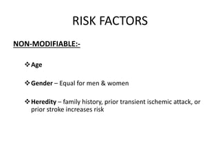
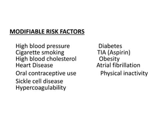
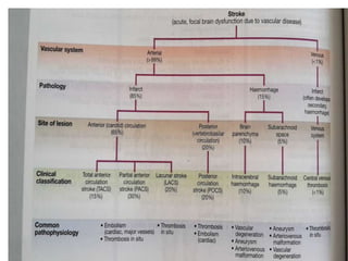
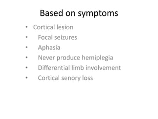
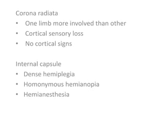
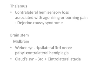
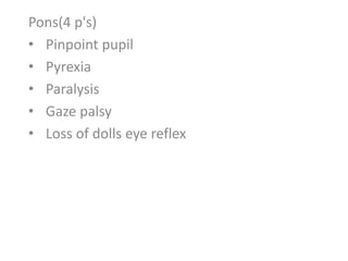
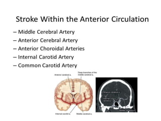
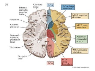
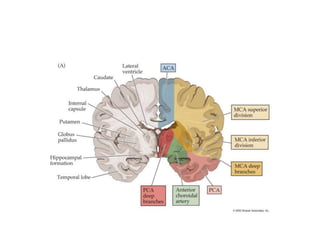
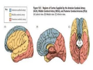
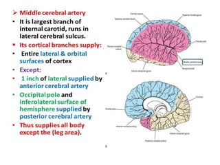
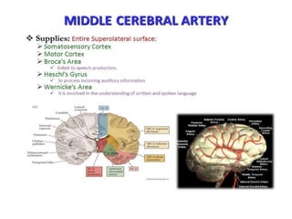
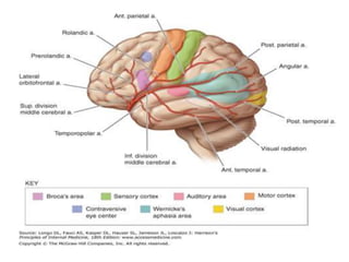
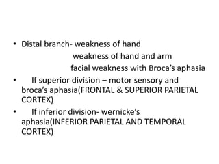
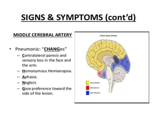
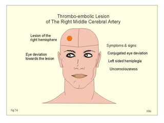
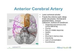
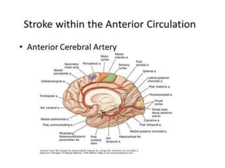
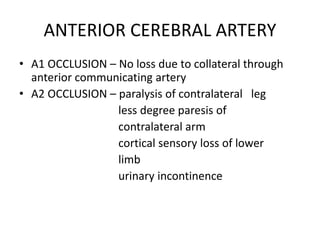
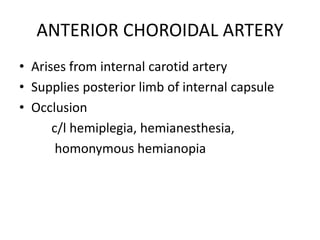
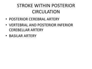
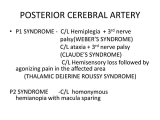
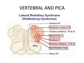
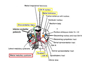
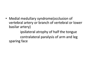
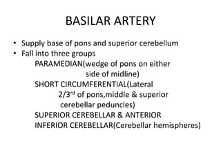
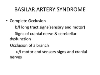
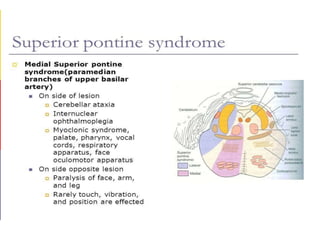
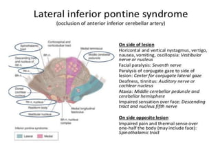
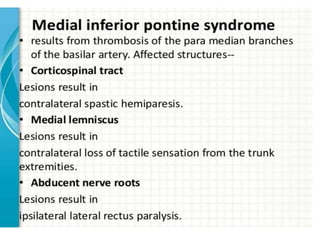
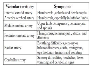
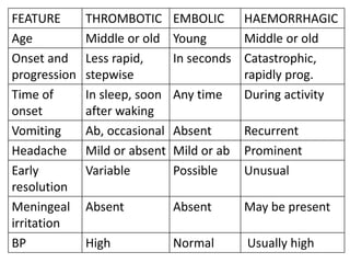
LOCALISATION OF STROKE
• Abrupt onset of neurological deficit that is
attributable to a focal vascular cause
• Signs and symptoms last more than 24 hours
RISK FACTORS
NON-MODIFIABLE:-
Age
Gender – Equal for men & women
Heredity – family history, prior transient ischemic attack, or
prior stroke increases risk
MODIFIABLE RISK FACTORS
High blood pressure Diabetes
Cigarette smoking TIA (Aspirin)
High blood cholesterol Obesity
Heart Disease Atrial fibrillation
Oral contraceptive use Physical inactivity
Sickle cell disease
Hypercoagulability
Based on symptoms
• Cortical lesion
• Focal seizures
• Aphasia
• Never produce hemiplegia
• Differential limb involvement
• Cortical senory loss
Corona radiata
• One limb more involved than other
• Cortical sensory loss
• No cortical signs
Internal capsule
• Dense hemiplegia
• Homonymous hemianopia
• Hemianesthesia
Thalamus
• Contralateral hemisensory loss
associated with agonising or burning pain
- Dejerine roussy syndrome
Brain stem
Midbrain
• Weber syn. -Ipsilateral 3rd nerve
palsy+contralateral hemiplegia
• Claud's syn - 3rd + Cintrolateral ataxia
Pons(4 p's)
• Pinpoint pupil
• Pyrexia
• Paralysis
• Gaze palsy
• Loss of dolls eye reflex
Stem occlusion
contralateral hemiplegia
contralateral hemianeasthesia
homonymous hemianopia
conjugate gaze palsy
dysarthria due to facial weakness
Dominant – global aphasia
Nondominant- constructional apraxia
• Distal branch- weakness of hand
weakness of hand and arm
facial weakness with Broca’s aphasia
• If superior division – motor sensory and
broca’s aphasia(FRONTAL & SUPERIOR PARIETAL
CORTEX)
• If inferior division- wernicke’s
aphasia(INFERIOR PARIETAL AND TEMPORAL
CORTEX)
ANTERIOR CEREBRAL ARTERY
• A1 OCCLUSION – No loss due to collateral through
anterior communicating artery
• A2 OCCLUSION – paralysis of contralateral leg
less degree paresis of
contralateral arm
cortical sensory loss of lower
limb
urinary incontinence
ANTERIOR CHOROIDAL ARTERY
• Arises from internal carotid artery
• Supplies posterior limb of internal capsule
• Occlusion
c/l hemiplegia, hemianesthesia,
homonymous hemianopia
STROKE WITHIN POSTERIOR
CIRCULATION
• POSTERIOR CEREBRAL ARTERY
• VERTEBRAL AND POSTERIOR INFERIOR
CEREBELLAR ARTERY
• BASILAR ARTERY
POSTERIOR CEREBRAL ARTERY
• P1 SYNDROME - C/L Hemiplegia + 3rd nerve
palsy(WEBER’S SYNDROME)
C/L ataxia + 3rd nerve palsy
(CLAUDE’S SYNDROME)
C/L Hemisensory loss followed by
agonizing pain in the affected area
(THALAMIC DEJERINE ROUSSY SYNDROME)
P2 SYNDROME -C/L homonymous
hemianopia with macula sparing
VERTEBRAL AND PICA
LATERAL MEDULLARY SYNDROME(occlusion of
vertebral, posterior inferior cerebellar,
superior, middle or inferior lateral medullary
artery)
Vertigo, Numbness of ipsilateral face
and c/l limb,diplopia, dysphagia, dysarthria,
hoarseness,ipsilateral horner’s syndrome
• Medial medullary syndrome(occlusion of
vertebral artery or branch of vertebral or lower
basilar artery)
ipsilateral atrophy of half the tongue
contralateral paralysis of arm and leg
sparing face
BASILAR ARTERY
• Supply base of pons and superior cerebellum
• Fall into three groups
PARAMEDIAN(wedge of pons on either
side of midline)
SHORT CIRCUMFERENTIAL(Lateral
2/3rd of pons,middle & superior
cerebellar peduncles)
SUPERIOR CEREBELLAR & ANTERIOR
INFERIOR CEREBELLAR(Cerebellar hemispheres)
BASILAR ARTERY SYNDROME
• Complete Occlusion
b/l long tract signs(sensory and motor)
Signs of cranial nerve & cerebellar
dysfunction
Occlusion of a branch
u/l motor and sensory signs and cranial
nerves
FEATURE THROMBOTIC EMBOLIC HAEMORRHAGIC
Age Middle or old Young Middle or old
Onset and
progression
Less rapid,
stepwise
In seconds Catastrophic,
rapidly prog.
Time of
onset
In sleep, soon
after waking
Any time During activity
Vomiting Ab, occasional Absent Recurrent
Headache Mild or absent Mild or ab Prominent
Early
resolution
Variable Possible Unusual
Meningeal
irritation
Absent Absent May be present
BP High Normal Usually high
FEATURE THROMBOTIC EMBOLIC HEMORRHAGIC
Carotid
bruit
Highly
supportive
Possible Not seen
CT Scan Pale infarct Pale infarct Haemorrhage
CSF Usually normal Normal Blood stained,
increased
presurre
THANK YOU

Localisation of stroke

  • 1.
  • 2.
    • Abrupt onsetof neurological deficit that is attributable to a focal vascular cause • Signs and symptoms last more than 24 hours
  • 3.
    RISK FACTORS NON-MODIFIABLE:- Age Gender –Equal for men & women Heredity – family history, prior transient ischemic attack, or prior stroke increases risk
  • 4.
    MODIFIABLE RISK FACTORS Highblood pressure Diabetes Cigarette smoking TIA (Aspirin) High blood cholesterol Obesity Heart Disease Atrial fibrillation Oral contraceptive use Physical inactivity Sickle cell disease Hypercoagulability
  • 6.
    Based on symptoms •Cortical lesion • Focal seizures • Aphasia • Never produce hemiplegia • Differential limb involvement • Cortical senory loss
  • 7.
    Corona radiata • Onelimb more involved than other • Cortical sensory loss • No cortical signs Internal capsule • Dense hemiplegia • Homonymous hemianopia • Hemianesthesia
  • 8.
    Thalamus • Contralateral hemisensoryloss associated with agonising or burning pain - Dejerine roussy syndrome Brain stem Midbrain • Weber syn. -Ipsilateral 3rd nerve palsy+contralateral hemiplegia • Claud's syn - 3rd + Cintrolateral ataxia
  • 9.
    Pons(4 p's) • Pinpointpupil • Pyrexia • Paralysis • Gaze palsy • Loss of dolls eye reflex
  • 17.
    Stem occlusion contralateral hemiplegia contralateralhemianeasthesia homonymous hemianopia conjugate gaze palsy dysarthria due to facial weakness Dominant – global aphasia Nondominant- constructional apraxia
  • 18.
    • Distal branch-weakness of hand weakness of hand and arm facial weakness with Broca’s aphasia • If superior division – motor sensory and broca’s aphasia(FRONTAL & SUPERIOR PARIETAL CORTEX) • If inferior division- wernicke’s aphasia(INFERIOR PARIETAL AND TEMPORAL CORTEX)
  • 23.
    ANTERIOR CEREBRAL ARTERY •A1 OCCLUSION – No loss due to collateral through anterior communicating artery • A2 OCCLUSION – paralysis of contralateral leg less degree paresis of contralateral arm cortical sensory loss of lower limb urinary incontinence
  • 24.
    ANTERIOR CHOROIDAL ARTERY •Arises from internal carotid artery • Supplies posterior limb of internal capsule • Occlusion c/l hemiplegia, hemianesthesia, homonymous hemianopia
  • 25.
    STROKE WITHIN POSTERIOR CIRCULATION •POSTERIOR CEREBRAL ARTERY • VERTEBRAL AND POSTERIOR INFERIOR CEREBELLAR ARTERY • BASILAR ARTERY
  • 27.
    POSTERIOR CEREBRAL ARTERY •P1 SYNDROME - C/L Hemiplegia + 3rd nerve palsy(WEBER’S SYNDROME) C/L ataxia + 3rd nerve palsy (CLAUDE’S SYNDROME) C/L Hemisensory loss followed by agonizing pain in the affected area (THALAMIC DEJERINE ROUSSY SYNDROME) P2 SYNDROME -C/L homonymous hemianopia with macula sparing
  • 28.
  • 29.
    LATERAL MEDULLARY SYNDROME(occlusionof vertebral, posterior inferior cerebellar, superior, middle or inferior lateral medullary artery) Vertigo, Numbness of ipsilateral face and c/l limb,diplopia, dysphagia, dysarthria, hoarseness,ipsilateral horner’s syndrome
  • 31.
    • Medial medullarysyndrome(occlusion of vertebral artery or branch of vertebral or lower basilar artery) ipsilateral atrophy of half the tongue contralateral paralysis of arm and leg sparing face
  • 32.
    BASILAR ARTERY • Supplybase of pons and superior cerebellum • Fall into three groups PARAMEDIAN(wedge of pons on either side of midline) SHORT CIRCUMFERENTIAL(Lateral 2/3rd of pons,middle & superior cerebellar peduncles) SUPERIOR CEREBELLAR & ANTERIOR INFERIOR CEREBELLAR(Cerebellar hemispheres)
  • 33.
    BASILAR ARTERY SYNDROME •Complete Occlusion b/l long tract signs(sensory and motor) Signs of cranial nerve & cerebellar dysfunction Occlusion of a branch u/l motor and sensory signs and cranial nerves
  • 42.
    FEATURE THROMBOTIC EMBOLICHAEMORRHAGIC Age Middle or old Young Middle or old Onset and progression Less rapid, stepwise In seconds Catastrophic, rapidly prog. Time of onset In sleep, soon after waking Any time During activity Vomiting Ab, occasional Absent Recurrent Headache Mild or absent Mild or ab Prominent Early resolution Variable Possible Unusual Meningeal irritation Absent Absent May be present BP High Normal Usually high
  • 43.
    FEATURE THROMBOTIC EMBOLICHEMORRHAGIC Carotid bruit Highly supportive Possible Not seen CT Scan Pale infarct Pale infarct Haemorrhage CSF Usually normal Normal Blood stained, increased presurre
  • 44.