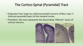
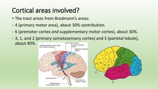
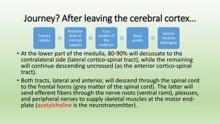
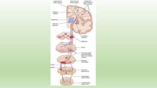
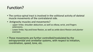
“Limb Weakness”
Part I
Osama Shukir Muhammed Amin
MBChB, MD, MRCP, FACP, FAHA, FCCP(USA), FRCP(Edin),
FRCP(Glasg), FRCP(Ire), FRCP(Lond)
Associate Professor of Neurology
School of Medicine, International Medical University,
Malaysia
Pepe: Neurology…I hate you!
Neurology: Good…
Move Your Limb!
We will discuss upper motor neuron
lesion’s weakness, i.e., central.
The Cortico-Spinal (Pyramidal) Tract
• Originates from large (so-called pyramidal) neurons of Betz, layer V
(internal pyramidal layer) of the cerebral cortex.
• Therefore, this tract represents the descending “efferent” axons of
cortical neurons.
Cortical areas involved?
• The tract arises from Brodmann’s areas:
- 4 (primary motor area), about 30% contribution.
- 6 (premotor cortex and supplementary motor cortex), about 30%.
- 3, 1, and 2 (primary somatosensory cortex) and 5 (parietal lobule),
about 40%.
Journey? After leaving the cerebral cortex…
• At the lower part of the medulla, 80-90% will decussate to the
contralateral side (lateral cortico-spinal tract), while the remaining
will continue descending uncrossed (as the anterior cortico-spinal
tract).
• Both tracts, lateral and anterior, will descend through the spinal cord
to the frontal horns (grey matter of the spinal cord). The latter will
send efferent fibers through the nerve roots (ventral rami), plexuses,
and peripheral nerves to supply skeletal muscles at the motor end-
plate (acetylcholine is the neurotransmitter).
Corona
radiata
Posterior
limb of
internal
capsule
Crus
cerebri of
the
midbrain
Basis
pontis
Ventral
medulla
oblongata
Function?
• The cortico-spinal tract is involved in the volitional activity of skeletal
muscle movements of the contralateral side.
• Antigravity muscles and movements?
- upper limbs: shoulder abduction, as well as elbow, wrist, and fingers
extensions.
- Lower limbs: hip and knee flexion, as well as ankle dorsi-flexion and planter
eversion.
• These movements are further controlled/modulated by the
extrapyramidal and cerebellar systems, with respect to initiation,
coordination, speed, tone, etc.
Cortical motor homunculus? Map?
Lesions of cortico-spinal tract?
• This would result in loss of function of that tract with secondary
dominance of extrapyramidal and other tracts’ functions, e.g.,
rubrospinal, vestibulospinal, etc.
• The resulting clinical features depend on the etiology of the lesion,
onset, severity, multiplicity, and association with other lesions within
and outside the CNS (i.e., peripheral nervous system).
Pyramidal system damage would result in…?
• Weakness or paresis of the targeted movement (there is no complete
paralysis).
• Reduced control of volitional movements, especially of fine dexterity, e.g.,
buttoning and unbuttoning.
• Hypertonia of clasp-knife spasticity and “sustained” clonus.
• Exaggerated deep tendon reflexes.
• Extensor planter reflex (Babinski sign), Hoffman sign, pronator drift, etc.
• No or very minimal atrophy. Disuse atrophy occurs in longstanding cases.
That is to say, signs of upper motor neuron lesion!
Any lesion from the cerebral cortex down to the anterior horn cells of the
spinal cord!
Clinically?
Muscle Power
The Medical Research Council (MRC) Scale of Muscle Strength/Power
6 Grades, from:
0 (no movement at all).
1 (flickering or trace of movement)
2 (active movement with gravity eliminated).
3 (active movement against gravity but not resistance).
4 (active movement against gravity but not full resistance)
5 (full movement against full resistance).
Distribution of weakness?
Whole limb. The weakness can be:
One limb: monoparesis.
Two limbs, same side: hemiparesis.
Both lower limbs: paraparesis.
All four limbs: quadriparesis (or tetrapartesis).
Two (contralateral to each other) or three limbs (upper
and lower limbs): e.g., right upper limb and left lower limb
or left arm and both legs, both arms and one leg.
Patterned weakness?
YES!
The pattern of pyramidal weakness is weakness of
upper limbs’ extensors and lower limbs’ flexors.
For instance, left-sided pyramidal weakness,
grade 3 power in both upper and lower limbs.
Other patterned weakness?
Yes, but non-pyramidal patterns!
Will be discussed in the 2nd lecture.
Factors influencing weakness?
In long-standing cases:
Spasticity (which is usually painful) may be
overshadow the weakness. Contractures and
“disuse atrophy” may further complicate the
picture.
Etiology, onset, course?
Any lesion to the pyramidal tract from the cerebral cortex
down to the ventral grey horns of the spinal cord can result in
weakness.
The resulting weakness can be:
• Acute, subacute, or chronic.
• Reversible, stable non-reversible, fluctuating, stuttering or
step-ladder, or progressive.
• Associated signs and symptoms (such as severe cerebellar
dysfunction or involuntary movements?
• All depend on where is the lesion, what is the lesion, and
why?
Onset?
• Acute: something of a sudden or abrupt onset,
typically vascular (stroke), inflammatory,
demyelination, trauma, etc.
• Subacute: usually few days to few weeks (the
definition depends on the etiology, e.g., subacute
subdural hematoma is from 3 days to 21 days).
• Chronic: usually more than few weeks (?etiology)
typically degenerative, benign tumors, etc.
Course?
•Variable degree of improvement in muscle
power may appear through out the course,
depending on the underlying etiology and
compensatory mechanisms.
•Or else, the course is relentlessly progressive.
Always look for other signs and symptoms?
•These help localize the lesion (hemispheric,
brainstem, and/or spinal cord).
•Choose the appropriate diagnostic
investigation(s) (e.g., brain versus spinal MRI;
cervical MRI versus dorsal MRI).
•Guide the priority in the managing plan (rapid
life-saving treatment, physiotherapy, follow-up,
etc.).
Localization of the weakness?
• Hemiparesis: depends on whether facial weakness is present or not.
• No facial weakness the lesion is below the lower pons.
(Remember, brainstem lesions can result in “crossed signs”)
Signs ipsilateral and contralateral to the lesion
(out of the scope for undergraduate students)
Upper and lower limbs on one side?
Say, left hemiparesis
Contralateral to the lesion Ipsilateral to the lesion
From the cerebral cortex down to Below pyramidal decussation to C5 cord
just before the medullary
(pyramidal) decussation
NB: Lesions at the level of the pyramidal decussation, e.g., foramen magnum
lesions, are beyond undergraduates!
Causes of hemiparesis?
Unilateral lesions:
Stroke, trauma, tumor, abscess, etc.
Both lower limbs, .e.g., spastic paraparesis
• Right and left halves of the spinal cord, from T2 downwards to L1/2.
Lesions within the cord from L2 downwards will cause more distal
weakness.
• The lesion could be symmetrical or asymmetrical.
• Etiology: demyelination, tumor, abscess, trauma, central disc
prolapse, anterior spinal artery occlusion, etc.
• Look for sphincter disturbances and sensory level on the trunk!
NB: Uncommon causes (beyond localization of T2 to L1/2): e.g.,
anterior midline frontal mass (both leg cortical areas will be damaged),
degenerative (e.g., hereditary spastic paraparesis), and spastic diplegia
(of cerebral palsy), vitamin B12 deficiency (subacute combined
degeneration of the cord).
Spastic monoparesis?
•The lesion is a small one, inflicting the upper or
lower limb’s cortical area (map). This will cause
contralateral spastic monoparesis.
•Hemisectioning of the spinal cord (Brown-
Séquard syndrome), due to trauma, tumors, etc.
There is ipsilateral spastic leg weakness.
•Other causes: yes, beyond you!
Spastic quadriparesis?
• Bilateral corticospinal lesions above C5 spinal cord.
• Most of these lesions damage the pyramidal tracts
when they become close to each other, e.g., at the
brainstem or high cervical cord.
• Etiology: Brainstem stroke and brainstem (posterior
fossa) tumors.
Two (apart from hemiparesis or paraparesis) or
three limbs?
Beyond undergraduates!
Investigations(s)?
•The 3 questions are, after taking a thorough
history and doing neurological examination?
Where is the lesion?
What is the lesion?
And why?
The list of “diagnostic investigations” in upper
motor neuron lesions?
• Depends on the overall clinical presentation and neuroligcal findings.
• Whether there is/was any prior neurological insult (e.g., old stroke).
• And your provisional diagnosis!
Some investigations :
- Are unsuitable for clinically unstable critical patients, e.g. brain MRI in
patients with delirium.
- Rapid access, availability, cost, etc.
- Remember contraindications of certain tests, e.g. contrast use in
brain CT scan in patients with mild renal impairment or cervical spine
MRI for a patient with hip prosthesis.
Paresis, where?
Hemiparesis with facial weakness: brain CT scan or brain MRI, with or
without contrast. Hemiparesis without facial weakness: Brain/upper
cervical MRI, with or without contrast.
NB: Urgent non-contrast brain CT scan is the most suitable imaging
modality in patients with acute hemiparesis because of its availability in
Emergency Departments (usually), and it is rapid (the period of the test
is very brief (compared to MRI). Drawback: poor visualization of
posterior fossa structures because of boney artifacts, except in
posterior fossa hemorrhages.
Paraparesis: dorsal spine MRI, with or without contrast or in some
cases, brain/cervical MRI (with or without contrast).
Other pareses?
Quadriparesis: brain/high cervical cord MRI, with or
without contrast. Brain CT scan can also be used in the
acute setting; depending on the etiology, it may reveal
a diagnosis, e.g., pontine hemorrhage.
Other pareses: Beyond undergraduates!
Other investigations to consolidate the
diagnosis?
• Brain MRV (magnetic resonance venography) and
MRA (magnetic resonance angiography).
• Conventional (4-vessel) cerebral angiography.
• CSF analysis.
• Evoked potentials (e.g., visual evoked potentials in
multiple sclerosis).
• …etc.
Treatment of upper motor neuron weakness?
• Depends on the etiology: specific or symptomatic.
• Treatment of spasticity and contractures: medications,
physiotherapy, surgical intervention, intrathecal
baclofen pump, etc.
• Treatment of associated features, e.g., hyper-reflexic
urinary bladder, dysthesic pain, dystonia/chorea, etc.
Examples…
Get ready!
True or false?
The cortico-spinal tract contains fibers from the premotor cerebral cortex
only?
The corticospinal tracts connect the cerebral cortex with the posterior grey
horn of the spinal cord?
Lesions of the corticospinal tracts result in early and prominent muscle
atrophy?
Early in the course of corticospinal tracts, muscle contractures overshadow
the accompanying weakness?
The resulting upper motor neuron signs should be contralateral to the
culprit lesion?
Quadriparesis indicates a lesion below T2 of the spinal cord?
MRI of the cervico-dorsal spine may be unremarkable in slowly progressive
spastic paraparesis?
Brain CT is the first-line investigation in patients with acute hemiparesis?
Cuneiform inscriptions on gold earrings stating that these earrings
were gifts from King Shulgi of Ur. Ur III Dynasty, circa 2100 BCE.
From Mesopotamia, Southern Iraq. The Sulaymaniyah Museum,
Iraq.

Limb Weakness Part I

  • 1.
    “Limb Weakness” Part I OsamaShukir Muhammed Amin MBChB, MD, MRCP, FACP, FAHA, FCCP(USA), FRCP(Edin), FRCP(Glasg), FRCP(Ire), FRCP(Lond) Associate Professor of Neurology School of Medicine, International Medical University, Malaysia
  • 2.
    Pepe: Neurology…I hateyou! Neurology: Good…
  • 3.
  • 4.
    We will discussupper motor neuron lesion’s weakness, i.e., central.
  • 5.
    The Cortico-Spinal (Pyramidal)Tract • Originates from large (so-called pyramidal) neurons of Betz, layer V (internal pyramidal layer) of the cerebral cortex. • Therefore, this tract represents the descending “efferent” axons of cortical neurons.
  • 6.
    Cortical areas involved? •The tract arises from Brodmann’s areas: - 4 (primary motor area), about 30% contribution. - 6 (premotor cortex and supplementary motor cortex), about 30%. - 3, 1, and 2 (primary somatosensory cortex) and 5 (parietal lobule), about 40%.
  • 7.
    Journey? After leavingthe cerebral cortex… • At the lower part of the medulla, 80-90% will decussate to the contralateral side (lateral cortico-spinal tract), while the remaining will continue descending uncrossed (as the anterior cortico-spinal tract). • Both tracts, lateral and anterior, will descend through the spinal cord to the frontal horns (grey matter of the spinal cord). The latter will send efferent fibers through the nerve roots (ventral rami), plexuses, and peripheral nerves to supply skeletal muscles at the motor end- plate (acetylcholine is the neurotransmitter). Corona radiata Posterior limb of internal capsule Crus cerebri of the midbrain Basis pontis Ventral medulla oblongata
  • 9.
    Function? • The cortico-spinaltract is involved in the volitional activity of skeletal muscle movements of the contralateral side. • Antigravity muscles and movements? - upper limbs: shoulder abduction, as well as elbow, wrist, and fingers extensions. - Lower limbs: hip and knee flexion, as well as ankle dorsi-flexion and planter eversion. • These movements are further controlled/modulated by the extrapyramidal and cerebellar systems, with respect to initiation, coordination, speed, tone, etc.
  • 10.
  • 11.
    Lesions of cortico-spinaltract? • This would result in loss of function of that tract with secondary dominance of extrapyramidal and other tracts’ functions, e.g., rubrospinal, vestibulospinal, etc. • The resulting clinical features depend on the etiology of the lesion, onset, severity, multiplicity, and association with other lesions within and outside the CNS (i.e., peripheral nervous system).
  • 12.
    Pyramidal system damagewould result in…? • Weakness or paresis of the targeted movement (there is no complete paralysis). • Reduced control of volitional movements, especially of fine dexterity, e.g., buttoning and unbuttoning. • Hypertonia of clasp-knife spasticity and “sustained” clonus. • Exaggerated deep tendon reflexes. • Extensor planter reflex (Babinski sign), Hoffman sign, pronator drift, etc. • No or very minimal atrophy. Disuse atrophy occurs in longstanding cases. That is to say, signs of upper motor neuron lesion! Any lesion from the cerebral cortex down to the anterior horn cells of the spinal cord!
  • 13.
    Clinically? Muscle Power The MedicalResearch Council (MRC) Scale of Muscle Strength/Power 6 Grades, from: 0 (no movement at all). 1 (flickering or trace of movement) 2 (active movement with gravity eliminated). 3 (active movement against gravity but not resistance). 4 (active movement against gravity but not full resistance) 5 (full movement against full resistance).
  • 14.
    Distribution of weakness? Wholelimb. The weakness can be: One limb: monoparesis. Two limbs, same side: hemiparesis. Both lower limbs: paraparesis. All four limbs: quadriparesis (or tetrapartesis). Two (contralateral to each other) or three limbs (upper and lower limbs): e.g., right upper limb and left lower limb or left arm and both legs, both arms and one leg.
  • 15.
    Patterned weakness? YES! The patternof pyramidal weakness is weakness of upper limbs’ extensors and lower limbs’ flexors. For instance, left-sided pyramidal weakness, grade 3 power in both upper and lower limbs.
  • 16.
    Other patterned weakness? Yes,but non-pyramidal patterns! Will be discussed in the 2nd lecture.
  • 17.
    Factors influencing weakness? Inlong-standing cases: Spasticity (which is usually painful) may be overshadow the weakness. Contractures and “disuse atrophy” may further complicate the picture.
  • 18.
    Etiology, onset, course? Anylesion to the pyramidal tract from the cerebral cortex down to the ventral grey horns of the spinal cord can result in weakness. The resulting weakness can be: • Acute, subacute, or chronic. • Reversible, stable non-reversible, fluctuating, stuttering or step-ladder, or progressive. • Associated signs and symptoms (such as severe cerebellar dysfunction or involuntary movements? • All depend on where is the lesion, what is the lesion, and why?
  • 19.
    Onset? • Acute: somethingof a sudden or abrupt onset, typically vascular (stroke), inflammatory, demyelination, trauma, etc. • Subacute: usually few days to few weeks (the definition depends on the etiology, e.g., subacute subdural hematoma is from 3 days to 21 days). • Chronic: usually more than few weeks (?etiology) typically degenerative, benign tumors, etc.
  • 20.
    Course? •Variable degree ofimprovement in muscle power may appear through out the course, depending on the underlying etiology and compensatory mechanisms. •Or else, the course is relentlessly progressive.
  • 21.
    Always look forother signs and symptoms? •These help localize the lesion (hemispheric, brainstem, and/or spinal cord). •Choose the appropriate diagnostic investigation(s) (e.g., brain versus spinal MRI; cervical MRI versus dorsal MRI). •Guide the priority in the managing plan (rapid life-saving treatment, physiotherapy, follow-up, etc.).
  • 22.
    Localization of theweakness? • Hemiparesis: depends on whether facial weakness is present or not. • No facial weakness the lesion is below the lower pons. (Remember, brainstem lesions can result in “crossed signs”) Signs ipsilateral and contralateral to the lesion (out of the scope for undergraduate students)
  • 23.
    Upper and lowerlimbs on one side? Say, left hemiparesis Contralateral to the lesion Ipsilateral to the lesion From the cerebral cortex down to Below pyramidal decussation to C5 cord just before the medullary (pyramidal) decussation NB: Lesions at the level of the pyramidal decussation, e.g., foramen magnum lesions, are beyond undergraduates!
  • 24.
    Causes of hemiparesis? Unilaterallesions: Stroke, trauma, tumor, abscess, etc.
  • 25.
    Both lower limbs,.e.g., spastic paraparesis • Right and left halves of the spinal cord, from T2 downwards to L1/2. Lesions within the cord from L2 downwards will cause more distal weakness. • The lesion could be symmetrical or asymmetrical. • Etiology: demyelination, tumor, abscess, trauma, central disc prolapse, anterior spinal artery occlusion, etc. • Look for sphincter disturbances and sensory level on the trunk! NB: Uncommon causes (beyond localization of T2 to L1/2): e.g., anterior midline frontal mass (both leg cortical areas will be damaged), degenerative (e.g., hereditary spastic paraparesis), and spastic diplegia (of cerebral palsy), vitamin B12 deficiency (subacute combined degeneration of the cord).
  • 26.
    Spastic monoparesis? •The lesionis a small one, inflicting the upper or lower limb’s cortical area (map). This will cause contralateral spastic monoparesis. •Hemisectioning of the spinal cord (Brown- Séquard syndrome), due to trauma, tumors, etc. There is ipsilateral spastic leg weakness. •Other causes: yes, beyond you!
  • 27.
    Spastic quadriparesis? • Bilateralcorticospinal lesions above C5 spinal cord. • Most of these lesions damage the pyramidal tracts when they become close to each other, e.g., at the brainstem or high cervical cord. • Etiology: Brainstem stroke and brainstem (posterior fossa) tumors.
  • 28.
    Two (apart fromhemiparesis or paraparesis) or three limbs? Beyond undergraduates!
  • 29.
    Investigations(s)? •The 3 questionsare, after taking a thorough history and doing neurological examination? Where is the lesion? What is the lesion? And why?
  • 30.
    The list of“diagnostic investigations” in upper motor neuron lesions? • Depends on the overall clinical presentation and neuroligcal findings. • Whether there is/was any prior neurological insult (e.g., old stroke). • And your provisional diagnosis! Some investigations : - Are unsuitable for clinically unstable critical patients, e.g. brain MRI in patients with delirium. - Rapid access, availability, cost, etc. - Remember contraindications of certain tests, e.g. contrast use in brain CT scan in patients with mild renal impairment or cervical spine MRI for a patient with hip prosthesis.
  • 31.
    Paresis, where? Hemiparesis withfacial weakness: brain CT scan or brain MRI, with or without contrast. Hemiparesis without facial weakness: Brain/upper cervical MRI, with or without contrast. NB: Urgent non-contrast brain CT scan is the most suitable imaging modality in patients with acute hemiparesis because of its availability in Emergency Departments (usually), and it is rapid (the period of the test is very brief (compared to MRI). Drawback: poor visualization of posterior fossa structures because of boney artifacts, except in posterior fossa hemorrhages. Paraparesis: dorsal spine MRI, with or without contrast or in some cases, brain/cervical MRI (with or without contrast).
  • 32.
    Other pareses? Quadriparesis: brain/highcervical cord MRI, with or without contrast. Brain CT scan can also be used in the acute setting; depending on the etiology, it may reveal a diagnosis, e.g., pontine hemorrhage. Other pareses: Beyond undergraduates!
  • 33.
    Other investigations toconsolidate the diagnosis? • Brain MRV (magnetic resonance venography) and MRA (magnetic resonance angiography). • Conventional (4-vessel) cerebral angiography. • CSF analysis. • Evoked potentials (e.g., visual evoked potentials in multiple sclerosis). • …etc.
  • 34.
    Treatment of uppermotor neuron weakness? • Depends on the etiology: specific or symptomatic. • Treatment of spasticity and contractures: medications, physiotherapy, surgical intervention, intrathecal baclofen pump, etc. • Treatment of associated features, e.g., hyper-reflexic urinary bladder, dysthesic pain, dystonia/chorea, etc.
  • 35.
  • 41.
    True or false? Thecortico-spinal tract contains fibers from the premotor cerebral cortex only? The corticospinal tracts connect the cerebral cortex with the posterior grey horn of the spinal cord? Lesions of the corticospinal tracts result in early and prominent muscle atrophy? Early in the course of corticospinal tracts, muscle contractures overshadow the accompanying weakness? The resulting upper motor neuron signs should be contralateral to the culprit lesion? Quadriparesis indicates a lesion below T2 of the spinal cord? MRI of the cervico-dorsal spine may be unremarkable in slowly progressive spastic paraparesis? Brain CT is the first-line investigation in patients with acute hemiparesis?
  • 42.
    Cuneiform inscriptions ongold earrings stating that these earrings were gifts from King Shulgi of Ur. Ur III Dynasty, circa 2100 BCE. From Mesopotamia, Southern Iraq. The Sulaymaniyah Museum, Iraq.