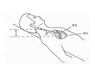
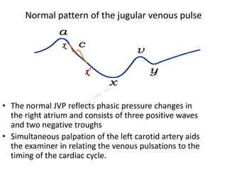
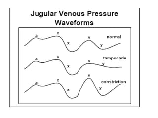
JVP
Definition
• Jugular Venous Pulse:
defined as the oscillating top of vertical
column of blood in right IJV that reflects
pressure changes in Right Atrium in cardiac
cycle.
• Jugular Venous Pressure:
Vertical height of oscillating column of
blood.
Why Internal Jugular Vein?
• IJV has a direct course to RA.
• IJV is anatomically closer to RA.
• IJV has no valves( Valves in EJV prevent
transmission of RA pressure)
• Vasoconstriction Secondary to hypotension
( in CCF) can make EJV small and barely visible.
Why Right Internal Jugular Vein?
• Right jugular veins extend in an almost straight
line to superior vena cava, thus favouring
transmission of the haemodynamic changes
from the right atrium.
• The left innominate vein is not in a straight line
and may be kinked or compressed between
Aortic Arch and sternum, by a dilated aorta, or
by an aneurysm.
Difference from Carotid Pulse
Venous Pulse Carotid Pulse
More lateral Medial
Wavy, Undulant Forceful, Brisk
Decrease with Inspiration No change
Increase in supine position No change
^with abdominal pressure No change
Double Peaked Single Peak
Obliterated with Pressure Cannot be Obliterated
Better Visible Better palpated
Better viewed from foot
end of bed
Method Of Examination
• The patient should lie comfortably during the examination.
• Clothing should be removed from the neck and upper thorax.
• Patient reclining with head elevated 45 °
• Neck should not be sharply flexed.
• Examined effectively by shining a light tangentially across the
neck.
• There should not be any tight bands around abdomen
Observations Made
• the level of venous pressure.
• the type of venous wave pattern.
The level of venous pressure
• Using a centimeter ruler, measure the vertical distance
between the angle of Louis (manubrio sternal joint) and the
highest level of jugular vein pulsation.
• The upper limit of normal is 4 cm above the sternal angle,.
• Add 5 cm to measure central venous pressure since right
atrium is 5 cm below the sternal angle.
• Normal CVP is < 9 cm H2O
Normal pattern of the jugular venous pulse
• The normal JVP reflects phasic pressure changes in
the right atrium and consists of three positive waves
and two negative troughs
• Simultaneous palpation of the left carotid artery aids
the examiner in relating the venous pulsations to the
timing of the cardiac cycle.
a WAVE
• Venous distension due to RA contraction
Retrograde blood flow into SVC and IJV
• Synchronous with S1, Follow P of ECG
• Precede Carotid pulse
• The x descent: is due to
X Atrial relaxation
X` Descent of the floor of the right atrium
during right ventricular systole.
Begins during systole and ends before S2
• The c wave:
Occurs simultaneously with the carotid pulse
Artifact by Carotid pulsation
Bulging of TV into RA during ICP
v WAVE
• Rising right atrial pressure when blood flows into the right
atrium during ventricular systole when the tricuspid valve is
shut.
• Synchronous with Carotid pulse
• Begins in early systole, Peaks after S2 and ends in early
diastole
y DESCENT
• The decline in right atrial pressure when the tricuspid valve
reopens
• Following the bottom of the y descent and before beginning of
the a wave is a period of relatively slow filling of the ventricle,
the diastases period, a wave termed the h wave.
Identifying Wave Forms
• The x descent occurs just prior to the second heart sound (
during systole), while the y descent occurs after the second
heart sound (during diastole).
• Normally X descent is more prominent than Y descent. Y
descent is only sometimes seen during diastole. Descents are
better seen than positive waves.
• The a wave occurs just before the first sound or carotid pulse
and has a sharp rise and fall.
• The v wave occurs just after the arterial pulse and has a slower
undulating pattern.
• The c wave is never seen normally.
Abnormalities of jugular venous pulse
A. Low jugular venous pressure
1. Hypovolaemia.
B. Elevated jugular venous pressure
1. Intravascular volume overload conditions
Right ventricular infarction
Left heart failure
Myocardial infarction.
Valvular Heart Disease
Cardiomyopathy
2. Constrictive pericarditis.
3. Pericardial effusion with tamponade
Elevated “a” wave
Increased Resistance
to RV Filling.
Tricuspid stenosis
R Heart Failure
PS
PAH
Cannon “a” wave
• Atrial-ventricular
Dissociation
(atria contract against
a closed tricuspid
valve)
Complete heart block
VPC
Ventricular
tachycardia
Ventricular pacing
Junctional rhythm
Junctional
tachycardia.
Absent “a” wave
• 1. Atrial fibrillation
Elevated “v” wave
1. Tricuspid regurgitation.
2. Right ventricular failure.
3. Restrictive cardiomyopathy.
4. Cor Pulmonale
Tricuspid regurgitation
• Absent X Decsent
• CV/ Regurgitant Wave
• Has a rounded contour
and a sustained peak
• Followed by a rapid deep
Y descent
• Amplitude of V increases
with inspiration.
• Cause subtle motion of
ear lobe with each heart
beat
“a” wave equal to “v” wave
ASD
Prominent X descent
followed by a large V
wave
M Configuration
Indicates a large L-R shunt
With PAH A wave
becomes more
prominent
If L JVP > R JVP indicates
associated PAPVC
Prominent “x” descent
1. Cardiac tamponade.
2. Constrictive Pericarditis
3. RVMI
4. Restrictive Cardiomyopathy
5. Atrial septal defect
Blunted “x” descent
1. Tricuspid regurgitation.
2. Right atrial ischaemia
Prominent “y” descent
1. Constrictive pericarditis.
2. Tricuspid regurgitation.
3. Atrial septal defect.
1. Cardiac tamponade.
2. Right ventricular infarction
3. Restrictive Cardiomyopathy
Absent “y” descent
Slow “y” descent
1. Tricuspid stenosis.
2. Right atrial myxoma.
Constrictive pericarditis.
• M shaped contour
• Prominent X and Y descent (FRIEDREICH`SIGN)
• Y descent is prominent as ventricular filling is
unimpeded during early diastole.
• This is interrupted by a rapid raise in pressure as the
filling is impeded by constricting Pericardium
• The Ventriclar pressure curve exhibit Square Root sign
Abdomino-jugular reflux
• Is positive when JVP increase after 10 sec of abdominal
pressure followed by a rapid drop in pressure of 4 cm on
release of compression.
• Most common cause of a positive test is RHF
• Positive test in: Borderline elevation of JVP
Silent TR
Latent RHF
• False positive: Fluid overload
• False Negative: SVC/IVC obstruction
Budd Chiari syndrome
• Positive Test imply SVC and IVC are patent
Kussmaul sign
Failure of decline in JVP occur during inspiration.
• Constrictive Pericarditis
• Severe RHF
• Restrictive Cardiomyopathy
• Tricuspid Stenosis
THANK YOU

JVP - A SHORT REVIEW

  • 1.
  • 2.
    Definition • Jugular VenousPulse: defined as the oscillating top of vertical column of blood in right IJV that reflects pressure changes in Right Atrium in cardiac cycle. • Jugular Venous Pressure: Vertical height of oscillating column of blood.
  • 3.
    Why Internal JugularVein? • IJV has a direct course to RA. • IJV is anatomically closer to RA. • IJV has no valves( Valves in EJV prevent transmission of RA pressure) • Vasoconstriction Secondary to hypotension ( in CCF) can make EJV small and barely visible.
  • 4.
    Why Right InternalJugular Vein? • Right jugular veins extend in an almost straight line to superior vena cava, thus favouring transmission of the haemodynamic changes from the right atrium. • The left innominate vein is not in a straight line and may be kinked or compressed between Aortic Arch and sternum, by a dilated aorta, or by an aneurysm.
  • 7.
    Difference from CarotidPulse Venous Pulse Carotid Pulse More lateral Medial Wavy, Undulant Forceful, Brisk Decrease with Inspiration No change Increase in supine position No change ^with abdominal pressure No change Double Peaked Single Peak Obliterated with Pressure Cannot be Obliterated Better Visible Better palpated Better viewed from foot end of bed
  • 8.
    Method Of Examination •The patient should lie comfortably during the examination. • Clothing should be removed from the neck and upper thorax. • Patient reclining with head elevated 45 ° • Neck should not be sharply flexed. • Examined effectively by shining a light tangentially across the neck. • There should not be any tight bands around abdomen
  • 9.
    Observations Made • thelevel of venous pressure. • the type of venous wave pattern.
  • 10.
    The level ofvenous pressure • Using a centimeter ruler, measure the vertical distance between the angle of Louis (manubrio sternal joint) and the highest level of jugular vein pulsation. • The upper limit of normal is 4 cm above the sternal angle,. • Add 5 cm to measure central venous pressure since right atrium is 5 cm below the sternal angle. • Normal CVP is < 9 cm H2O
  • 12.
    Normal pattern ofthe jugular venous pulse • The normal JVP reflects phasic pressure changes in the right atrium and consists of three positive waves and two negative troughs • Simultaneous palpation of the left carotid artery aids the examiner in relating the venous pulsations to the timing of the cardiac cycle.
  • 13.
    a WAVE • Venousdistension due to RA contraction Retrograde blood flow into SVC and IJV • Synchronous with S1, Follow P of ECG • Precede Carotid pulse
  • 14.
    • The xdescent: is due to X Atrial relaxation X` Descent of the floor of the right atrium during right ventricular systole. Begins during systole and ends before S2 • The c wave: Occurs simultaneously with the carotid pulse Artifact by Carotid pulsation Bulging of TV into RA during ICP
  • 15.
    v WAVE • Risingright atrial pressure when blood flows into the right atrium during ventricular systole when the tricuspid valve is shut. • Synchronous with Carotid pulse • Begins in early systole, Peaks after S2 and ends in early diastole
  • 16.
    y DESCENT • Thedecline in right atrial pressure when the tricuspid valve reopens • Following the bottom of the y descent and before beginning of the a wave is a period of relatively slow filling of the ventricle, the diastases period, a wave termed the h wave.
  • 18.
    Identifying Wave Forms •The x descent occurs just prior to the second heart sound ( during systole), while the y descent occurs after the second heart sound (during diastole). • Normally X descent is more prominent than Y descent. Y descent is only sometimes seen during diastole. Descents are better seen than positive waves. • The a wave occurs just before the first sound or carotid pulse and has a sharp rise and fall. • The v wave occurs just after the arterial pulse and has a slower undulating pattern. • The c wave is never seen normally.
  • 19.
    Abnormalities of jugularvenous pulse A. Low jugular venous pressure 1. Hypovolaemia.
  • 20.
    B. Elevated jugularvenous pressure 1. Intravascular volume overload conditions Right ventricular infarction Left heart failure Myocardial infarction. Valvular Heart Disease Cardiomyopathy 2. Constrictive pericarditis. 3. Pericardial effusion with tamponade
  • 21.
    Elevated “a” wave IncreasedResistance to RV Filling. Tricuspid stenosis R Heart Failure PS PAH
  • 22.
    Cannon “a” wave •Atrial-ventricular Dissociation (atria contract against a closed tricuspid valve) Complete heart block VPC Ventricular tachycardia Ventricular pacing Junctional rhythm Junctional tachycardia.
  • 23.
    Absent “a” wave •1. Atrial fibrillation
  • 24.
    Elevated “v” wave 1.Tricuspid regurgitation. 2. Right ventricular failure. 3. Restrictive cardiomyopathy. 4. Cor Pulmonale
  • 25.
    Tricuspid regurgitation • AbsentX Decsent • CV/ Regurgitant Wave • Has a rounded contour and a sustained peak • Followed by a rapid deep Y descent • Amplitude of V increases with inspiration. • Cause subtle motion of ear lobe with each heart beat
  • 26.
    “a” wave equalto “v” wave ASD Prominent X descent followed by a large V wave M Configuration Indicates a large L-R shunt With PAH A wave becomes more prominent If L JVP > R JVP indicates associated PAPVC
  • 27.
    Prominent “x” descent 1.Cardiac tamponade. 2. Constrictive Pericarditis 3. RVMI 4. Restrictive Cardiomyopathy 5. Atrial septal defect Blunted “x” descent 1. Tricuspid regurgitation. 2. Right atrial ischaemia
  • 28.
    Prominent “y” descent 1.Constrictive pericarditis. 2. Tricuspid regurgitation. 3. Atrial septal defect. 1. Cardiac tamponade. 2. Right ventricular infarction 3. Restrictive Cardiomyopathy Absent “y” descent Slow “y” descent 1. Tricuspid stenosis. 2. Right atrial myxoma.
  • 29.
    Constrictive pericarditis. • Mshaped contour • Prominent X and Y descent (FRIEDREICH`SIGN) • Y descent is prominent as ventricular filling is unimpeded during early diastole. • This is interrupted by a rapid raise in pressure as the filling is impeded by constricting Pericardium • The Ventriclar pressure curve exhibit Square Root sign
  • 31.
    Abdomino-jugular reflux • Ispositive when JVP increase after 10 sec of abdominal pressure followed by a rapid drop in pressure of 4 cm on release of compression. • Most common cause of a positive test is RHF • Positive test in: Borderline elevation of JVP Silent TR Latent RHF • False positive: Fluid overload • False Negative: SVC/IVC obstruction Budd Chiari syndrome • Positive Test imply SVC and IVC are patent
  • 32.
    Kussmaul sign Failure ofdecline in JVP occur during inspiration. • Constrictive Pericarditis • Severe RHF • Restrictive Cardiomyopathy • Tricuspid Stenosis
  • 33.