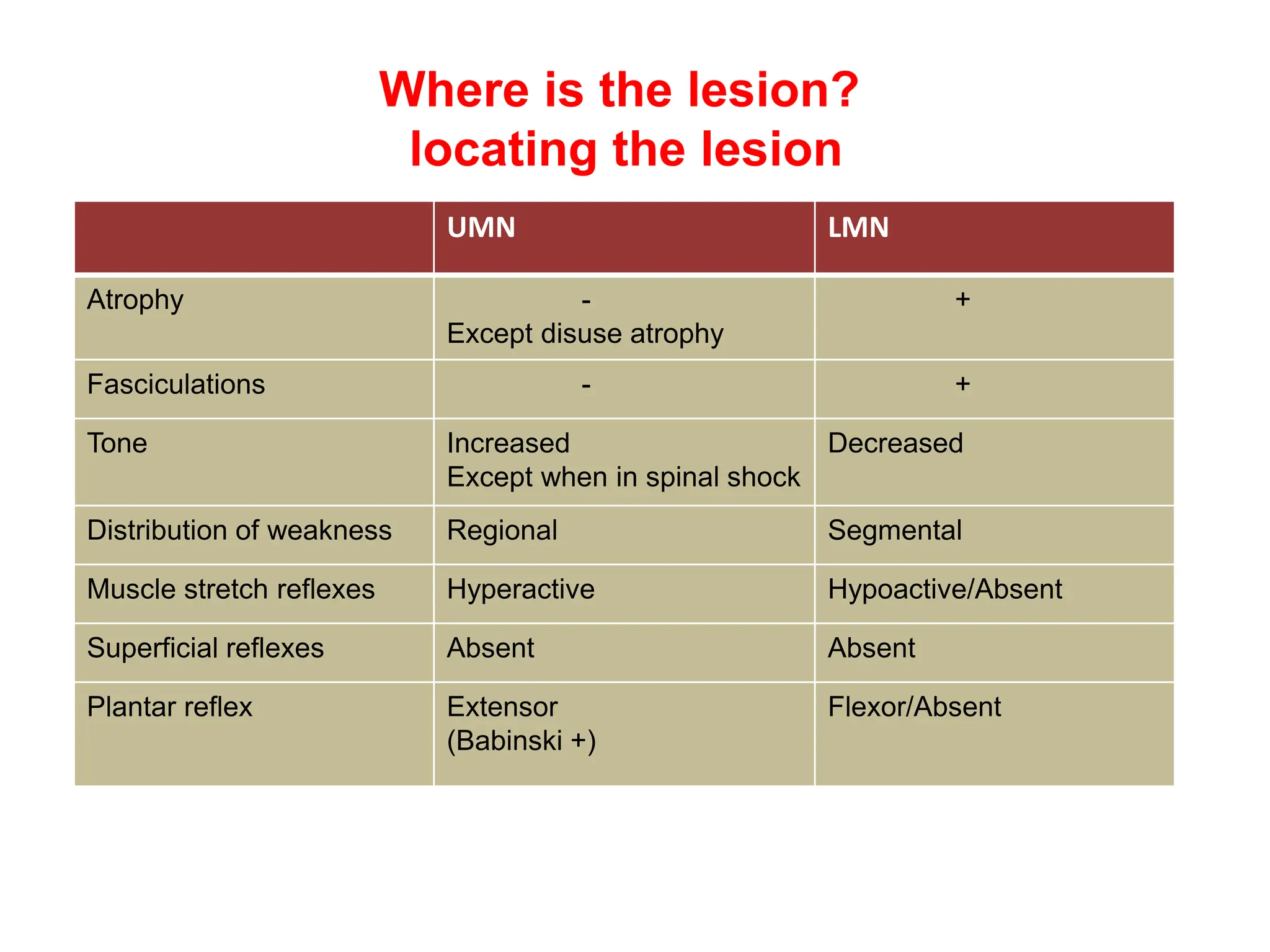
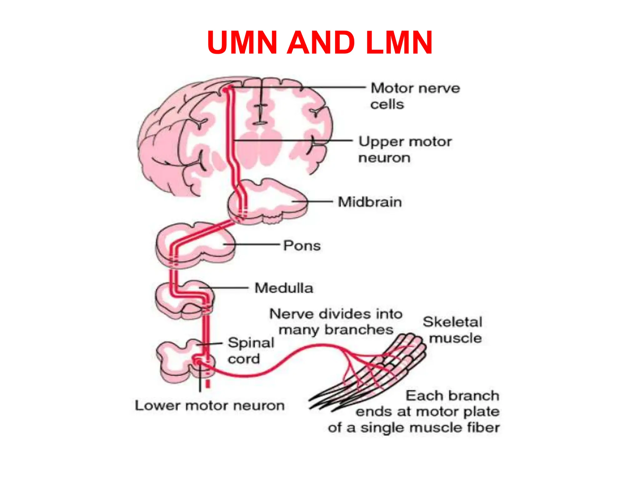
APPROACH TO NEUROLOGICAL
WEAKNESS
Dr.G.VENKATA RAMANA
MBBS DNB FAMILY MEDICINE
• Weakness
• Reduction in the power that can be exerted by one or
more muscles
• Fatigability
• Inability to sustain the performance of an activity that
should be normal for a person of the same age, sex, and
size
• Bradykinesia
• Increased time is required for full power to be exerted
• Apraxia
• A disorder of planning and initiating a skilled or learned
movement unrelated to a significant motor or sensory
deficit
• Paralysis or Plegia  Weakness so severe that a
muscle cannot be contracted at all
• Paresis  Less severe weakness
• Hemi  One-half of the body
• Para  Both legs
• Quadri  All four limbs
• Distribution of weakness
• Weakness from involvement of upper motor neurons
occurs particularly in the extensors and abductors of the
upper limb and the flexors of the lower limb
• Lower motor neuron weakness depends on whether
involvement is at the level of the anterior horn cells,
nerve root, limb plexus, or peripheral nerve
• Myopathic weakness is generally most marked in
proximal muscles
• Weakness from impaired neuromuscular transmission
has no specific pattern of involvement
• Muscle bulk
• Not affected by upper motor neuron lesions,mild disuse atrophy
eventually may occur
• Atrophy seen in lower motor neuron lesion , Advanced muscle disease
• Tone
• Resistance of a muscle to passive stretch
• Increased in UMN lesions and decreased in LMN lesions
• Types
• Spasticity
• Rigidity
• Paratonia
• Flaccidity
• Muscle stretch (tendon) reflexes
• Increased with upper motor neuron lesions
• Decreased or absent for a variable period immediately after
onset of an acute lesion
• Hyperreflexia is usually,but not invariably accompanied by
loss of cutaneous reflexes (such as superficial abdominals)
and, in particular, by an extensor plantar (Babinski) response
• The muscle stretch reflexes are depressed with lower motor
neuron lesions directly involving specific reflex arcs
• Preserved in patients with myopathic weakness except in
advanced stages, when they sometimes are attenuated
• In disorders of the neuromuscular junction, reflex responses
may be affected by preceding voluntary activity of affected
muscles
• Such activity may lead to enhancement of initially depressed
reflexes in Lambert-Eaton myasthenic syndrome and,
conversely, to depression of initially normal reflexes in
myasthenia gravis
• Weakness
• Reduction in the power that can be exerted by one or more muscles
• Grading
• Grade 5 Normal power
• Grade 4 Movement against resistance
• Grade 3 Movement against gravity
• Grade 2 Gravity eliminated movement (lateral movements in bed)
• Grade 1 There is a visible or palpable flicker of contraction, but no
resultant movement of joint
• Grade 0 Total paralysis
• Grade 4 power covers a broad range – (4 –, 4, and 4+) denoting
movement against slight, moderate and stronger resistance
Where is the lesion?
locating the lesion
UMN LMN
Atrophy -
Except disuse atrophy
+
Fasciculations - +
Tone Increased
Except when in spinal shock
Decreased
Distribution of weakness Regional Segmental
Muscle stretch reflexes Hyperactive Hypoactive/Absent
Superficial reflexes Absent Absent
Plantar reflex Extensor
(Babinski +)
Flexor/Absent
UMN AND LMN
UMN
Lesion Features
Cortex C/L Hemiplegia/Monoplegia
C/L 7th CN UMN
Seizure/Language/Apraxia
Subcortex C/L Hemiplegia/Monoplegia
C/L 7th CN UMN
Transcortical aphasia
Internal capsule C/L Dense Hemiplegia
C/L Hemisensory loss
C/L 7th CN UMN
Homonymous hemianopia
Brain stem
• Mid brain
• Pons
• Medulla
Crossed hemiplegia
Discussed in next slide
Spinal cord I/L Hemiplegia
I/L loss of Posterior column sensation
C/L pain and temperature loss
No Cranial nerve involvement
UMN
Brain stem Features
Mid brain C/L Hemiplegia
C/L 7th CN UMN
I/L Opthalmoplegia
Pons C/L Hemiplegia
I/L 7th CN LMN
+/- 6th nerve palsy
Medulla - Medial Hemiplegia I/L or C/L or Cruciate
I/L LMN 12th cranial nerve palsy
MLF – Internuclear opthalmoplegia
C/L posterior column sensory loss
Medulla- Lateral Crossed sensory (I/L face and C/L body)
Horner’s syndrome
Ataxia 9th and 10th cranial nerve palsy
Hiccups
No hemiplegia
LMN Lesion Features
Anterior horn cells Asymmetrical
Wasting++ > Weakness
Distal > Proximal
Fasciculations ++
Reflexes variable
No sensory/ANS
Spinal nerve root/Plexus Asymmetrical weakness
Proximal > Distal
Reflexes absent
Sensory +,Atrophy +
Peripheral nerve Symmetrical
Weakness +/ - sensory +/-ANS
Distal to proximal
Reflexes absent
Atrophy/Fasciculations+
Neuromuscular junction Generalised
Fatiguability/fluctuating
(affected by preceding muscle activity)
No sensory
Muscle Symmetrical
Weakness (esp.LL),Proximal > distal
No sensory, Reflexes present
No wasting( until late)
No fasciculations
What is the lesion?
• Etiology
• Acute
• Vascular
• Traumatic
• Metabolic
• Demyelinating
• Subacute
• Inflammatory (Demyelinating)
• Infection
• Neoplasm
• Metabolic
• Chronic/Insidious onset
• Degeneration
Patterns/Distribution of weakness
• Hemiparesis
• Paraparesis
• Quadriparesis
• Monoparesis
• Distal weakness
• Proximal weakness
• Restricted distribution
• Ascending vs Descending
Hemiparesis
• Always UMN
• Results from an upper motor neuron lesion above the midcervical
spinal cord
• The presence of other neurologic deficits helps localize the lesion
• C/L Cortex / Subcortex / Internal capsule / Brain stem / I/L Upper
cervical cord
• Acute
• Vascular - Stroke
• Trauma – Head injury
• Todd’s paresis
• Sub acute
• Infection- Meningoencephalitis,Brain abscess
• Demyelination- MS,ADEM
• Tumour,Subdural hematoma
Hemiparesis
• Chronic
• Tumour
• Vascular malformation
• Mill’s hemiplegic variant of MND
• Investigation of hemiparesis of acute origin usually starts with a CT
scan of the brain and laboratory studies
• If the CT is normal, or in subacute or chronic cases of hemiparesis,
MRI of the brain and/or cervical spine (including the foramen
magnum) is performed, depending on the clinical accompaniments
Paraparesis
• Investigations typically begin with spinal MRI, but when upper motor neuron signs are
associated with drowsiness, confusion, seizures, or other hemispheric signs, brain MRI
should also be performed
• Electrophysiologic studies - when clinical findings suggest an underlying neuromuscular
disorder
UMN type LMN type
Acute
• Acute hydrocephalus
• Ant.cerebral artery/SSS thrombosis
• Spinal fracture,Spinal abscess
• Spinal hematoma
• Acute transverse myelitis
• Anterior spinal artery thrombosis
Acute
• UMN in shock
• GBS
• Polio
• Psoas abscess
• hematoma
Sub acute –Chronic
• Parasagittal meningioma
• Hydrocephalus
• MND (ALS)
• Multiple sclerosis
• Friedrich ataxia
• Subacute combined degeneration
Sub acute –Chronic
• CIDP
• Infective radiculitis(HIV,CMV,HTLV)
• MND
• Cauda equina
• Tabes dorsalis
• Myopathies
• Peripheral neuropathies
Quadriparesis
UMN type LMN type
• B/L Brainstem lesion
• CV junction anomaly
• High cervical cord compression
• MS
• MND
• GBS/CIDP
• Polio
• Peripheral neuropathies
• Myopathy
• Myasthenia gravis
• Snake bite
• OPC poisoning
• In obtunded patients, evaluation begins with a CT or MRI scan of the brain
• If upper motor neuron signs are present but the patient is alert, the initial test is
usually an MRI of the cervical cord
• If weakness is lower motor neuron, myopathic, or uncertain in origin, the clinical
approach begins with blood studies to determine the level of muscle enzymes
and electrolytes and with EMG and nerve conduction studies
Monoparesis
Lower limb Upper limb
UMN
• ACA territory stroke
• SSS thrombosis
• Trauma – Frontal lobe
contusion
• Infection – Granuloma in frontal
lobe
UMN
Stroke – Superior division of MCA
Head injury – Parietal contusion
LMN
• Lumbosacral plexopathy
LMN
Trauma – Brachial plexus/injury to
multiple cervical roots
Investigations
UMN signs: Brain CT or MRI
LMN signs: EMG,NCS
Distal weakness
• Anterior horn cells
Asymmetrical
Wasting++ > Weakness
Distal > Proximal
Fasciculations ++
Reflexes variable
No sensory/ANS
• Peripheral nerve
Symmetrical
Weakness +/- Sensory +/- ANS
Distal to Proximal
Reflexes absent
Atrophy/Fasciculations+
Proximal weakness
• Root/Plexus
• Asymmetrical weakness
• Proximal > Distal
• Reflexes absent
• Sensory +
• Atrophy +
• Muscle
• Symmetrical
• Weakness (esp.LL)
• Proximal > Distal
• No sensory
• Reflexes present
• No wasting( until late)/No fasciculations
• Restricted distribution
• Neuropathy
Entrapment
Mononeuritis multiplex
• Intermittent weakness
• Electrolyte disturbances
• Channelopathies
• Metabolic disorders of muscles
• Ascending
• GBS
• Extra medullary compression
• Descending
• Botulism
• Diptheria
• Snake bite
• Intramedullary compression
• Ells berg phenomenon: Anticlock wise
• Compressive myelopathy
Anterior horn cell disease/MND
• Chronic:
• UMN+LMN:Amyotrophic lateral sclerosis
• Pure LMN :X-linked spinobulbar muscular
atrophy(kennedy’s),Adult Tac-Sachs,Spinal muscular
atrophy
• Pure UMN:Primary lateral sclerosis,Hereditary spastic
paraplegia
• Acute:
• Virus (Polio,Herpes Zoster,Coxsackie)
Plexopathy
• Brachial Plexopathy – Neoplasm, Trauma, Brachial
neuritis
• Lumbosacral Plexopathy – Neoplasm, Psoas abscess
Neuromuscular junction disorders
• Myasthenia Gravis (Post synaptic)- Autoimmune
• Lambert Eaton Myasthenic Syndrome (Pre synaptic) –
Paraneoplastic
• Snake bite (History, Descending)
Cobra - Post synaptic
Krait - Pre synaptic
• Botulism (Descending)
• OPC – Intermediate syndrome
APPROACH TO NEUROLOGICAL WEAKNESS.pptx

APPROACH TO NEUROLOGICAL WEAKNESS.pptx

  • 1.
    APPROACH TO NEUROLOGICAL WEAKNESS Dr.G.VENKATARAMANA MBBS DNB FAMILY MEDICINE
  • 2.
    • Weakness • Reductionin the power that can be exerted by one or more muscles • Fatigability • Inability to sustain the performance of an activity that should be normal for a person of the same age, sex, and size • Bradykinesia • Increased time is required for full power to be exerted • Apraxia • A disorder of planning and initiating a skilled or learned movement unrelated to a significant motor or sensory deficit
  • 3.
    • Paralysis orPlegia  Weakness so severe that a muscle cannot be contracted at all • Paresis  Less severe weakness • Hemi  One-half of the body • Para  Both legs • Quadri  All four limbs
  • 4.
    • Distribution ofweakness • Weakness from involvement of upper motor neurons occurs particularly in the extensors and abductors of the upper limb and the flexors of the lower limb • Lower motor neuron weakness depends on whether involvement is at the level of the anterior horn cells, nerve root, limb plexus, or peripheral nerve • Myopathic weakness is generally most marked in proximal muscles • Weakness from impaired neuromuscular transmission has no specific pattern of involvement
  • 5.
    • Muscle bulk •Not affected by upper motor neuron lesions,mild disuse atrophy eventually may occur • Atrophy seen in lower motor neuron lesion , Advanced muscle disease • Tone • Resistance of a muscle to passive stretch • Increased in UMN lesions and decreased in LMN lesions • Types • Spasticity • Rigidity • Paratonia • Flaccidity
  • 6.
    • Muscle stretch(tendon) reflexes • Increased with upper motor neuron lesions • Decreased or absent for a variable period immediately after onset of an acute lesion • Hyperreflexia is usually,but not invariably accompanied by loss of cutaneous reflexes (such as superficial abdominals) and, in particular, by an extensor plantar (Babinski) response • The muscle stretch reflexes are depressed with lower motor neuron lesions directly involving specific reflex arcs • Preserved in patients with myopathic weakness except in advanced stages, when they sometimes are attenuated • In disorders of the neuromuscular junction, reflex responses may be affected by preceding voluntary activity of affected muscles • Such activity may lead to enhancement of initially depressed reflexes in Lambert-Eaton myasthenic syndrome and, conversely, to depression of initially normal reflexes in myasthenia gravis
  • 7.
    • Weakness • Reductionin the power that can be exerted by one or more muscles • Grading • Grade 5 Normal power • Grade 4 Movement against resistance • Grade 3 Movement against gravity • Grade 2 Gravity eliminated movement (lateral movements in bed) • Grade 1 There is a visible or palpable flicker of contraction, but no resultant movement of joint • Grade 0 Total paralysis • Grade 4 power covers a broad range – (4 –, 4, and 4+) denoting movement against slight, moderate and stronger resistance
  • 8.
    Where is thelesion? locating the lesion UMN LMN Atrophy - Except disuse atrophy + Fasciculations - + Tone Increased Except when in spinal shock Decreased Distribution of weakness Regional Segmental Muscle stretch reflexes Hyperactive Hypoactive/Absent Superficial reflexes Absent Absent Plantar reflex Extensor (Babinski +) Flexor/Absent
  • 9.
  • 10.
    UMN Lesion Features Cortex C/LHemiplegia/Monoplegia C/L 7th CN UMN Seizure/Language/Apraxia Subcortex C/L Hemiplegia/Monoplegia C/L 7th CN UMN Transcortical aphasia Internal capsule C/L Dense Hemiplegia C/L Hemisensory loss C/L 7th CN UMN Homonymous hemianopia Brain stem • Mid brain • Pons • Medulla Crossed hemiplegia Discussed in next slide Spinal cord I/L Hemiplegia I/L loss of Posterior column sensation C/L pain and temperature loss No Cranial nerve involvement
  • 11.
    UMN Brain stem Features Midbrain C/L Hemiplegia C/L 7th CN UMN I/L Opthalmoplegia Pons C/L Hemiplegia I/L 7th CN LMN +/- 6th nerve palsy Medulla - Medial Hemiplegia I/L or C/L or Cruciate I/L LMN 12th cranial nerve palsy MLF – Internuclear opthalmoplegia C/L posterior column sensory loss Medulla- Lateral Crossed sensory (I/L face and C/L body) Horner’s syndrome Ataxia 9th and 10th cranial nerve palsy Hiccups No hemiplegia
  • 12.
    LMN Lesion Features Anteriorhorn cells Asymmetrical Wasting++ > Weakness Distal > Proximal Fasciculations ++ Reflexes variable No sensory/ANS Spinal nerve root/Plexus Asymmetrical weakness Proximal > Distal Reflexes absent Sensory +,Atrophy + Peripheral nerve Symmetrical Weakness +/ - sensory +/-ANS Distal to proximal Reflexes absent Atrophy/Fasciculations+ Neuromuscular junction Generalised Fatiguability/fluctuating (affected by preceding muscle activity) No sensory Muscle Symmetrical Weakness (esp.LL),Proximal > distal No sensory, Reflexes present No wasting( until late) No fasciculations
  • 13.
    What is thelesion? • Etiology • Acute • Vascular • Traumatic • Metabolic • Demyelinating • Subacute • Inflammatory (Demyelinating) • Infection • Neoplasm • Metabolic • Chronic/Insidious onset • Degeneration
  • 14.
    Patterns/Distribution of weakness •Hemiparesis • Paraparesis • Quadriparesis • Monoparesis • Distal weakness • Proximal weakness • Restricted distribution • Ascending vs Descending
  • 15.
    Hemiparesis • Always UMN •Results from an upper motor neuron lesion above the midcervical spinal cord • The presence of other neurologic deficits helps localize the lesion • C/L Cortex / Subcortex / Internal capsule / Brain stem / I/L Upper cervical cord • Acute • Vascular - Stroke • Trauma – Head injury • Todd’s paresis • Sub acute • Infection- Meningoencephalitis,Brain abscess • Demyelination- MS,ADEM • Tumour,Subdural hematoma
  • 16.
    Hemiparesis • Chronic • Tumour •Vascular malformation • Mill’s hemiplegic variant of MND • Investigation of hemiparesis of acute origin usually starts with a CT scan of the brain and laboratory studies • If the CT is normal, or in subacute or chronic cases of hemiparesis, MRI of the brain and/or cervical spine (including the foramen magnum) is performed, depending on the clinical accompaniments
  • 17.
    Paraparesis • Investigations typicallybegin with spinal MRI, but when upper motor neuron signs are associated with drowsiness, confusion, seizures, or other hemispheric signs, brain MRI should also be performed • Electrophysiologic studies - when clinical findings suggest an underlying neuromuscular disorder UMN type LMN type Acute • Acute hydrocephalus • Ant.cerebral artery/SSS thrombosis • Spinal fracture,Spinal abscess • Spinal hematoma • Acute transverse myelitis • Anterior spinal artery thrombosis Acute • UMN in shock • GBS • Polio • Psoas abscess • hematoma Sub acute –Chronic • Parasagittal meningioma • Hydrocephalus • MND (ALS) • Multiple sclerosis • Friedrich ataxia • Subacute combined degeneration Sub acute –Chronic • CIDP • Infective radiculitis(HIV,CMV,HTLV) • MND • Cauda equina • Tabes dorsalis • Myopathies • Peripheral neuropathies
  • 18.
    Quadriparesis UMN type LMNtype • B/L Brainstem lesion • CV junction anomaly • High cervical cord compression • MS • MND • GBS/CIDP • Polio • Peripheral neuropathies • Myopathy • Myasthenia gravis • Snake bite • OPC poisoning • In obtunded patients, evaluation begins with a CT or MRI scan of the brain • If upper motor neuron signs are present but the patient is alert, the initial test is usually an MRI of the cervical cord • If weakness is lower motor neuron, myopathic, or uncertain in origin, the clinical approach begins with blood studies to determine the level of muscle enzymes and electrolytes and with EMG and nerve conduction studies
  • 19.
    Monoparesis Lower limb Upperlimb UMN • ACA territory stroke • SSS thrombosis • Trauma – Frontal lobe contusion • Infection – Granuloma in frontal lobe UMN Stroke – Superior division of MCA Head injury – Parietal contusion LMN • Lumbosacral plexopathy LMN Trauma – Brachial plexus/injury to multiple cervical roots Investigations UMN signs: Brain CT or MRI LMN signs: EMG,NCS
  • 20.
    Distal weakness • Anteriorhorn cells Asymmetrical Wasting++ > Weakness Distal > Proximal Fasciculations ++ Reflexes variable No sensory/ANS • Peripheral nerve Symmetrical Weakness +/- Sensory +/- ANS Distal to Proximal Reflexes absent Atrophy/Fasciculations+
  • 21.
    Proximal weakness • Root/Plexus •Asymmetrical weakness • Proximal > Distal • Reflexes absent • Sensory + • Atrophy + • Muscle • Symmetrical • Weakness (esp.LL) • Proximal > Distal • No sensory • Reflexes present • No wasting( until late)/No fasciculations
  • 22.
    • Restricted distribution •Neuropathy Entrapment Mononeuritis multiplex • Intermittent weakness • Electrolyte disturbances • Channelopathies • Metabolic disorders of muscles
  • 23.
    • Ascending • GBS •Extra medullary compression • Descending • Botulism • Diptheria • Snake bite • Intramedullary compression • Ells berg phenomenon: Anticlock wise • Compressive myelopathy
  • 26.
    Anterior horn celldisease/MND • Chronic: • UMN+LMN:Amyotrophic lateral sclerosis • Pure LMN :X-linked spinobulbar muscular atrophy(kennedy’s),Adult Tac-Sachs,Spinal muscular atrophy • Pure UMN:Primary lateral sclerosis,Hereditary spastic paraplegia • Acute: • Virus (Polio,Herpes Zoster,Coxsackie)
  • 27.
    Plexopathy • Brachial Plexopathy– Neoplasm, Trauma, Brachial neuritis • Lumbosacral Plexopathy – Neoplasm, Psoas abscess
  • 29.
    Neuromuscular junction disorders •Myasthenia Gravis (Post synaptic)- Autoimmune • Lambert Eaton Myasthenic Syndrome (Pre synaptic) – Paraneoplastic • Snake bite (History, Descending) Cobra - Post synaptic Krait - Pre synaptic • Botulism (Descending) • OPC – Intermediate syndrome