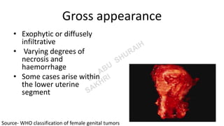
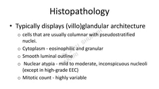
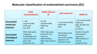
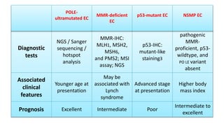
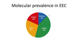
Endometrioid carcinoma
of the uterine corpus
Based on WHO Classification of female genital tumors, 5th edition
Dr Abu Suraih Sakhri
Definition
• Malignant epithelial neoplasm
• varying proportions of glandular, papillary, and
solid architecture
• Neoplastic cells showing endometrioid
differentiation
Subtypes
Four molecular subtypes
1. POLE-ultramutated endometrioid carcinoma
2. Mismatch repair-deficient endometrioid
carcinoma
3. p53-mutant endometrioid carcinoma
4. No specific molecular profile (NSMP) endometrioid
carcinoma.
Clinical features
• Postmenopausal women (90% > 50 years)
• Median patient age - 63 years
• Typically present with abnormal/postmenopausal
bleeding.
• Advanced disease presents with pelvic/
abdominal symptoms resemble ovarian
carcinoma !
Epidemiology
• Sixth most commonly diagnosed cancer in women
• Second most commonly diagnosed female genital
organ cancer.
• The highest rates were found in very high Human
Development Index (HDI) countries (more than half
of the cases)
• Post-menopausal bleeding- Most common
symptom leading to diagnosis
• High-income countries-5-year survival rate is high
(-80% in the USA)
• Worldwide- 14th leading cause of cancer death.
• Close to 90,000 deaths/ year
Etiology
• Prolonged exposure to unopposed estrogen-
Mainly
Pathogenesis
• Shared molecular alterations with endometrial atypical hyperplasia /
endometrioid intraepithelial neoplasia.
• Association with Lynch syndrome and Cowden syndrome
• Exposure to higher total concentrations of estrogens
o early menarche
o Late menopause
o Nulliparity
o Obesity
o Tamoxifen
o Polycystic ovary syndrome
o Estrogen-producing ovarian tumours
Gross appearance
• Exophytic or diffusely
infiltrative
• Varying degrees of
necrosis and
haemorrhage
• Some cases arise within
the lower uterine
segment
Source- WHO classification of female genital tumors
Histopathology
• Typically displays (villo)glandular architecture
o cells that are usually columnar with pseudostratified
nuclei.
o Cytoplasm - eosinophilic and granular
o Smooth luminal outline
o Nuclear atypia - mild to moderate, inconspicuous nucleoli
(except in high-grade EEC)
o Mitotic count - highly variable
o Squamous differentiation is frequent (10-25%)
⁻ As morules or solid sheets of eosinophilic cells
⁻ Keratinization +
Histological patterns, which are not associated
with different prognosis
1. Secretory patterns
Resemble early secretory endometrium, either focal or diffuse,
mimicking clear cell carcinoma
Which are
o Small non-villous papillae
o Microglandular pattern
o Spindle cell pattern
o Sertoliform pattern
o sex cord-like formations and hyalinization
Stromal Invasion
• Differentiate from Hyperplasia and invasive cancer
• Defined as
oLoss of intervening stroma (a confluent glandular, cribriform, maze-like
pattern)
oor An altered endometrial stroma (desmoplastic stromal
reaction)
oor Complex (mostly villoglandular) architecture.
Endometrioid endometrial cancer with mucinous
differentiation –difficult to distinguish from atypical
mucinous glandular proliferations
How to differentiate?
cribriform or confluent architecture and cytological
atypia in the latter.
Grading
• FIGO grading criteria
Grade 1 < 5%
Grade 2 6-50% Solid non-glandular, Non-squamous growth
Grade 3 > 50%
• Binary Grading
------ Low Grade
------ High Grade
• Severe cytological atypia in the majority of
cells (> 50%) increases the grade by one level
• Exclude Serous carcinoma - nuclear atypia out
of proportion to the architecture!
• In low-grade EEC- Usually endometrioid
differentiation
• In high-grade - squamous differentiation
strongly favours EEC
Immunohistochemistry
Low Grade EEC
Endocervical
adenocarcinoma
ER/ PR
Diffuse strong
immunoreactivity
Negative
p16 Patchy positivity Diffuse immunoreactivity
may be difficult to distinguish
from serous endometrial carcinoma, but it is usually solid and
shows less-pronounced nuclear pleomorphism. raises
serous carcinoma as a possibility. Loss of immunoreactivity for
ARID1A, PTEN, or one of the mismatch repair proteins favours
high-grade EEC. Abnormal p53 expression is reported in 2-5%
of low-grade and 20% of high-grade EECs {274,1465B,1465D}.
High Grade EEC
Serous Endometrial
carcinoma
 solid and shows less-pronounced
nuclear pleomorphism
 Loss of immunoreactivity for
ARID1A, PTEN, or one of the
mismatch repair proteins favours
high-grade EEC
 Diffuse high grade atypia out of
proportion to architectural features
EEC- Endometrioid endometrial carcinoma
Lymphovascular invasion
 Present in 5-15% of cases
 Associated with the microcystic, elongated, and
fragmented (MELF) pattern of invasion and with
mismatch repair
deficiency.
 Particular artefacts, such as displacement of
tumour cells in spaces and retraction of tissue due to
delayed fixation, may mimic lymphovascular invasion
and should be excluded
POLE-
ultramutated EC
MMR-deficient
EC
p53-mutant EC
NSMP EC
Associated
molecular
features
> 100
mutations/Mb,
SCNA very
low, MSS
10-100
mutations/Mb,
SCNA low,
MSI
< 10 mutations/Mb,
SCNA high,
MSS
< 10 mutations/Mb,
SCNA low,
MSS, 30-40% with
CTNNB1
mutations
Associated
histological
features
Often high-grade,
ambiguous
morphology with
scattered tumour
giant cells,
prominent TILs
Often high-grade,
prominent TILs,
mucinous
differentiation,
MELF-type invasion,
LVSI
Mostly high-grade
with diffuse
cytonuclear atypia;
glandular and
solid forms exist
Mostly low-grade
with frequent
squamous
differentiation or
morule,
absence of TILs
Molecular classification of endometrioid carcinoma (EC)
Diagnostic
tests
NGS / Sanger
sequencing /
hotspot
analysis
MMR-IHC:
MLH1, MSH2,
MSH6,
and PMS2; MSI
assay; NGS
p53-IHC:
mutant-like
staining3
pathogenic
MMR-
proficient, p53-
wildtype, and
PO LE variant
absent
Associated
clinical
features
Younger age at
presentation
May be
associated with
Lynch
syndrome
Advanced stage
at presentation
Higher body
mass index
Prognosis Excellent Intermediate Poor
Intermediate to
excellent
POLE-
ultramutated EC
MMR-deficient
EC
p53-mutant EC NSMP EC
Cytology
Not clinically relevant
Essential and desirable diagnostic criteria
Essential: invasive endometrial carcinoma with
endometrioid differentiation.
Desirable: some degree of squamous, secretory,
or mucinous differentiation.
Staging
TNM classification
FIGO staging system.
Molecular prevalence in EEC
Prognosis and prediction
• FIGO and UICC staging is based on depth of myometrial
invasion (< 50% or > 50%) and on endocervical stromal,
adnexal, and lymph node involvement.
• Unequivocal lymphovascular invasion is used in treatment
algorithms
• Distinction between focal and extensive lymphovascular
invasion (> 5 vessels) have prognostic significance.
Synchronous endometrioid carcinomas of
endometrium and ovaries
• Most are clonally related
• Conservative management when the following four
criteria are met:
(1) both tumours are low-grade
(2) < 50% myometrial invasion
(3) no involvement of any other site
(4) absence of extensive lymphovascular invasion.
• For treatment purposes, these neoplasm
should be managed as independent tumours.
 The efficacy of conservative treatment of grade 1
endometrioid carcinoma or endometrial atypical
hyperplasia / endometrioid intraepithelial neoplasia
with hormonal agents may be monitored by
histology, but this is not yet standard clinical practice
Endometrioid carcinoma. High-grade endometrioid endometrial carcinoma
(FIGO grade 3), p53-mutant
Endometrioid carcinoma. A High-grade endometrioid endometrial carcinoma (EEC) (FIGO grade 3), mismatch
repair-deficient. Note the abundance of tumour-infiltrating lymphocytes and the substantial lymphovascular
space invasion frequently observed in mismatch repair-deficient EEC. B Low-grade EEC (FIGO grade 1), no specific
molecular profile (NSMP). This tumour was mismatch repair-proficient and p53-wildtype and did not carry a
pathogenic PO LE variant; it was therefore designated NSMP. Note the squamous differentiation frequently
observed in low-grade EEC.
The p53 immunohistochemistry of this tumor shows abnormal diffuse nuclear overexpression
associated with the presence of TP53 mutation.
High-grade endometrioid endometrial carcinoma (FIGO grade 3),
POLE-mutant. Note the solid non-squamous growth
Thank you
Send feedbacks at abusuraihsakhri@gmail.com

Pathology of Endometrial cancer 2022.pptx

  • 1.
    Endometrioid carcinoma of theuterine corpus Based on WHO Classification of female genital tumors, 5th edition Dr Abu Suraih Sakhri
  • 2.
    Definition • Malignant epithelialneoplasm • varying proportions of glandular, papillary, and solid architecture • Neoplastic cells showing endometrioid differentiation
  • 3.
    Subtypes Four molecular subtypes 1.POLE-ultramutated endometrioid carcinoma 2. Mismatch repair-deficient endometrioid carcinoma 3. p53-mutant endometrioid carcinoma 4. No specific molecular profile (NSMP) endometrioid carcinoma.
  • 4.
    Clinical features • Postmenopausalwomen (90% > 50 years) • Median patient age - 63 years • Typically present with abnormal/postmenopausal bleeding. • Advanced disease presents with pelvic/ abdominal symptoms resemble ovarian carcinoma !
  • 5.
    Epidemiology • Sixth mostcommonly diagnosed cancer in women • Second most commonly diagnosed female genital organ cancer. • The highest rates were found in very high Human Development Index (HDI) countries (more than half of the cases)
  • 6.
    • Post-menopausal bleeding-Most common symptom leading to diagnosis • High-income countries-5-year survival rate is high (-80% in the USA) • Worldwide- 14th leading cause of cancer death. • Close to 90,000 deaths/ year
  • 7.
    Etiology • Prolonged exposureto unopposed estrogen- Mainly
  • 8.
    Pathogenesis • Shared molecularalterations with endometrial atypical hyperplasia / endometrioid intraepithelial neoplasia. • Association with Lynch syndrome and Cowden syndrome • Exposure to higher total concentrations of estrogens o early menarche o Late menopause o Nulliparity o Obesity o Tamoxifen o Polycystic ovary syndrome o Estrogen-producing ovarian tumours
  • 9.
    Gross appearance • Exophyticor diffusely infiltrative • Varying degrees of necrosis and haemorrhage • Some cases arise within the lower uterine segment Source- WHO classification of female genital tumors
  • 10.
    Histopathology • Typically displays(villo)glandular architecture o cells that are usually columnar with pseudostratified nuclei. o Cytoplasm - eosinophilic and granular o Smooth luminal outline o Nuclear atypia - mild to moderate, inconspicuous nucleoli (except in high-grade EEC) o Mitotic count - highly variable
  • 11.
    o Squamous differentiationis frequent (10-25%) ⁻ As morules or solid sheets of eosinophilic cells ⁻ Keratinization +
  • 12.
    Histological patterns, whichare not associated with different prognosis 1. Secretory patterns Resemble early secretory endometrium, either focal or diffuse, mimicking clear cell carcinoma Which are o Small non-villous papillae o Microglandular pattern o Spindle cell pattern o Sertoliform pattern o sex cord-like formations and hyalinization
  • 13.
    Stromal Invasion • Differentiatefrom Hyperplasia and invasive cancer • Defined as oLoss of intervening stroma (a confluent glandular, cribriform, maze-like pattern) oor An altered endometrial stroma (desmoplastic stromal reaction) oor Complex (mostly villoglandular) architecture.
  • 14.
    Endometrioid endometrial cancerwith mucinous differentiation –difficult to distinguish from atypical mucinous glandular proliferations How to differentiate? cribriform or confluent architecture and cytological atypia in the latter.
  • 15.
    Grading • FIGO gradingcriteria Grade 1 < 5% Grade 2 6-50% Solid non-glandular, Non-squamous growth Grade 3 > 50% • Binary Grading ------ Low Grade ------ High Grade
  • 16.
    • Severe cytologicalatypia in the majority of cells (> 50%) increases the grade by one level • Exclude Serous carcinoma - nuclear atypia out of proportion to the architecture! • In low-grade EEC- Usually endometrioid differentiation • In high-grade - squamous differentiation strongly favours EEC
  • 17.
    Immunohistochemistry Low Grade EEC Endocervical adenocarcinoma ER/PR Diffuse strong immunoreactivity Negative p16 Patchy positivity Diffuse immunoreactivity
  • 18.
    may be difficultto distinguish from serous endometrial carcinoma, but it is usually solid and shows less-pronounced nuclear pleomorphism. raises serous carcinoma as a possibility. Loss of immunoreactivity for ARID1A, PTEN, or one of the mismatch repair proteins favours high-grade EEC. Abnormal p53 expression is reported in 2-5% of low-grade and 20% of high-grade EECs {274,1465B,1465D}.
  • 19.
    High Grade EEC SerousEndometrial carcinoma  solid and shows less-pronounced nuclear pleomorphism  Loss of immunoreactivity for ARID1A, PTEN, or one of the mismatch repair proteins favours high-grade EEC  Diffuse high grade atypia out of proportion to architectural features EEC- Endometrioid endometrial carcinoma
  • 20.
    Lymphovascular invasion  Presentin 5-15% of cases  Associated with the microcystic, elongated, and fragmented (MELF) pattern of invasion and with mismatch repair deficiency.  Particular artefacts, such as displacement of tumour cells in spaces and retraction of tissue due to delayed fixation, may mimic lymphovascular invasion and should be excluded
  • 21.
    POLE- ultramutated EC MMR-deficient EC p53-mutant EC NSMPEC Associated molecular features > 100 mutations/Mb, SCNA very low, MSS 10-100 mutations/Mb, SCNA low, MSI < 10 mutations/Mb, SCNA high, MSS < 10 mutations/Mb, SCNA low, MSS, 30-40% with CTNNB1 mutations Associated histological features Often high-grade, ambiguous morphology with scattered tumour giant cells, prominent TILs Often high-grade, prominent TILs, mucinous differentiation, MELF-type invasion, LVSI Mostly high-grade with diffuse cytonuclear atypia; glandular and solid forms exist Mostly low-grade with frequent squamous differentiation or morule, absence of TILs Molecular classification of endometrioid carcinoma (EC)
  • 22.
    Diagnostic tests NGS / Sanger sequencing/ hotspot analysis MMR-IHC: MLH1, MSH2, MSH6, and PMS2; MSI assay; NGS p53-IHC: mutant-like staining3 pathogenic MMR- proficient, p53- wildtype, and PO LE variant absent Associated clinical features Younger age at presentation May be associated with Lynch syndrome Advanced stage at presentation Higher body mass index Prognosis Excellent Intermediate Poor Intermediate to excellent POLE- ultramutated EC MMR-deficient EC p53-mutant EC NSMP EC
  • 23.
  • 24.
    Essential and desirablediagnostic criteria Essential: invasive endometrial carcinoma with endometrioid differentiation. Desirable: some degree of squamous, secretory, or mucinous differentiation.
  • 25.
  • 26.
  • 27.
    Prognosis and prediction •FIGO and UICC staging is based on depth of myometrial invasion (< 50% or > 50%) and on endocervical stromal, adnexal, and lymph node involvement. • Unequivocal lymphovascular invasion is used in treatment algorithms • Distinction between focal and extensive lymphovascular invasion (> 5 vessels) have prognostic significance.
  • 28.
    Synchronous endometrioid carcinomasof endometrium and ovaries • Most are clonally related • Conservative management when the following four criteria are met: (1) both tumours are low-grade (2) < 50% myometrial invasion (3) no involvement of any other site (4) absence of extensive lymphovascular invasion.
  • 29.
    • For treatmentpurposes, these neoplasm should be managed as independent tumours.
  • 30.
     The efficacyof conservative treatment of grade 1 endometrioid carcinoma or endometrial atypical hyperplasia / endometrioid intraepithelial neoplasia with hormonal agents may be monitored by histology, but this is not yet standard clinical practice
  • 31.
    Endometrioid carcinoma. High-gradeendometrioid endometrial carcinoma (FIGO grade 3), p53-mutant
  • 32.
    Endometrioid carcinoma. AHigh-grade endometrioid endometrial carcinoma (EEC) (FIGO grade 3), mismatch repair-deficient. Note the abundance of tumour-infiltrating lymphocytes and the substantial lymphovascular space invasion frequently observed in mismatch repair-deficient EEC. B Low-grade EEC (FIGO grade 1), no specific molecular profile (NSMP). This tumour was mismatch repair-proficient and p53-wildtype and did not carry a pathogenic PO LE variant; it was therefore designated NSMP. Note the squamous differentiation frequently observed in low-grade EEC.
  • 33.
    The p53 immunohistochemistryof this tumor shows abnormal diffuse nuclear overexpression associated with the presence of TP53 mutation.
  • 34.
    High-grade endometrioid endometrialcarcinoma (FIGO grade 3), POLE-mutant. Note the solid non-squamous growth
  • 35.
    Thank you Send feedbacksat abusuraihsakhri@gmail.com