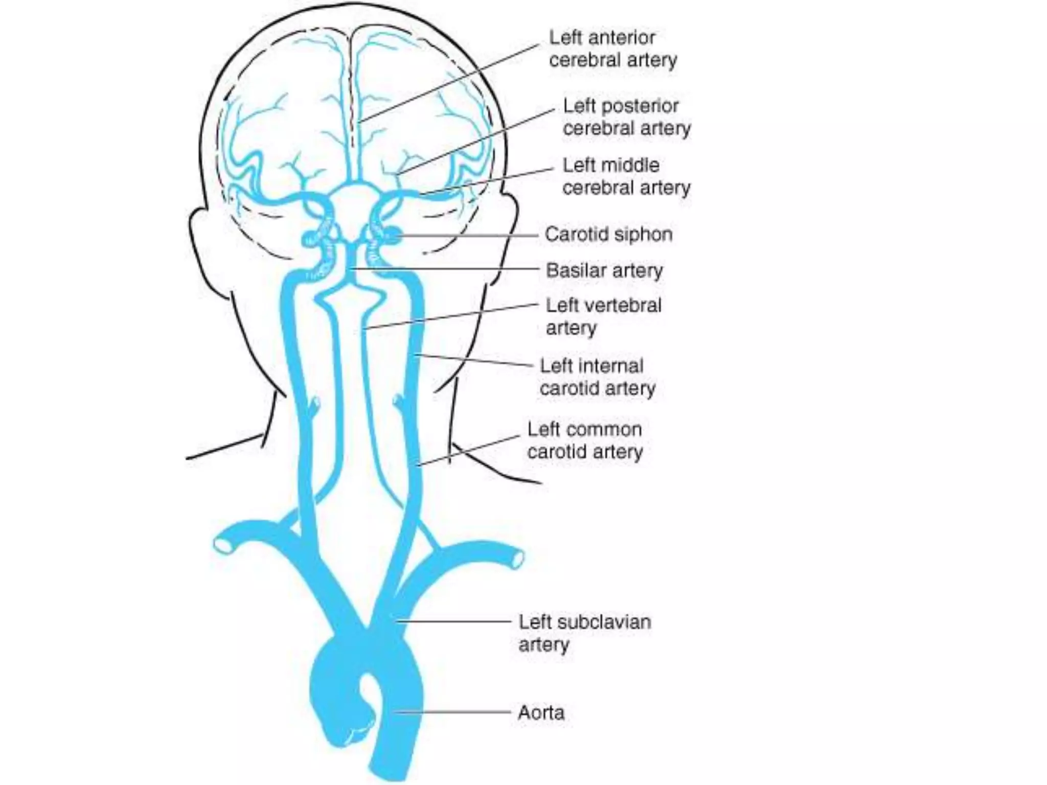
Blood supply of Brain
Vascular Supply
• About 18% of the total blood volume in the body circulates
in the brain, which accounts for about 2% of the body
weight.
• The blood transports oxygen, nutrients, and other
substances necessary for proper functioning of the brain
tissues and carries away metabolites.
• Loss of consciousness occurs in less than 15 seconds after
blood flow to the brain has stopped, and irreparable
damage to the brain tissue occurs within 5 minutes.
• Cerebrovascular disease, or stroke, occurs as a result of
vascular compromise or haemorrhage and is one of the
most frequent sources of neurologic disability.
• Nearly half of the admissions are because of strokes.
• Cerebrovascular disease is the third most common cause of
death in industrialized societies.
BLOOD SUPPLY TO THE BRAIN
• High demand for oxygen and nutrients
• Arterial blood through: internal carotid and
vertebral arteries
• Venous blood from brain in the internal
jugular veins
• Cerebrovascular accidents (CVA): stroke,
shutting off blood supply to brain
Arterial supply
• The circle of Willis (after the English
neuroanatomist Sir Thomas Willis) is a
confluence of vessels that gives rise to all of
the major cerebral arteries.
• Supplied by the paired internal carotid
arteries and the basilar artery.
• Contains a paired posterior communicating
artery and an unpaired anterior
communicating artery.
• The circle of Willis shows many variations
among individuals.
• Occlusion of major cerebral arteries
produces a characteristic clinical picture.
VENOUS DRAINAGE
• The venous drainage of the brain and
coverings includes:
– the veins of the brain itself,
– the dural venous sinuses,
– the dura's meningeal veins,
– and the diploic veins between the tables of the
skull.
CRANIAL MENINGES
• Dura mater: outer and inner layers
• Arachnoid mater: epithelial layer and
arachnoid trabeculae
• Pia mater: sticks to brain surface
• Pia and arachnoid = leptomeninges
• Dural folds hold the brain in position -
Falx cerebri
Tentorium cerebelli
• Dural sinuses (veins located within the
folds)
VENTRICLES OF THE BRAIN
• Lined with ependymal cells
• Lateral ventricle (each hemisphere)
• Third ventricle (diencephalon)
• Cerebral aqueduct (midbrain)
• Fourth ventricle: (btw pons and cerebellum,
continuous with central canal of spinal cord)
• CSF flows within ventricles, central canal and
into subarachnoid space
Ventricles
CEREBROSPINAL FLUID
Surrounds and bathes the CNS
Functions:
1. Supporting of brain and spinal cord
2. Transport of nutrients, chemical
messengers, and waste products
THE FORMATION OF CSF
• Choroid plexus: contains specialised
ependymal cells and capillaries (500ml/day),
total volume: 150ml
• Choroid plexuses secrete CSF into ventricles
• Circulation: from choroid plexus to ventricles
and central canal of spinal cord to
subarachnoid space to sinuses
Circulation (cont.):
• CSF reaches subarachnoid space through two
lateral apertures and a single medial aperture in
the 4th ventricle
• Arachnoid granulations: penetrate dura
mater meningeal layer of venous sinuses, CSF
absorbed into the venous circulation
• Hydrocephalus: “water in the brain”
Thank you

Blood supply of brain

  • 1.
  • 2.
    Vascular Supply • About18% of the total blood volume in the body circulates in the brain, which accounts for about 2% of the body weight. • The blood transports oxygen, nutrients, and other substances necessary for proper functioning of the brain tissues and carries away metabolites. • Loss of consciousness occurs in less than 15 seconds after blood flow to the brain has stopped, and irreparable damage to the brain tissue occurs within 5 minutes. • Cerebrovascular disease, or stroke, occurs as a result of vascular compromise or haemorrhage and is one of the most frequent sources of neurologic disability. • Nearly half of the admissions are because of strokes. • Cerebrovascular disease is the third most common cause of death in industrialized societies.
  • 3.
    BLOOD SUPPLY TOTHE BRAIN • High demand for oxygen and nutrients • Arterial blood through: internal carotid and vertebral arteries • Venous blood from brain in the internal jugular veins • Cerebrovascular accidents (CVA): stroke, shutting off blood supply to brain
  • 4.
    Arterial supply • Thecircle of Willis (after the English neuroanatomist Sir Thomas Willis) is a confluence of vessels that gives rise to all of the major cerebral arteries. • Supplied by the paired internal carotid arteries and the basilar artery. • Contains a paired posterior communicating artery and an unpaired anterior communicating artery. • The circle of Willis shows many variations among individuals. • Occlusion of major cerebral arteries produces a characteristic clinical picture.
  • 11.
    VENOUS DRAINAGE • Thevenous drainage of the brain and coverings includes: – the veins of the brain itself, – the dural venous sinuses, – the dura's meningeal veins, – and the diploic veins between the tables of the skull.
  • 16.
    CRANIAL MENINGES • Duramater: outer and inner layers • Arachnoid mater: epithelial layer and arachnoid trabeculae • Pia mater: sticks to brain surface • Pia and arachnoid = leptomeninges • Dural folds hold the brain in position - Falx cerebri Tentorium cerebelli • Dural sinuses (veins located within the folds)
  • 20.
    VENTRICLES OF THEBRAIN • Lined with ependymal cells • Lateral ventricle (each hemisphere) • Third ventricle (diencephalon) • Cerebral aqueduct (midbrain) • Fourth ventricle: (btw pons and cerebellum, continuous with central canal of spinal cord) • CSF flows within ventricles, central canal and into subarachnoid space
  • 21.
  • 22.
    CEREBROSPINAL FLUID Surrounds andbathes the CNS Functions: 1. Supporting of brain and spinal cord 2. Transport of nutrients, chemical messengers, and waste products
  • 23.
    THE FORMATION OFCSF • Choroid plexus: contains specialised ependymal cells and capillaries (500ml/day), total volume: 150ml • Choroid plexuses secrete CSF into ventricles • Circulation: from choroid plexus to ventricles and central canal of spinal cord to subarachnoid space to sinuses
  • 24.
    Circulation (cont.): • CSFreaches subarachnoid space through two lateral apertures and a single medial aperture in the 4th ventricle • Arachnoid granulations: penetrate dura mater meningeal layer of venous sinuses, CSF absorbed into the venous circulation • Hydrocephalus: “water in the brain”
  • 26.