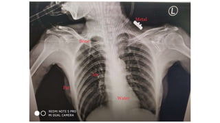
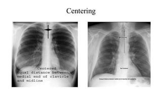
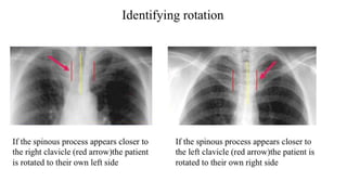
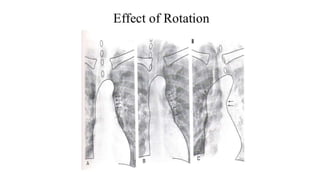
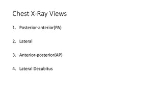
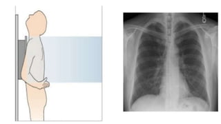
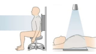
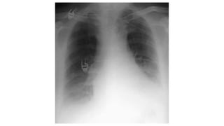
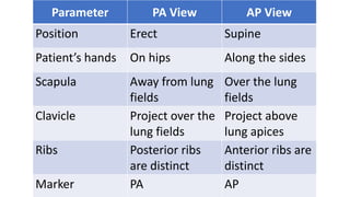
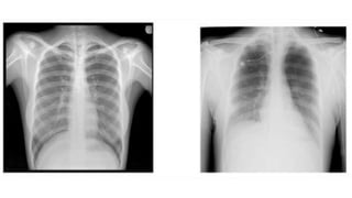
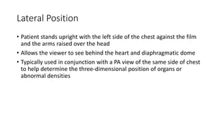
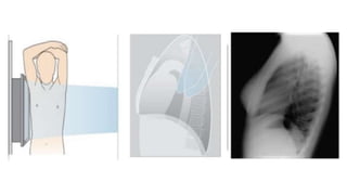
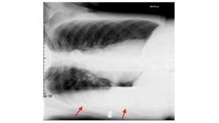
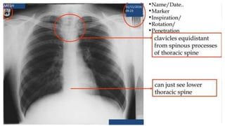
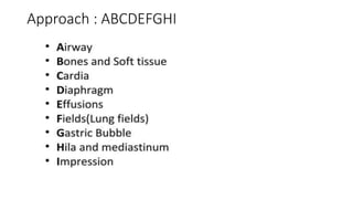
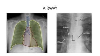
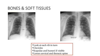
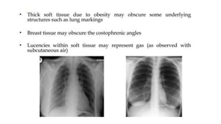
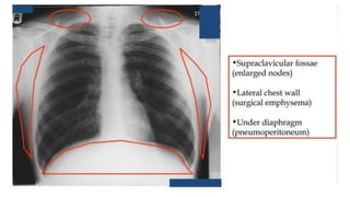
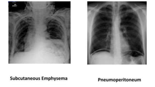
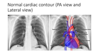
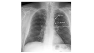
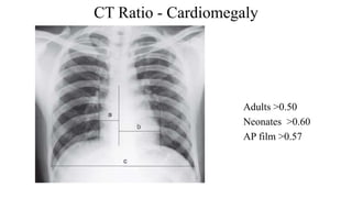
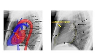
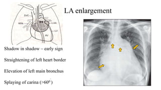
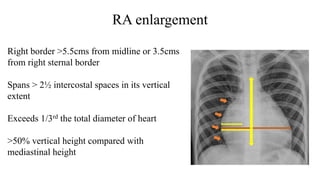
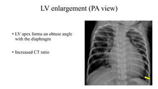
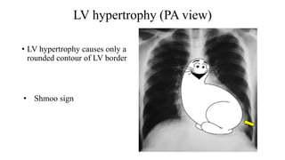
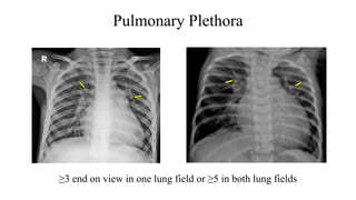
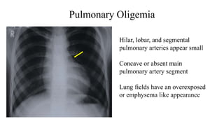
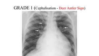
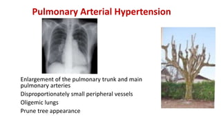
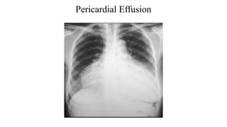
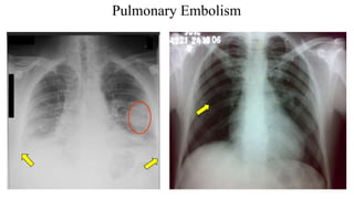
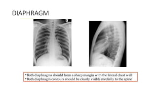
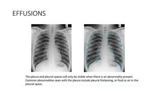
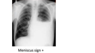
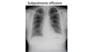
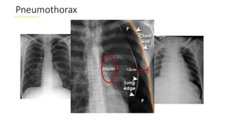
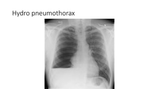
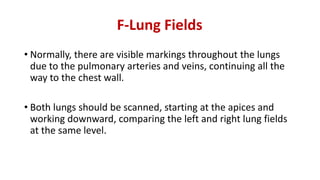
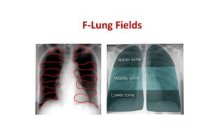
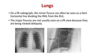
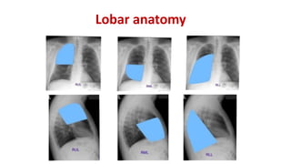
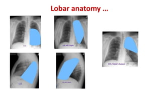
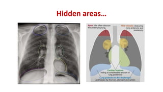
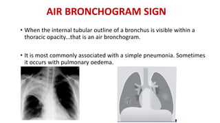
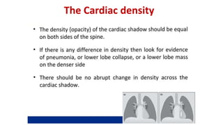
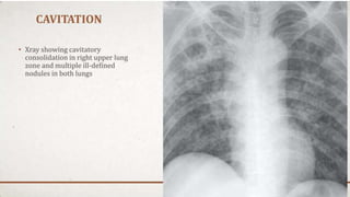
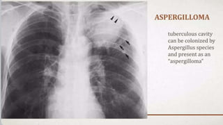
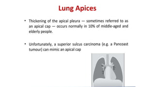
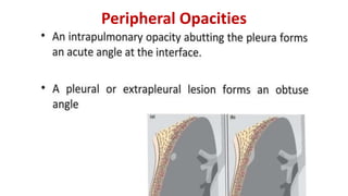
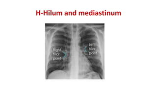
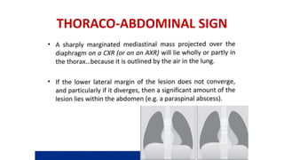
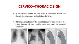
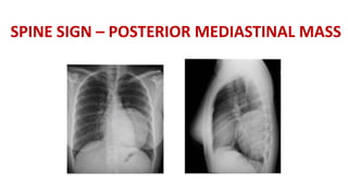
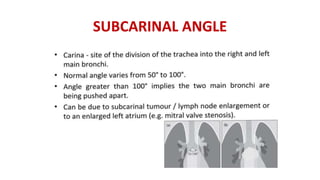
Chest x-rays are commonly used to image the chest. They should be interpreted in the context of clinical findings and compared to prior x-rays. Tissues have different densities: gas is least dense while bone is most dense. Inspiration, penetration, and rotation impact image quality. Standard views include PA, AP, lateral, and decubitus. When interpreting, examine the airway, bones, cardiomegaly, vascular patterns, diaphragm, effusions, and lung fields for abnormalities. Prior films aid in detecting subtle changes over time.