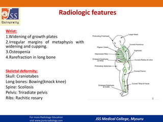
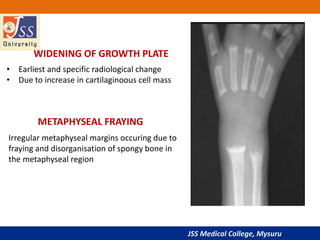
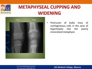
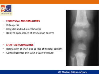
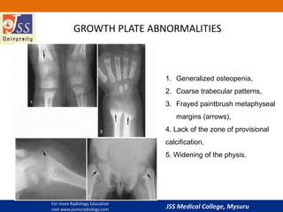
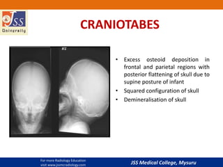
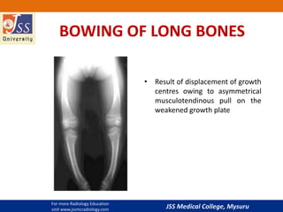
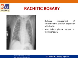
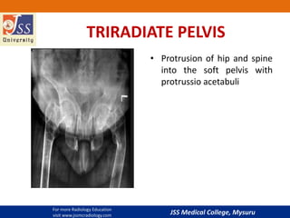
JSS Medical College, Mysuru
RICKETS
-Dr.Shilpa
Resident,
JSS Medical College ,Mysuru
Please visit www.jssmcradiology.com for more Radiology education
For more Radiology Education
visit www.jssmcradiology.com
JSS Medical College, Mysuru
Introduction
 It is the childhood equivalent of osteomalacia in the mature skeleton.
 Symptoms consist of muscle tetany, irritability, weakness, delayed
development, small stature, bone deformities and pain.
 Soft tissue swellings occurring around the growth plates owing to
hypertrophied cartilage ex: costochondral bumps (rachitic rosary).
 Earliest sign : distal ends of radius and ulna. Ulnar growth plate grows more
rapidly so manifestations are seen earlier in ulnar growth plate.*
*Silvermanfn. dysostoses,miscellaneous syndromes and metabolic abnormalities. Caffey’s padiatric
radiology.1993;2:1746
JSS Medical College, Mysuru
 In rickets - disordered proliferation of cartilage cells and defective mineralization in
zone of provisional calcification.
 Decreased quantity of calcified osteoid.
 Owing to a lack of osteoid mineralization the metaphyseal zone of provisional
calcification will also be absent radiographically.
 Bones appear more radiolucent, with coarsened trabecular patterns
 widened growth plate with irregular, frayed, and cupped metaphyseal margins
(paintbrush metaphyses).
 Bowing deformities, fractures, decreased bony length, scoliosis, and pseudo-
fractures are other notable findings. Bulbous enlargement of the costochondral
junctions (rachitic rosary)
Patho-radiological features:
JSS Medical College, Mysuru
Radiologic features
Wrist:
1.Widening of growth plates
2.Irregular margins of metaphysis with
widening and cupping.
3.Osteopenia
4.Rarefraction in long bone
Skeletal deformity:
Skull: Craniotabes
Long bones: Bowing(knock knee)
Spine: Scoliosis
Pelvis: Triradiate pelvis
Ribs: Rachitic rosary
For more Radiology Education
visit www.jssmcradiology.com
JSS Medical College, Mysuru
WIDENING OF GROWTH PLATE
• Earliest and specific radiological change
• Due to increase in cartilaginoous cell mass
Irregular metaphyseal margins occuring due to
fraying and disorganisation of spongy bone in
the metaphyseal region
METAPHYSEAL FRAYING
JSS Medical College, Mysuru
METAPHYSEAL CUPPING AND
WIDENING
• Protrusion of bulky mass of
cartilageneous cells in the zone of
hypertrophy into the poorly
mineralized metaphysis
For more Radiology Education
visit www.jssmcradiology.com
JSS Medical College, Mysuru
• EPIPHYSEAL ABNORMALITIES
• Osteopenia
• Irregular and indistinct borders
• Delayed appearance of ossification centres
• SHAFT ABNORMALITIES
• Rarefaction of shaft due to loss of mineral content
• Cortex becomes thin with a coarse texture
JSS Medical College, Mysuru
1. Generalized osteopenia,
2. Coarse trabecular patterns,
3. Frayed paintbrush metaphyseal
margins (arrows),
4. Lack of the zone of provisional
calcification,
5. Widening of the physis.
GROWTH PLATE ABNORMALITIES
For more Radiology Education
visit www.jssmcradiology.com
JSS Medical College, Mysuru
CRANIOTABES
• Excess osteoid deposition in
frontal and parietal regions with
posterior flattening of skull due to
supine posture of infant
• Squared configuration of skull
• Demineralisation of skull
For more Radiology Education
visit www.jssmcradiology.com
JSS Medical College, Mysuru
BOWING OF LONG BONES
• Result of displacement of growth
centres owing to asymmetrical
musculotendinous pull on the
weakened growth plate
For more Radiology Education
visit www.jssmcradiology.com
JSS Medical College, Mysuru
RACHITIC ROSARY
• Bulbous enlargement of
costochondral junction especially
middle ribs
• May indent pleural surface or
thymic shadow
For more Radiology Education
visit www.jssmcradiology.com
JSS Medical College, Mysuru
TRIRADIATE PELVIS
• Protrusion of hip and spine
into the soft pelvis with
protrussio acetabuli
For more Radiology Education
visit www.jssmcradiology.com
JSS Medical College, Mysuru
Thank You
For more Radiology Education visit
www.jssmcradiology.com

Imaging in rickets

  • 1.
    JSS Medical College,Mysuru RICKETS -Dr.Shilpa Resident, JSS Medical College ,Mysuru Please visit www.jssmcradiology.com for more Radiology education For more Radiology Education visit www.jssmcradiology.com
  • 2.
    JSS Medical College,Mysuru Introduction  It is the childhood equivalent of osteomalacia in the mature skeleton.  Symptoms consist of muscle tetany, irritability, weakness, delayed development, small stature, bone deformities and pain.  Soft tissue swellings occurring around the growth plates owing to hypertrophied cartilage ex: costochondral bumps (rachitic rosary).  Earliest sign : distal ends of radius and ulna. Ulnar growth plate grows more rapidly so manifestations are seen earlier in ulnar growth plate.* *Silvermanfn. dysostoses,miscellaneous syndromes and metabolic abnormalities. Caffey’s padiatric radiology.1993;2:1746
  • 3.
    JSS Medical College,Mysuru  In rickets - disordered proliferation of cartilage cells and defective mineralization in zone of provisional calcification.  Decreased quantity of calcified osteoid.  Owing to a lack of osteoid mineralization the metaphyseal zone of provisional calcification will also be absent radiographically.  Bones appear more radiolucent, with coarsened trabecular patterns  widened growth plate with irregular, frayed, and cupped metaphyseal margins (paintbrush metaphyses).  Bowing deformities, fractures, decreased bony length, scoliosis, and pseudo- fractures are other notable findings. Bulbous enlargement of the costochondral junctions (rachitic rosary) Patho-radiological features:
  • 4.
    JSS Medical College,Mysuru Radiologic features Wrist: 1.Widening of growth plates 2.Irregular margins of metaphysis with widening and cupping. 3.Osteopenia 4.Rarefraction in long bone Skeletal deformity: Skull: Craniotabes Long bones: Bowing(knock knee) Spine: Scoliosis Pelvis: Triradiate pelvis Ribs: Rachitic rosary For more Radiology Education visit www.jssmcradiology.com
  • 5.
    JSS Medical College,Mysuru WIDENING OF GROWTH PLATE • Earliest and specific radiological change • Due to increase in cartilaginoous cell mass Irregular metaphyseal margins occuring due to fraying and disorganisation of spongy bone in the metaphyseal region METAPHYSEAL FRAYING
  • 6.
    JSS Medical College,Mysuru METAPHYSEAL CUPPING AND WIDENING • Protrusion of bulky mass of cartilageneous cells in the zone of hypertrophy into the poorly mineralized metaphysis For more Radiology Education visit www.jssmcradiology.com
  • 7.
    JSS Medical College,Mysuru • EPIPHYSEAL ABNORMALITIES • Osteopenia • Irregular and indistinct borders • Delayed appearance of ossification centres • SHAFT ABNORMALITIES • Rarefaction of shaft due to loss of mineral content • Cortex becomes thin with a coarse texture
  • 8.
    JSS Medical College,Mysuru 1. Generalized osteopenia, 2. Coarse trabecular patterns, 3. Frayed paintbrush metaphyseal margins (arrows), 4. Lack of the zone of provisional calcification, 5. Widening of the physis. GROWTH PLATE ABNORMALITIES For more Radiology Education visit www.jssmcradiology.com
  • 9.
    JSS Medical College,Mysuru CRANIOTABES • Excess osteoid deposition in frontal and parietal regions with posterior flattening of skull due to supine posture of infant • Squared configuration of skull • Demineralisation of skull For more Radiology Education visit www.jssmcradiology.com
  • 10.
    JSS Medical College,Mysuru BOWING OF LONG BONES • Result of displacement of growth centres owing to asymmetrical musculotendinous pull on the weakened growth plate For more Radiology Education visit www.jssmcradiology.com
  • 11.
    JSS Medical College,Mysuru RACHITIC ROSARY • Bulbous enlargement of costochondral junction especially middle ribs • May indent pleural surface or thymic shadow For more Radiology Education visit www.jssmcradiology.com
  • 12.
    JSS Medical College,Mysuru TRIRADIATE PELVIS • Protrusion of hip and spine into the soft pelvis with protrussio acetabuli For more Radiology Education visit www.jssmcradiology.com
  • 13.
    JSS Medical College,Mysuru Thank You For more Radiology Education visit www.jssmcradiology.com