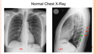
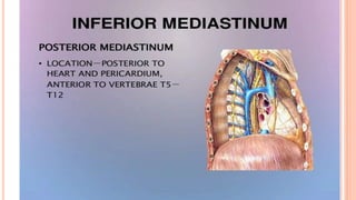
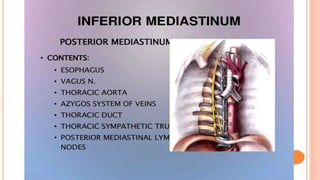
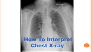
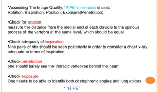
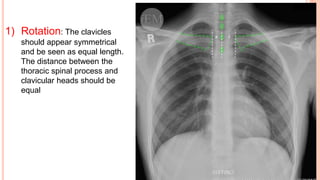
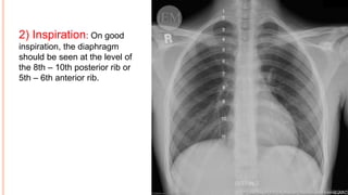
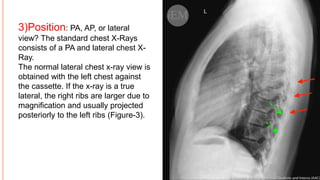
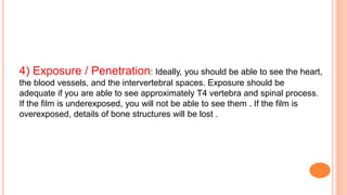
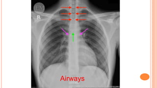
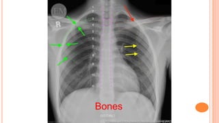
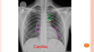
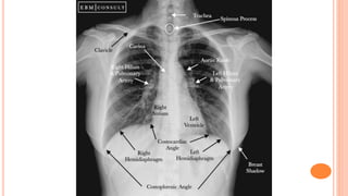
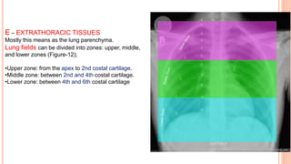
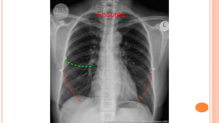
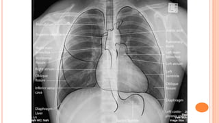
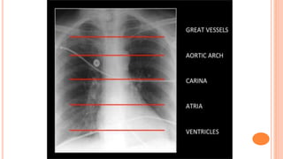
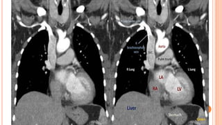
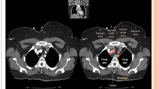
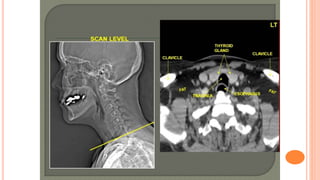
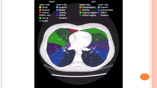
This document provides an overview of the normal anatomy of the thorax as seen on chest x-rays and CT scans. It describes the three types of chest x-rays and divides the mediastinum into upper and lower regions. It also provides details on how to properly interpret a chest x-ray, including checking for adequate rotation, inspiration, position and exposure using the "RIPE" mnemonic. A systematic approach to interpretation is outlined using the "ABCDEF" method to evaluate the airways, bones, cardiac structure, diaphragm, extra-thoracic tissues and lung fields/fissures. Key anatomical structures are labeled on sample chest x-rays and CT scans.