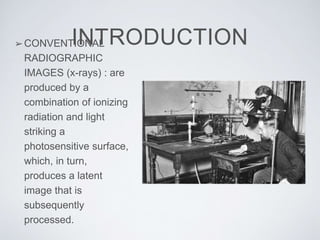
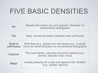
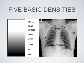
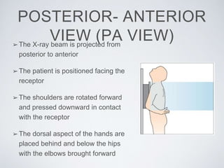
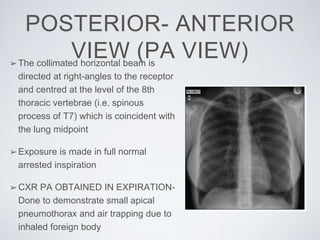
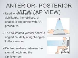
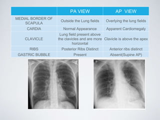
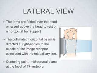
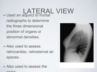
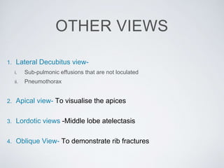
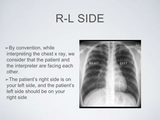
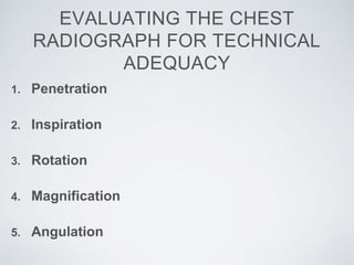
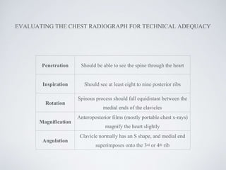
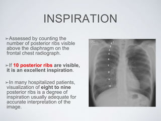
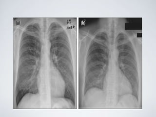
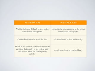
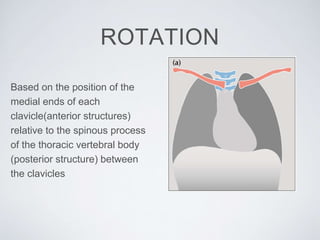
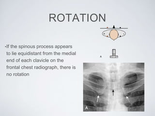
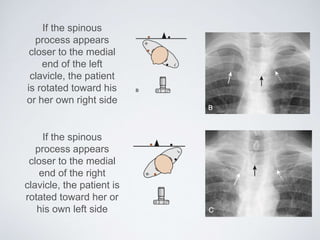
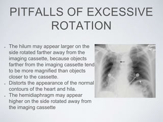
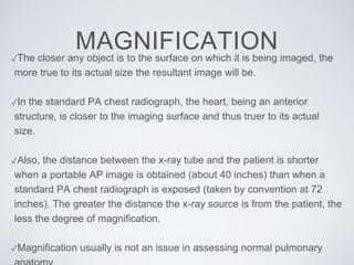
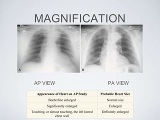
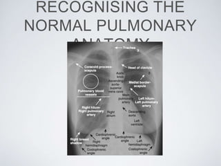
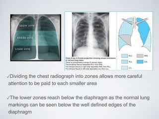
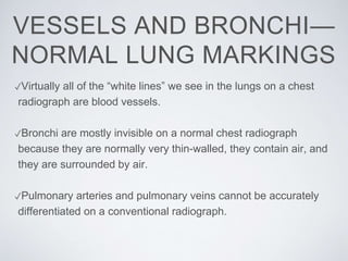
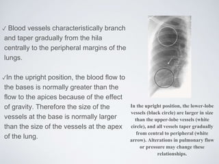
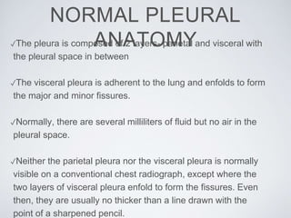
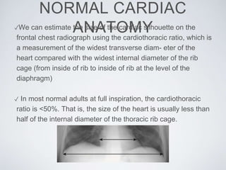
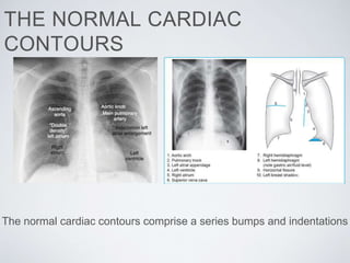
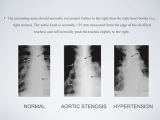
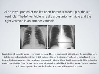
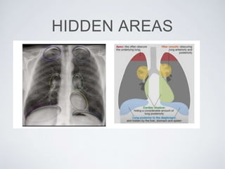
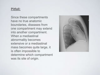
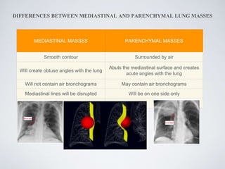
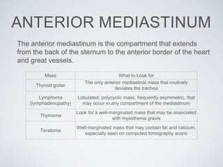
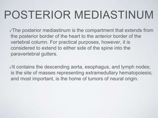
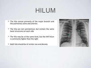
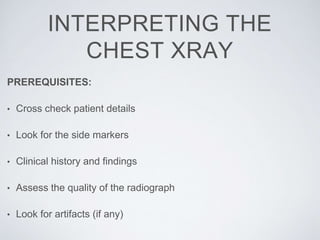
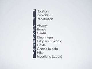
INTRODUCTION TO
CHEST RADIOGRAPH
PRESENTER: DR ANUSHA
MODERATOR : DR SURESH
CONTENTS• History
• Introduction
• Basic densities
• Different views
• R-L side
• Evaluating the chest X-ray for technical adequacy
• Normal pulmonary anatomy
• Normal pleural anatomy
• Normal cardiac anatomy
• Hidden areas
• Mediastinum
• Hilum
• Interpreting the chest X-ray
HISTORY
Hand mit Ringen (Hand with
Rings)
Wilhelm Röntgen's first "medical"
X-ray, of his wife's hand, taken on
22 December 1895
One of the first chest radiographs taken by
Francis Henry Williams, the Father of Chest
Radiology in North America
INTRODUCTION➢CONVENTIONAL
RADIOGRAPHIC
IMAGES (x-rays) : are
produced by a
combination of ionizing
radiation and light
striking a
photosensitive surface,
which, in turn,
produces a latent
image that is
subsequently
processed.
INTRODUCTION
➢At first, the processing of film was carried out in a darkroom - the
films were then hung out to dry.
➢DIGITAL RADIOGRAPHY: the photographic film replaced by a
photosensitive cassette or plate that is processed by an electronic
reader and the image stored in a digital format.
➢PACS( Picture Archiving, Communications, and Storage): images
stored and archived for posterity and communicated to others on
computer servers
Air
Absorbs the least x-ray and appears “blackest” on
conventional radiographs
Fat Gray, somewhat darker (blacker) than soft tissue
Fluid or
soft tissue
Both fluid (e.g., blood) and soft tissue (e.g., muscle)
have the same densities on conventional radiographs
Calcium
The most dense, naturally occurring material (e.g.,
bones); absorbs most x-rays
Metal
Usually absorbs all x-rays and appears the “whitest”
(e.g., bullets, barium)
FIVE BASIC DENSITIES
POSTERIOR- ANTERIOR
VIEW (PA VIEW)➢The X-ray beam is projected from
posterior to anterior
➢The patient is positioned facing the
receptor
➢The shoulders are rotated forward
and pressed downward in contact
with the receptor
➢The dorsal aspect of the hands are
placed behind and below the hips
with the elbows brought forward
POSTERIOR- ANTERIOR
VIEW (PA VIEW)➢The collimated horizontal beam is
directed at right-angles to the receptor
and centred at the level of the 8th
thoracic vertebrae (i.e. spinous
process of T7) which is coincident with
the lung midpoint
➢Exposure is made in full normal
arrested inspiration
➢CXR PA OBTAINED IN EXPIRATION-
Done to demonstrate small apical
pneumothorax and air trapping due to
inhaled foreign body
ANTERIOR- POSTERIOR
VIEW (AP VIEW)➢Used when the patient is
debilitated, immobilised, or
unable to cooperate with PA
procedure.
➢The collimated vertical beam is
angled caudally at right-angles
to the sternum .
➢Centred midway between the
sternal notch and the
xiphisternum.
PA VIEW AP VIEW
MEDIAL BORDER OF
SCAPULA
Outside the Lung fields Overlying the lung fields
CARDIA Normal Appearance Apparent Cardiomegaly
CLAVICLE
Lung field present above
the clavicles and are more
horizontal
Clavicle is above the apex
RIBS Posterior Ribs Distinct Anterior ribs distinct
GASTRIC BUBBLE Present Absent(Supine AP)
LATERAL VIEW
➢The arms are folded over the head
or raised above the head to rest on
a horizontal bar support
➢The collimated horizontal beam is
directed at right-angles to the
middle of the image receptor
coincident with the midaxillary line.
➢Centering point- mid coronal plane
at the level of T7 vertebra
LATERAL VIEW➢Used an adjunct to frontal
radiographs to determine
the three dimensional
position of organs or
abnormal densities.
➢Also used to assess
retrocardiac, retrosternal air
spaces.
➢Also used to assess the
OTHER VIEWS
1. Lateral Decubitus view-
i. Sub-pulmonic effusions that are not loculated
ii. Pneumothorax
2. Apical view- To visualise the apices
3. Lordotic views -Middle lobe atelectasis
4. Oblique View- To demonstrate rib fractures
R-L SIDE
➢By convention, while
interpreting the chest x ray, we
consider that the patient and
the interpreter are facing each
other.
➢The patient’s right side is on
your left side, and the patient’s
left side should be on your
right side
RIGHT LEFT
EVALUATING THE CHEST
RADIOGRAPH FOR TECHNICAL
ADEQUACY
1. Penetration
2. Inspiration
3. Rotation
4. Magnification
5. Angulation
EVALUATING THE CHEST RADIOGRAPH FOR TECHNICAL ADEQUACY
Penetration Should be able to see the spine through the heart
Inspiration Should see at least eight to nine posterior ribs
Rotation
Spinous process should fall equidistant between the
medial ends of the clavicles
Magnification
Anteroposterior films (mostly portable chest x-rays)
magnify the heart slightly
Angulation
Clavicle normally has an S shape, and medial end
superimposes onto the 3rd or 4th rib
PENETRATION/EXPOSURE
❑ The thoracic spine should be just visible through the cardiac
shadow
❑ Pulmonary vessels can be traced nearly to the edges of the lungs
PENETRATION
UNDER PENETRATED FILM
❖ Hemi diaphragms are
obscured- may mimic or hide
true disease
❖ Pulmonary markings more
prominent than they actually
are.- may be mistaken for CCF/
pulmonary fibrosis
❖ Soft tissue appears brighter.
PENETRATION
OVER PENETRATED FILM
❖ Lung fields darker than
normal - may obscure subtle
pathologies( pulmonary
nodules)
❖ Spine seen well beyond the
diaphragms
❖ Inadequate lung detail-
may be mistaken for
emphysema/
pneumothorax
INSPIRATION
➢Assessed by counting the
number of posterior ribs visible
above the diaphragm on the
frontal chest radiograph.
➢If 10 posterior ribs are visible,
it is an excellent inspiration.
➢In many hospitalized patients,
visualization of eight to nine
posterior ribs is a degree of
inspiration usually adequate for
accurate interpretation of the
image.
INSPIRATION
➢A poor inspiratory effort will
compress and crowd the lung
markings, especially at the
bases of the lungs near the
diaphragm which can be
mistaken for a lower lobe
pneumonia.
GOOD INSPIRATORY FILM
ANTERIOR RIBS POSTERIOR RIBS
Visible, but more difficult to see, on the
frontal chest radiograph.
Immediately more apparent to the eye on
frontal chest radiographs.
Oriented downward toward the feet. Oriented more or less horizontally
Attach to the sternum or to each other with
cartilage that usually is not visible until
later in life, when the cartilage may
calcify.
Attach to a thoracic vertebral body.
ROTATION
Based on the position of the
medial ends of each
clavicle(anterior structures)
relative to the spinous process
of the thoracic vertebral body
(posterior structure) between
the clavicles
ROTATION
•If the spinous process appears
to lie equidistant from the medial
end of each clavicle on the
frontal chest radiograph, there is
no rotation
If the spinous
process appears
closer to the medial
end of the left
clavicle, the patient
is rotated toward his
or her own right side
If the spinous
process appears
closer to the medial
end of the right
clavicle, the patient is
rotated toward her or
his own left side
PITFALLS OF EXCESSIVE
ROTATION
The hilum may appear larger on the
side rotated farther away from the
imaging cassette, because objects
farther from the imaging cassette tend
to be more magnified than objects
closer to the cassette.
Distorts the appearance of the normal
contours of the heart and hila.
The hemidiaphragm may appear
higher on the side rotated away from
the imaging cassette
MAGNIFICATION✓The closer any object is to the surface on which it is being imaged, the
more true to its actual size the resultant image will be.
✓In the standard PA chest radiograph, the heart, being an anterior
structure, is closer to the imaging surface and thus truer to its actual
size.
✓Also, the distance between the x-ray tube and the patient is shorter
when a portable AP image is obtained (about 40 inches) than when a
standard PA chest radiograph is exposed (taken by convention at 72
inches). The greater the distance the x-ray source is from the patient, the
less the degree of magnification.
✓Magnification usually is not an issue in assessing normal pulmonary
MAGNIFICATION
AP VIEW PA VIEW
Appearance of Heart on AP Study Probable Heart Size
Borderline enlarged Normal size
Significantly enlarged Enlarged
Touching, or almost touching, the left lateral
chest wall
Definitely enlarged
ANGULATION
✓Normally, the x-ray beam passes horizontally (parallel to the floor)
for an upright chest study, and in that position, the plane of the
thorax is perpendicular to the x-ray beam.
✓ Hospitalized patients in particular may not be able to sit
completely upright in bed in order that the x-ray beam may enter
the thorax with the patient’s head and thorax tilted backward.
✓This has the same effect as angling the x-ray beam toward the
patient’s head, and the image obtained thus is called an apical
lordotic view of the chest.
✓On apical lordotic views, anterior structures in the chest (such as
the clavicles) are projected higher on the resultant radiographic
image than posterior structures in the chest, which are projected
lower.
Pitfall of excessive angulation:
In an apical lordotic chest study
the clavicles project at or above
the 1st ribs on the frontal image.
An apical lordotic view distorts the
appearance of the clavicles,
straightening their normal S-
shaped appearance.
Apical lordotic views may also
distort the appearance of other
structures in the thorax. The heart
may have an unusual shape,
which sometimes mimics
cardiomegaly and distorts the
normal appearance of the cardiac
borders.
The sharp border of the left
hemidiaphragm may be lost, which
could be mistaken as a sign of a
left pleural effusion or left lower
lobe pneumonia.
The clavicles are projected above the
1st ribs and that their usual S shape
is now straight (white arrows). The
lordotic view also distorts the shape
of the heart and produces spurious
obscuration of the left
hemidiaphragm (black arrow)
RECOGNISING THE
NORMAL PULMONARY
ANATOMY
✓Dividing the chest radiograph into zones allows more careful
attention to be paid to each smaller area
✓The lower zones reach below the diaphragm as the normal lung
markings can be seen below the well defined edges of the
diaphragm
✓Virtually all of the “white lines” we see in the lungs on a chest
radiograph are blood vessels.
✓Bronchi are mostly invisible on a normal chest radiograph
because they are normally very thin-walled, they contain air, and
they are surrounded by air.
✓Pulmonary arteries and pulmonary veins cannot be accurately
differentiated on a conventional radiograph.
VESSELS AND BRONCHI—
NORMAL LUNG MARKINGS
✓ Blood vessels characteristically branch
and taper gradually from the hila
centrally to the peripheral margins of the
lungs.
✓In the upright position, the blood flow to
the bases is normally greater than the
flow to the apices because of the effect
of gravity. Therefore the size of the
vessels at the base is normally larger
than the size of the vessels at the apex
of the lung.
In the upright position, the lower-lobe
vessels (black circle) are larger in size
than the upper-lobe vessels (white
circle), and all vessels taper gradually
from central to peripheral (white
arrow). Alterations in pulmonary flow
or pressure may change these
relationships.
✓The pleura is composed of 2 layers- parietal and visceral with
the pleural space in between
✓The visceral pleura is adherent to the lung and enfolds to form
the major and minor fissures.
✓Normally, there are several milliliters of fluid but no air in the
pleural space.
✓Neither the parietal pleura nor the visceral pleura is normally
visible on a conventional chest radiograph, except where the
two layers of visceral pleura enfold to form the fissures. Even
then, they are usually no thicker than a line drawn with the
point of a sharpened pencil.
NORMAL PLEURAL
ANATOMY
NORMAL CARDIAC
ANATOMY✓We can estimate the size of the cardiac silhouette on the
frontal chest radiograph using the cardiothoracic ratio, which is
a measurement of the widest transverse diam- eter of the
heart compared with the widest internal diameter of the rib
cage (from inside of rib to inside of rib at the level of the
diaphragm)
✓ In most normal adults at full inspiration, the cardiothoracic
ratio is <50%. That is, the size of the heart is usually less than
half of the internal diameter of the thoracic rib cage.
THE NORMAL CARDIAC
CONTOURS
The normal cardiac contours comprise a series bumps and indentations
• The ascending aorta should normally not project farther to the right than the right heart border (i.e.,
right atrium). The aortic knob is normally <35 mm (measured from the edge of the air-filled
trachea) and will normally push the trachea slightly to the right.
NORMAL AORTIC STENOSIS HYPERTENSION
✓The main pulmonary artery segment is usually concave or flat.
In younger females, it may normally be convex outward.
✓The normal left atrium does not contribute to the border of the
heart on a nonrotated frontal chest radiograph.
✓An enlarged left atrium “fills-in” and straightens the normal
concavity just inferior to the main pulmonary artery segment and
may sometimes be visible on the right side of the heart.
✓Normally, the descending aorta parallels the spine and is barely
visible on the frontal radiograph of the chest. When it becomes
tortuous or uncoiled, it swings farther away from the thoracic
spine towards the patient’s left
✓The lower portion of the left heart border is made up of the left
ventricle. The left ventricle is really a posterior ventricle and the
right ventricle is an anterior ventricle.
Heart size with stenotic versus regurgitant valve. A, There is poststenotic dilatation of the ascending aorta
(white arrow) from turbulent flow in this patient with aortic stenosis. The heart is not enlarged even
though this lesion produces left ventricular hypertrophy (dotted black double arrows). B, This patient has
aortic regurgitation. Note the extremely large left ventricle (solid black double arrows). Volume overload
will cause a greater increase in chamber size than will increased pressure.
HIDDEN AREAS
MEDIASTINUM
✓ The mediastinum is an area whose lateral margins are defined
by the medial borders of each lung, whose anterior margin is the
sternum and anterior chest wall, and whose posterior margin is
the spine, usually including the paravertebral gutters.
✓The mediastinum can be arbitrarily subdivided into three
compartments: the anterior, middle, and posterior
compartments, and each contains its favorite set of diseases.
✓The superior mediastinum, roughly the area above the plane of
the aortic arch, is a division which is now usually combined with
one of the other three compartments mentioned above.
Pitfall:
Since these compartments
have no true anatomic
boundaries, diseases from
one compartment may extend
into another compartment.
When a mediastinal
abnormality becomes
extensive or a mediastinal
mass becomes quite large, it
is often impossible to
determine which compartment
was its site of origin.
DIFFERENCES BETWEEN MEDIASTINAL AND PARENCHYMAL LUNG MASSES
MEDIASTINAL MASSES PARENCHYMAL MASSES
Smooth contour Surrounded by air
Will create obtuse angles with the lung
Abuts the mediastinal surface and creates
acute angles with the lung
Will not contain air bronchograms May contain air bronchograms
Mediastinal lines will be disrupted Will be on one side only
The anterior mediastinum is the compartment that extends
from the back of the sternum to the anterior border of the heart
and great vessels.
Mass What to Look for
Thyroid goiter
The only anterior mediastinal mass that routinely
deviates the trachea
Lymphoma
(lymphadenopathy)
Lobulated, polycyclic mass, frequently asymmetric, that
may occur in any compartment of the mediastinum
Thymoma
Look for a well-marginated mass that may be associated
with myasthenia gravis
Teratoma
Well-marginated mass that may contain fat and calcium,
especially seen on computed tomography scans
ANTERIOR MEDIASTINUM
MIDDLE MEDIASTINUM
✓The middle mediastinum is the compartment that extends from the
anterior border of the heart and aorta to the posterior border of the
heart and contains the heart, the origins of the great vessels,
trachea, and main bronchi, along with lymph nodes.
✓ Lymphadenopathy produces the most common mass in this
compartment.
POSTERIOR MEDIASTINUM
✓The posterior mediastinum is the compartment that extends from
the posterior border of the heart to the anterior border of the
vertebral column. For practical purposes, however, it is
considered to extend to either side of the spine into the
paravertebral gutters.
✓It contains the descending aorta, esophagus, and lymph nodes;
is the site of masses representing extramedullary hematopoiesis;
and most important, is the home of tumors of neural origin.
HILUM
INTERPRETING THE
CHEST XRAY
PREREQUISITES:
• Cross check patient details
• Look for the side markers
• Clinical history and findings
• Assess the quality of the radiograph
• Look for artifacts (if any)
R
I
P
A
B
C
D
E
F
G
H
I
Rotation
Inspiration
Penetration
Airway
Bones
Cardia
Diaphragm
Edges/ effusions
Fields
Gastric bubble
Hila
Insertions (tubes)
THANK YOU

Basics of cxr modified ppt

  • 1.
    INTRODUCTION TO CHEST RADIOGRAPH PRESENTER:DR ANUSHA MODERATOR : DR SURESH
  • 2.
    CONTENTS• History • Introduction •Basic densities • Different views • R-L side • Evaluating the chest X-ray for technical adequacy • Normal pulmonary anatomy • Normal pleural anatomy • Normal cardiac anatomy • Hidden areas • Mediastinum • Hilum • Interpreting the chest X-ray
  • 3.
  • 4.
    Hand mit Ringen(Hand with Rings) Wilhelm Röntgen's first "medical" X-ray, of his wife's hand, taken on 22 December 1895 One of the first chest radiographs taken by Francis Henry Williams, the Father of Chest Radiology in North America
  • 5.
    INTRODUCTION➢CONVENTIONAL RADIOGRAPHIC IMAGES (x-rays) :are produced by a combination of ionizing radiation and light striking a photosensitive surface, which, in turn, produces a latent image that is subsequently processed.
  • 6.
    INTRODUCTION ➢At first, theprocessing of film was carried out in a darkroom - the films were then hung out to dry. ➢DIGITAL RADIOGRAPHY: the photographic film replaced by a photosensitive cassette or plate that is processed by an electronic reader and the image stored in a digital format. ➢PACS( Picture Archiving, Communications, and Storage): images stored and archived for posterity and communicated to others on computer servers
  • 7.
    Air Absorbs the leastx-ray and appears “blackest” on conventional radiographs Fat Gray, somewhat darker (blacker) than soft tissue Fluid or soft tissue Both fluid (e.g., blood) and soft tissue (e.g., muscle) have the same densities on conventional radiographs Calcium The most dense, naturally occurring material (e.g., bones); absorbs most x-rays Metal Usually absorbs all x-rays and appears the “whitest” (e.g., bullets, barium) FIVE BASIC DENSITIES
  • 8.
  • 9.
    POSTERIOR- ANTERIOR VIEW (PAVIEW)➢The X-ray beam is projected from posterior to anterior ➢The patient is positioned facing the receptor ➢The shoulders are rotated forward and pressed downward in contact with the receptor ➢The dorsal aspect of the hands are placed behind and below the hips with the elbows brought forward
  • 10.
    POSTERIOR- ANTERIOR VIEW (PAVIEW)➢The collimated horizontal beam is directed at right-angles to the receptor and centred at the level of the 8th thoracic vertebrae (i.e. spinous process of T7) which is coincident with the lung midpoint ➢Exposure is made in full normal arrested inspiration ➢CXR PA OBTAINED IN EXPIRATION- Done to demonstrate small apical pneumothorax and air trapping due to inhaled foreign body
  • 11.
    ANTERIOR- POSTERIOR VIEW (APVIEW)➢Used when the patient is debilitated, immobilised, or unable to cooperate with PA procedure. ➢The collimated vertical beam is angled caudally at right-angles to the sternum . ➢Centred midway between the sternal notch and the xiphisternum.
  • 12.
    PA VIEW APVIEW MEDIAL BORDER OF SCAPULA Outside the Lung fields Overlying the lung fields CARDIA Normal Appearance Apparent Cardiomegaly CLAVICLE Lung field present above the clavicles and are more horizontal Clavicle is above the apex RIBS Posterior Ribs Distinct Anterior ribs distinct GASTRIC BUBBLE Present Absent(Supine AP)
  • 13.
    LATERAL VIEW ➢The armsare folded over the head or raised above the head to rest on a horizontal bar support ➢The collimated horizontal beam is directed at right-angles to the middle of the image receptor coincident with the midaxillary line. ➢Centering point- mid coronal plane at the level of T7 vertebra
  • 14.
    LATERAL VIEW➢Used anadjunct to frontal radiographs to determine the three dimensional position of organs or abnormal densities. ➢Also used to assess retrocardiac, retrosternal air spaces. ➢Also used to assess the
  • 15.
    OTHER VIEWS 1. LateralDecubitus view- i. Sub-pulmonic effusions that are not loculated ii. Pneumothorax 2. Apical view- To visualise the apices 3. Lordotic views -Middle lobe atelectasis 4. Oblique View- To demonstrate rib fractures
  • 16.
    R-L SIDE ➢By convention,while interpreting the chest x ray, we consider that the patient and the interpreter are facing each other. ➢The patient’s right side is on your left side, and the patient’s left side should be on your right side RIGHT LEFT
  • 17.
    EVALUATING THE CHEST RADIOGRAPHFOR TECHNICAL ADEQUACY 1. Penetration 2. Inspiration 3. Rotation 4. Magnification 5. Angulation
  • 18.
    EVALUATING THE CHESTRADIOGRAPH FOR TECHNICAL ADEQUACY Penetration Should be able to see the spine through the heart Inspiration Should see at least eight to nine posterior ribs Rotation Spinous process should fall equidistant between the medial ends of the clavicles Magnification Anteroposterior films (mostly portable chest x-rays) magnify the heart slightly Angulation Clavicle normally has an S shape, and medial end superimposes onto the 3rd or 4th rib
  • 19.
    PENETRATION/EXPOSURE ❑ The thoracicspine should be just visible through the cardiac shadow ❑ Pulmonary vessels can be traced nearly to the edges of the lungs
  • 20.
    PENETRATION UNDER PENETRATED FILM ❖Hemi diaphragms are obscured- may mimic or hide true disease ❖ Pulmonary markings more prominent than they actually are.- may be mistaken for CCF/ pulmonary fibrosis ❖ Soft tissue appears brighter.
  • 21.
    PENETRATION OVER PENETRATED FILM ❖Lung fields darker than normal - may obscure subtle pathologies( pulmonary nodules) ❖ Spine seen well beyond the diaphragms ❖ Inadequate lung detail- may be mistaken for emphysema/ pneumothorax
  • 22.
    INSPIRATION ➢Assessed by countingthe number of posterior ribs visible above the diaphragm on the frontal chest radiograph. ➢If 10 posterior ribs are visible, it is an excellent inspiration. ➢In many hospitalized patients, visualization of eight to nine posterior ribs is a degree of inspiration usually adequate for accurate interpretation of the image.
  • 23.
    INSPIRATION ➢A poor inspiratoryeffort will compress and crowd the lung markings, especially at the bases of the lungs near the diaphragm which can be mistaken for a lower lobe pneumonia.
  • 24.
  • 25.
    ANTERIOR RIBS POSTERIORRIBS Visible, but more difficult to see, on the frontal chest radiograph. Immediately more apparent to the eye on frontal chest radiographs. Oriented downward toward the feet. Oriented more or less horizontally Attach to the sternum or to each other with cartilage that usually is not visible until later in life, when the cartilage may calcify. Attach to a thoracic vertebral body.
  • 26.
    ROTATION Based on theposition of the medial ends of each clavicle(anterior structures) relative to the spinous process of the thoracic vertebral body (posterior structure) between the clavicles
  • 27.
    ROTATION •If the spinousprocess appears to lie equidistant from the medial end of each clavicle on the frontal chest radiograph, there is no rotation
  • 28.
    If the spinous processappears closer to the medial end of the left clavicle, the patient is rotated toward his or her own right side If the spinous process appears closer to the medial end of the right clavicle, the patient is rotated toward her or his own left side
  • 29.
    PITFALLS OF EXCESSIVE ROTATION Thehilum may appear larger on the side rotated farther away from the imaging cassette, because objects farther from the imaging cassette tend to be more magnified than objects closer to the cassette. Distorts the appearance of the normal contours of the heart and hila. The hemidiaphragm may appear higher on the side rotated away from the imaging cassette
  • 30.
    MAGNIFICATION✓The closer anyobject is to the surface on which it is being imaged, the more true to its actual size the resultant image will be. ✓In the standard PA chest radiograph, the heart, being an anterior structure, is closer to the imaging surface and thus truer to its actual size. ✓Also, the distance between the x-ray tube and the patient is shorter when a portable AP image is obtained (about 40 inches) than when a standard PA chest radiograph is exposed (taken by convention at 72 inches). The greater the distance the x-ray source is from the patient, the less the degree of magnification. ✓Magnification usually is not an issue in assessing normal pulmonary
  • 31.
    MAGNIFICATION AP VIEW PAVIEW Appearance of Heart on AP Study Probable Heart Size Borderline enlarged Normal size Significantly enlarged Enlarged Touching, or almost touching, the left lateral chest wall Definitely enlarged
  • 32.
    ANGULATION ✓Normally, the x-raybeam passes horizontally (parallel to the floor) for an upright chest study, and in that position, the plane of the thorax is perpendicular to the x-ray beam. ✓ Hospitalized patients in particular may not be able to sit completely upright in bed in order that the x-ray beam may enter the thorax with the patient’s head and thorax tilted backward.
  • 33.
    ✓This has thesame effect as angling the x-ray beam toward the patient’s head, and the image obtained thus is called an apical lordotic view of the chest. ✓On apical lordotic views, anterior structures in the chest (such as the clavicles) are projected higher on the resultant radiographic image than posterior structures in the chest, which are projected lower.
  • 34.
    Pitfall of excessiveangulation: In an apical lordotic chest study the clavicles project at or above the 1st ribs on the frontal image. An apical lordotic view distorts the appearance of the clavicles, straightening their normal S- shaped appearance. Apical lordotic views may also distort the appearance of other structures in the thorax. The heart may have an unusual shape, which sometimes mimics cardiomegaly and distorts the normal appearance of the cardiac borders. The sharp border of the left hemidiaphragm may be lost, which could be mistaken as a sign of a left pleural effusion or left lower lobe pneumonia. The clavicles are projected above the 1st ribs and that their usual S shape is now straight (white arrows). The lordotic view also distorts the shape of the heart and produces spurious obscuration of the left hemidiaphragm (black arrow)
  • 35.
  • 36.
    ✓Dividing the chestradiograph into zones allows more careful attention to be paid to each smaller area ✓The lower zones reach below the diaphragm as the normal lung markings can be seen below the well defined edges of the diaphragm
  • 37.
    ✓Virtually all ofthe “white lines” we see in the lungs on a chest radiograph are blood vessels. ✓Bronchi are mostly invisible on a normal chest radiograph because they are normally very thin-walled, they contain air, and they are surrounded by air. ✓Pulmonary arteries and pulmonary veins cannot be accurately differentiated on a conventional radiograph. VESSELS AND BRONCHI— NORMAL LUNG MARKINGS
  • 38.
    ✓ Blood vesselscharacteristically branch and taper gradually from the hila centrally to the peripheral margins of the lungs. ✓In the upright position, the blood flow to the bases is normally greater than the flow to the apices because of the effect of gravity. Therefore the size of the vessels at the base is normally larger than the size of the vessels at the apex of the lung. In the upright position, the lower-lobe vessels (black circle) are larger in size than the upper-lobe vessels (white circle), and all vessels taper gradually from central to peripheral (white arrow). Alterations in pulmonary flow or pressure may change these relationships.
  • 39.
    ✓The pleura iscomposed of 2 layers- parietal and visceral with the pleural space in between ✓The visceral pleura is adherent to the lung and enfolds to form the major and minor fissures. ✓Normally, there are several milliliters of fluid but no air in the pleural space. ✓Neither the parietal pleura nor the visceral pleura is normally visible on a conventional chest radiograph, except where the two layers of visceral pleura enfold to form the fissures. Even then, they are usually no thicker than a line drawn with the point of a sharpened pencil. NORMAL PLEURAL ANATOMY
  • 40.
    NORMAL CARDIAC ANATOMY✓We canestimate the size of the cardiac silhouette on the frontal chest radiograph using the cardiothoracic ratio, which is a measurement of the widest transverse diam- eter of the heart compared with the widest internal diameter of the rib cage (from inside of rib to inside of rib at the level of the diaphragm) ✓ In most normal adults at full inspiration, the cardiothoracic ratio is <50%. That is, the size of the heart is usually less than half of the internal diameter of the thoracic rib cage.
  • 41.
    THE NORMAL CARDIAC CONTOURS Thenormal cardiac contours comprise a series bumps and indentations
  • 42.
    • The ascendingaorta should normally not project farther to the right than the right heart border (i.e., right atrium). The aortic knob is normally <35 mm (measured from the edge of the air-filled trachea) and will normally push the trachea slightly to the right. NORMAL AORTIC STENOSIS HYPERTENSION
  • 43.
    ✓The main pulmonaryartery segment is usually concave or flat. In younger females, it may normally be convex outward. ✓The normal left atrium does not contribute to the border of the heart on a nonrotated frontal chest radiograph. ✓An enlarged left atrium “fills-in” and straightens the normal concavity just inferior to the main pulmonary artery segment and may sometimes be visible on the right side of the heart. ✓Normally, the descending aorta parallels the spine and is barely visible on the frontal radiograph of the chest. When it becomes tortuous or uncoiled, it swings farther away from the thoracic spine towards the patient’s left
  • 44.
    ✓The lower portionof the left heart border is made up of the left ventricle. The left ventricle is really a posterior ventricle and the right ventricle is an anterior ventricle. Heart size with stenotic versus regurgitant valve. A, There is poststenotic dilatation of the ascending aorta (white arrow) from turbulent flow in this patient with aortic stenosis. The heart is not enlarged even though this lesion produces left ventricular hypertrophy (dotted black double arrows). B, This patient has aortic regurgitation. Note the extremely large left ventricle (solid black double arrows). Volume overload will cause a greater increase in chamber size than will increased pressure.
  • 45.
  • 46.
    MEDIASTINUM ✓ The mediastinumis an area whose lateral margins are defined by the medial borders of each lung, whose anterior margin is the sternum and anterior chest wall, and whose posterior margin is the spine, usually including the paravertebral gutters. ✓The mediastinum can be arbitrarily subdivided into three compartments: the anterior, middle, and posterior compartments, and each contains its favorite set of diseases. ✓The superior mediastinum, roughly the area above the plane of the aortic arch, is a division which is now usually combined with one of the other three compartments mentioned above.
  • 47.
    Pitfall: Since these compartments haveno true anatomic boundaries, diseases from one compartment may extend into another compartment. When a mediastinal abnormality becomes extensive or a mediastinal mass becomes quite large, it is often impossible to determine which compartment was its site of origin.
  • 48.
    DIFFERENCES BETWEEN MEDIASTINALAND PARENCHYMAL LUNG MASSES MEDIASTINAL MASSES PARENCHYMAL MASSES Smooth contour Surrounded by air Will create obtuse angles with the lung Abuts the mediastinal surface and creates acute angles with the lung Will not contain air bronchograms May contain air bronchograms Mediastinal lines will be disrupted Will be on one side only
  • 49.
    The anterior mediastinumis the compartment that extends from the back of the sternum to the anterior border of the heart and great vessels. Mass What to Look for Thyroid goiter The only anterior mediastinal mass that routinely deviates the trachea Lymphoma (lymphadenopathy) Lobulated, polycyclic mass, frequently asymmetric, that may occur in any compartment of the mediastinum Thymoma Look for a well-marginated mass that may be associated with myasthenia gravis Teratoma Well-marginated mass that may contain fat and calcium, especially seen on computed tomography scans ANTERIOR MEDIASTINUM
  • 50.
    MIDDLE MEDIASTINUM ✓The middlemediastinum is the compartment that extends from the anterior border of the heart and aorta to the posterior border of the heart and contains the heart, the origins of the great vessels, trachea, and main bronchi, along with lymph nodes. ✓ Lymphadenopathy produces the most common mass in this compartment.
  • 51.
    POSTERIOR MEDIASTINUM ✓The posteriormediastinum is the compartment that extends from the posterior border of the heart to the anterior border of the vertebral column. For practical purposes, however, it is considered to extend to either side of the spine into the paravertebral gutters. ✓It contains the descending aorta, esophagus, and lymph nodes; is the site of masses representing extramedullary hematopoiesis; and most important, is the home of tumors of neural origin.
  • 52.
  • 53.
    INTERPRETING THE CHEST XRAY PREREQUISITES: •Cross check patient details • Look for the side markers • Clinical history and findings • Assess the quality of the radiograph • Look for artifacts (if any)
  • 54.
  • 55.

Editor's Notes

  • #4 In 1895 darkened laboratory in Germany noticed that a screen painted with a fluorescent material in the same room, but a few feet from a cathode ray tube he had energized and made lightproof, started to glow (fluoresce). Sensing something important had happened, he recognized that the screen was responding to the nearby production of an unknown ray transmitted invisibly through the room. He named the new rays “x-rays,” using the mathematical symbol “x” for something unknown.
  • #18 help in determining if a chest radiograph is adequate for interpretation or whether certain artifacts may have been introduced that can lead us astray.
  • #21 The spine (solid black arrow) is not visible through the cardiac shadow. The left hemidiaphragm is also not visible (dotted black arrows)
  • #25 PA CXRs of the same patient taken minutes apart: (a) was obtained during a good inspiration, and (b) was obtained during a poor inspiration. In (b) the heart appears enlarged. A shallow inspiration can cause spurious cardiomegaly and also crowding of vessels at the lung bases. And can mimic infection.
  • #43 NORMAL The ascending aorta is a low-density, almost straight edge (solid white arrow) and does not project beyond the right heart border (dotted white arrow). The aortic knob is not enlarged (double arrow), and the descending aorta (solid black arrow) almost disappears with the shadow of the thoracic spine. AORTIC STENOSIS The ascending aorta is abnormal as it projects convex outward (solid white arrow) almost as far as the right heart border (dotted white arrow). This is due to poststenotic dilatation. The aortic knob (double arrow) and descending aorta (solid black arrow) remain normal. HYPERTENSION Both the ascending (solid white arrow) and descending aorta (solid black arrow) project too far to the right and left, respectively. The aortic knob is enlarged (double black arrows).