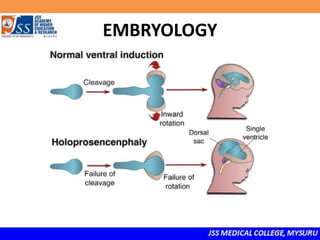
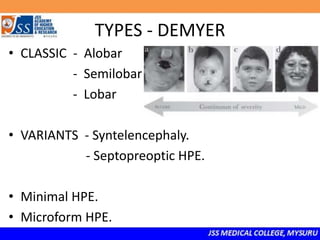
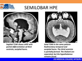
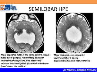
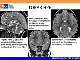
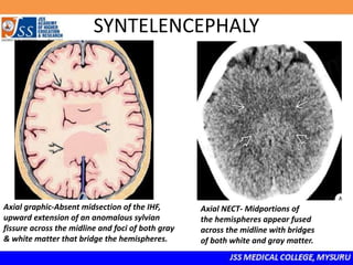
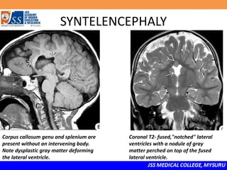
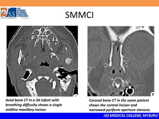
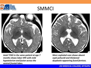
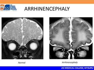
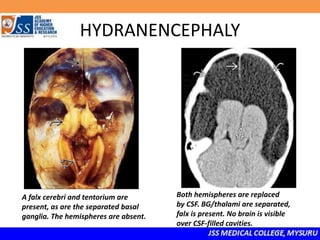
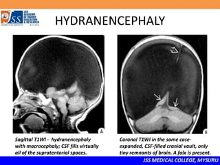
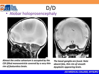
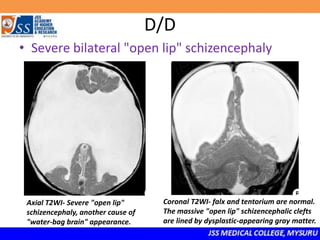
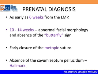
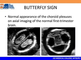
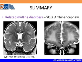
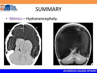
SlideShare a Scribd company logo
HOLOPROSENCEPHALIES,
RELATED DISORDERS & MIMICS
Dr. Vishnu Dakshin
Dr. Rudresh Hiremath
HOLOPROSENCEPHALY
• Rare congenital brain malformation resulting
from incomplete separation of the two
hemispheres.
• “Holo” – Single ventricle
• “pros” – Prosencephalon
• “encephaly” - Brain
EMBRYOLOGY
TYPES - DEMYER
• CLASSIC - Alobar
- Semilobar
- Lobar
• VARIANTS - Syntelencephaly.
- Septopreoptic HPE.
• Minimal HPE.
• Microform HPE.
CLASSIC
• Alobar HPE.
• Semilobar HPE.
• Lobar HPE.
• The distinction between these 3 forms is
based primarily on the presence or absence of
a midline fissure separating the hemispheres.
ALOBAR HPE
• Most severe.
• No midline fissure, no identifiable lobes.
• Fused basal ganglia.
• Agyric /disordered gyri /shallow sulci.
• Single crescent monoventricle - opening dorsally into
a large CSF filled dorsal cyst.
ALOBAR HPE
Small rim of cortex surrounds "horseshoe"
central monoventricle. Thalami are fused.
More cephalad scan in the same patient shows
a large dorsal cyst and central monoventricle
with thin rim of surrounding brain.
SEMILOBAR HPE
• Intermediate form.
• Primitive ventricular horns, third ventricle.
• Fused basal ganglia&hypothalami, thalami often separated.
• Rudimentary falx, posterior IHF.
• Posterior CC forms while anterior aspects are absent.
SEMILOBAR HPE
H-shaped central ventricle with primitive-
appearing temporal horns, fused basal ganglia,
and rudimentary interhemispheric fissure.
Axial T2WI - Rudimentary posterior
interhemispheric fissure, primitive
ventricular horns, and
anterior midline fusion.
SEMILOBAR HPE
Sagittal T1WI shows sHPE with
partial differentiation of third
ventricle, occipital horns.
Axial T2WI in the same patient-
Rudimentary temporal and
occipital horns. The third ventricle
is partially formed. The thalami are
separated, but the hypothalamus
remains fused.
SEMILOBAR HPE
More cephalad T2WI in the same patient shows
fused basal ganglia, rudimentary posterior
interhemispheric fissure, and absence of
anterior interhemispheric fissure with the brain
fused across the midline.
More cephalad scan shows the
upper aspect of a poorly
differentiated central monoventricle
LOBAR HPE
• Best-differentiated.
• IHF & Falx are clearly developed, with shallow and
dysplastic-appearing anterior aspects.
• Third & lateral ventricles – well formed.
• Cavum septum pellucidum is always absent.
• Rudimentary frontal horns are typical , and only the most
inferior portions of the frontal lobes are fused.
LOBAR HPE
Sagittal T2WI of lobar HPE
shows well-differentiated
brain, nearly normal-appearing
third ventricle & azygous ACA
Axial T2WI shows well-
developed occipital horns,
third ventricle, and
minimal anterior midline
fusion.
Coronal T2WI shows that the
anteroinferior frontal cortex is
fused across the midline.
VARIANTS
• Syntelencephaly aka middle interhemispheric
variant of HPE.
• Septopreoptic HPE
- Solitary median maxillary central incisor syndrome.
- Congenital nasal pyriform aperture stenosis.
SYNTELENCEPHALY
• CC genu, splenium present, middle absent.
(Only brain malformation with that morphology)
• Mid-sections of falx, interhemispheric fissure
absent.
• Posterior frontal gray/white matter fused across
midline.
SYNTELENCEPHALY
Axial graphic-Absent midsection of the IHF,
upward extension of an anomalous sylvian
fissure across the midline and foci of both gray
& white matter that bridge the hemispheres.
Axial NECT- Midportions of
the hemispheres appear fused
across the midline with bridges
of both white and gray matter.
SYNTELENCEPHALY
Corpus callosum genu and splenium are
present without an intervening body.
Note dysplastic gray matter deforming
the lateral ventricle.
Coronal T2- fused,"notched" lateral
ventricles with a nodule of gray
matter perched on top of the fused
lateral ventricle.
SOLITARY MEDIAN MAXILLARY
CENTRAL INCISOR SYNDROME.
• Single midline incisor.
• Often coexists with nasal anomalies.
• Brain anomalies of fornix, septi pellucidi, CC.
common.
SMMCI
Axial bone CT in a 3d infant with
breathing difficulty shows a single
midline maxillary incisor.
Coronal bone CT in the same patient
shows the central incisor and
narrowed pyriform aperture stenosis.
SMMCI
Axial T2WI in the same patient at age 7
months shows lobar HPE with mild
hypotelorism and fusion across the
ventral frontal lobes.
More cephalad scan shows absent
septi pellucidi and thickened
dysplastic-appearing fused fornices.
CONGENITAL NASAL PYRIFORM
APERTURE STENOSIS
• Choanal atresia, mid-nasal stenosis, pyriform
aperture stenosis.
• Often coexists with SMMCI.
• HPA axis dysfunction common.
CRANIOFACIAL
RELATED MIDLINE DISORDERS
• Septooptic dysplasia.
- SOD plus – associated with other
anomalies ( Schizencephaly / callosal agenesis)
• Arrhinencephaly.
SEPTOOPTIC DYSPLASIA
• Aka De Morsier syndrome.
• Absence of septi pellucidi.
- Squared-off frontal horns, pointed
inferiorly on coronal T2WI
• Hypoplastic optic nerves, chiasm.
• Look for
○ Malformations of cortical development
○ Thin stalk, small gland, ectopic posterior pituitary
SEPTOOPTIC DYSPLASIA
Absent cavum septi pellucidi
with flat-roofed anterior
horns and small optic chiasm.
Cavum septi pellucidi are
absent ; boxlike lateral
ventricles with inferiorly
pointed frontal horns.
Coronal T2WI in newborn-
absent cavum septi pellucidi,
schizencephaly,extensive
polymicrogyria, fused
fornices.
SEPTOOPTIC DYSPLASIA
Empty-appearing lateral ventricle with
low-lying fornix. The optic chiasm
appears small.
Hypoplastic optic chiasm, absent septi
pellucidi & the peculiar box-like or squared-
off" appearance of the frontal horns.
SEPTOOPTIC DYSPLASIA
Extreme hypoplasia of the optic chiasm,
small pituitary gland with inapparent stalk,
and low-lying fornices that give a striking
"empty“ appearance to the lateral ventricle.
Coronal IR in the same case- unilateral
schizencephaly with dysplastic gray
matter lining the cleft. Note contralateral
polymicrogyria.
ARRHINENCEPHALY
• Absent olfactory bulb and tracts.
• Often seen in association with other midline
facial abnormalities (cleft lip/palate,…)
• Kallman syndrome.
• CHARGE syndrome – in 25% with olfactory
agenesis.
ARRHINENCEPHALY
Normal Arrhinencephaly
MIMICS
• HYDRANENCEPHALY
- Consequence of severe brain destruction in utero.
- “Water bag" brain.
- Small nubbins of remnant brain with a normal falx
cerebri and posterior fossa.
- Hemihydranencephaly.
HYDRANENCEPHALY
Large head with striking
transillumination indicating that most
of the cranium is water-filled.
The hemispheres are absent ("water-bag
brain"), and only the basal ganglia are
present. Note separation.
HYDRANENCEPHALY
A falx cerebri and tentorium are
present, as are the separated basal
ganglia. The hemispheres are absent.
Both hemispheres are replaced
by CSF. BG/thalami are separated,
falx is present. No brain is visible
over CSF-filled cavities.
HYDRANENCEPHALY
Sagittal T1WI - hydranencephaly
with macrocephaly; CSF fills virtually
all of the supratentorial spaces.
Coronal T1WI in the same case-
expanded, CSF-filled cranial vault, only
tiny remnants of brain. A falx is present.
D/D
• Severe obstructive hydrocephalus (OH)
Sagittal T1–massively enlarged lateral ventricles
& tectal dysplasia causing aqueductal stenosis.
Thin rim of compressed but normally formed
cortex and subcortical WM lying under the
calvarium.
D/D
• Alobar holoprosencephaly
Almost the entire calvarium is occupied by the
CSF-filled monoventricle covered by a very thin
rim of featureless brain.
The basal ganglia are fused. Note
absent falx, thin rim of smooth
dysplastic appearing brain.
D/D
• Severe bilateral "open lip" schizencephaly
Axial T2WI- Severe "open lip"
schizencephaly, another cause of
"water-bag brain" appearance.
Coronal T2WI- falx and tentorium are normal.
The massive "open lip" schizencephalic clefts
are lined by dysplastic-appearing gray matter.
PRENATAL DIAGNOSIS
• As early as 6 weeks from the LMP.
• 10 - 14 weeks -- abnormal facial morphology
and absence of the “butterfly” sign.
• Early closure of the metopic suture.
• Absence of the cavum septum pellucidum –
Hallmark.
BUTTERFLY SIGN
• Normal appearance of the choroid plexuses
on axial imaging of the normal first-trimester
brain.
SUMMARY
• Incomplete separation of the two hemispheres.
• Types - Classic – Alobar, Semilobar, Lobar.
D/D – Hydranencephaly.
D/D – Alobar/ Lobar HPE,
depending on severity of sHPE.
D/D - SOD,Arrhinencephaly ,
Syntelencephaly
SUMMARY
• Variants- mivHPE, Septopreoptic HPE.
SUMMARY
• Related midline disorders – SOD, Arrhinencephaly.
D/D – Well-differentiated Lobar HPE.
SUMMARY
• Mimics – Hydranencephaly.
CONCLUSION
• HPE is not uniformly lethal, as is commonly
thought.
• Survival depends on the severity of the brain
and facial malformations, the presence of
chromosomal abnormalities, the involvement
of other organs, and the presence of a
multiple anomaly syndrome.
REFERENCES
• Osborn’s Brain Imaging, Pathology, and
anatomy – Second edition.
• Holoprosencephaly: A Survey of the Entity,
with Embryology and Fetal Imaging - Thomas
C. Winter , Anne M. Kennedy, Paula J.
Woodward.
• Holoprosencephaly: A Guide to Diagnosis and
Clinical Management - Manu S Raam,
Benjamin D Solomon and Maximilian Muenke.
THANK YOU!
Visit www.jssmcradiology.com for
more Radiology Education

More Related Content

What's hot

Intracranial ultrasound
Intracranial ultrasoundIntracranial ultrasound
Intracranial ultrasound
Dr. Mohit Goel
 
Anatomy of hippocampus ( radiology )
Anatomy of hippocampus ( radiology )Anatomy of hippocampus ( radiology )
Anatomy of hippocampus ( radiology )Sajith Selvaganesan
 
Imaging in holoprosencephaly and related disorders
Imaging in holoprosencephaly and related disordersImaging in holoprosencephaly and related disorders
Imaging in holoprosencephaly and related disorders
Khalaf Saba
 
Radiology Spotters
Radiology Spotters Radiology Spotters
Radiology Spotters
Anish Choudhary
 
Radiological anatomy of the brain
Radiological anatomy of the brainRadiological anatomy of the brain
Radiological anatomy of the brain
Mohamed M.A. Zaitoun
 
Ultrasound of thyroid nodules
Ultrasound of thyroid nodulesUltrasound of thyroid nodules
Ultrasound of thyroid nodules
Samir Haffar
 
Orbit imaging anatomy
Orbit imaging anatomyOrbit imaging anatomy
Orbit imaging anatomy
Anish Choudhary
 
The Radiology of Malrotation
The Radiology of MalrotationThe Radiology of Malrotation
The Radiology of Malrotationtboulden
 
Renal doppler
Renal dopplerRenal doppler
Renal doppler
Anish Choudhary
 
1 ultrasound diagnosis of fetal anomalies
1 ultrasound diagnosis of fetal anomalies1 ultrasound diagnosis of fetal anomalies
1 ultrasound diagnosis of fetal anomalies
Dr. Muhammad Bin Zulfiqar
 
Doppler ultrasound of carotid arteries
Doppler ultrasound of carotid arteriesDoppler ultrasound of carotid arteries
Doppler ultrasound of carotid arteriesSamir Haffar
 
Cisterns of brain
Cisterns of brainCisterns of brain
Cisterns of brain
suresh Bishokarma
 
Radiological anatomy & Techniques of the Ventricular system
Radiological anatomy & Techniques of the Ventricular systemRadiological anatomy & Techniques of the Ventricular system
Radiological anatomy & Techniques of the Ventricular system
Mohamed M.A. Zaitoun
 
Presentation1.pptx, ultrasound examination of the neonatal head.
Presentation1.pptx, ultrasound examination of the neonatal head.Presentation1.pptx, ultrasound examination of the neonatal head.
Presentation1.pptx, ultrasound examination of the neonatal head.Abdellah Nazeer
 
Larynx anatomy ct and mri
Larynx anatomy ct and mriLarynx anatomy ct and mri
Larynx anatomy ct and mri
Anish Choudhary
 
Presentation1.pptx, radiological imaging of intra cranial calcification.
Presentation1.pptx, radiological imaging of intra cranial calcification.Presentation1.pptx, radiological imaging of intra cranial calcification.
Presentation1.pptx, radiological imaging of intra cranial calcification.
Abdellah Nazeer
 
Spotters ppt 1
Spotters ppt 1Spotters ppt 1
Spotters ppt 1
Chandni Wadhwani
 
Presentation1.pptx, ultrasound examination of the 2nd & 3rd trimester pregnancy.
Presentation1.pptx, ultrasound examination of the 2nd & 3rd trimester pregnancy.Presentation1.pptx, ultrasound examination of the 2nd & 3rd trimester pregnancy.
Presentation1.pptx, ultrasound examination of the 2nd & 3rd trimester pregnancy.Abdellah Nazeer
 
Radiological features of intracranial tumors 1
Radiological features of intracranial tumors 1Radiological features of intracranial tumors 1
Radiological features of intracranial tumors 1
Dr Praveen kumar tripathi
 
Radiologic anatomy of the cranial nerves
Radiologic anatomy of the cranial nervesRadiologic anatomy of the cranial nerves
Radiologic anatomy of the cranial nerves
hazem youssef
 

What's hot (20)

Intracranial ultrasound
Intracranial ultrasoundIntracranial ultrasound
Intracranial ultrasound
 
Anatomy of hippocampus ( radiology )
Anatomy of hippocampus ( radiology )Anatomy of hippocampus ( radiology )
Anatomy of hippocampus ( radiology )
 
Imaging in holoprosencephaly and related disorders
Imaging in holoprosencephaly and related disordersImaging in holoprosencephaly and related disorders
Imaging in holoprosencephaly and related disorders
 
Radiology Spotters
Radiology Spotters Radiology Spotters
Radiology Spotters
 
Radiological anatomy of the brain
Radiological anatomy of the brainRadiological anatomy of the brain
Radiological anatomy of the brain
 
Ultrasound of thyroid nodules
Ultrasound of thyroid nodulesUltrasound of thyroid nodules
Ultrasound of thyroid nodules
 
Orbit imaging anatomy
Orbit imaging anatomyOrbit imaging anatomy
Orbit imaging anatomy
 
The Radiology of Malrotation
The Radiology of MalrotationThe Radiology of Malrotation
The Radiology of Malrotation
 
Renal doppler
Renal dopplerRenal doppler
Renal doppler
 
1 ultrasound diagnosis of fetal anomalies
1 ultrasound diagnosis of fetal anomalies1 ultrasound diagnosis of fetal anomalies
1 ultrasound diagnosis of fetal anomalies
 
Doppler ultrasound of carotid arteries
Doppler ultrasound of carotid arteriesDoppler ultrasound of carotid arteries
Doppler ultrasound of carotid arteries
 
Cisterns of brain
Cisterns of brainCisterns of brain
Cisterns of brain
 
Radiological anatomy & Techniques of the Ventricular system
Radiological anatomy & Techniques of the Ventricular systemRadiological anatomy & Techniques of the Ventricular system
Radiological anatomy & Techniques of the Ventricular system
 
Presentation1.pptx, ultrasound examination of the neonatal head.
Presentation1.pptx, ultrasound examination of the neonatal head.Presentation1.pptx, ultrasound examination of the neonatal head.
Presentation1.pptx, ultrasound examination of the neonatal head.
 
Larynx anatomy ct and mri
Larynx anatomy ct and mriLarynx anatomy ct and mri
Larynx anatomy ct and mri
 
Presentation1.pptx, radiological imaging of intra cranial calcification.
Presentation1.pptx, radiological imaging of intra cranial calcification.Presentation1.pptx, radiological imaging of intra cranial calcification.
Presentation1.pptx, radiological imaging of intra cranial calcification.
 
Spotters ppt 1
Spotters ppt 1Spotters ppt 1
Spotters ppt 1
 
Presentation1.pptx, ultrasound examination of the 2nd & 3rd trimester pregnancy.
Presentation1.pptx, ultrasound examination of the 2nd & 3rd trimester pregnancy.Presentation1.pptx, ultrasound examination of the 2nd & 3rd trimester pregnancy.
Presentation1.pptx, ultrasound examination of the 2nd & 3rd trimester pregnancy.
 
Radiological features of intracranial tumors 1
Radiological features of intracranial tumors 1Radiological features of intracranial tumors 1
Radiological features of intracranial tumors 1
 
Radiologic anatomy of the cranial nerves
Radiologic anatomy of the cranial nervesRadiologic anatomy of the cranial nerves
Radiologic anatomy of the cranial nerves
 

Similar to Holoprosencephaly

FETAL CENTRAL NERVOUS SYSTEM ANAOMALIES PRESENTATION
FETAL CENTRAL NERVOUS SYSTEM ANAOMALIES PRESENTATIONFETAL CENTRAL NERVOUS SYSTEM ANAOMALIES PRESENTATION
FETAL CENTRAL NERVOUS SYSTEM ANAOMALIES PRESENTATION
kumarramalakshmi
 
Imaging of congenital cns lesions
Imaging of congenital cns lesionsImaging of congenital cns lesions
Imaging of congenital cns lesions
Gobardhan Thapa
 
Imaging of congenital CNS lesions
Imaging of congenital CNS lesionsImaging of congenital CNS lesions
Imaging of congenital CNS lesions
Milan Silwal
 
Congenital CNS lesions
Congenital CNS lesionsCongenital CNS lesions
Congenital CNS lesions
Milan Silwal
 
HYDROCEPHALUS
HYDROCEPHALUSHYDROCEPHALUS
HYDROCEPHALUS
BHAWANAHAJARE1
 
Brain anomalies
Brain anomaliesBrain anomalies
Brain anomalies
Ahmed Koriesh
 
1.schizencephaly 2.holoprosencephaly 3.porencephaly
1.schizencephaly 2.holoprosencephaly 3.porencephaly1.schizencephaly 2.holoprosencephaly 3.porencephaly
1.schizencephaly 2.holoprosencephaly 3.porencephaly
airwave12
 
Fetal brain usg 2
Fetal brain usg 2Fetal brain usg 2
Fetal brain usg 2
Vrishit Saraswat
 
Hydrocephalus.pptx
Hydrocephalus.pptxHydrocephalus.pptx
Hydrocephalus.pptx
Pradeep Pande
 
cns ANAMOLIES-3.pptx
cns ANAMOLIES-3.pptxcns ANAMOLIES-3.pptx
cns ANAMOLIES-3.pptx
PranaviSagar1
 
Neural tube defects and Craniosynostosis
Neural tube defects and CraniosynostosisNeural tube defects and Craniosynostosis
Neural tube defects and CraniosynostosisThe Medical Post
 
Paediatric imaging rapid review of radiology
Paediatric imaging rapid review of radiologyPaediatric imaging rapid review of radiology
Paediatric imaging rapid review of radiology
Double M
 
Neonatal ultrasound overview
Neonatal ultrasound overviewNeonatal ultrasound overview
Neonatal ultrasound overview
Ahmed Bahnassy
 
Fundus examination
Fundus examinationFundus examination
Fundus examination
akhil deshmukh
 
Cranial usg final.pptx
Cranial usg final.pptxCranial usg final.pptx
Cranial usg final.pptx
Nishitha Ashok
 
Nervous system exam part 1
Nervous system exam part 1Nervous system exam part 1
Nervous system exam part 1
MUHAMMAD HOSSAIN
 
USMLE NEUROANATOMY 013 Blood Supply of CNS Blood Supply .pdf
USMLE   NEUROANATOMY 013 Blood Supply of CNS Blood Supply .pdfUSMLE   NEUROANATOMY 013 Blood Supply of CNS Blood Supply .pdf
USMLE NEUROANATOMY 013 Blood Supply of CNS Blood Supply .pdf
AHMED ASHOUR
 
Craniofacial anomalies
Craniofacial anomaliesCraniofacial anomalies
Craniofacial anomalies
Masuma Ryzvee
 
Case record... Septo-optic dysplasia
Case record... Septo-optic dysplasiaCase record... Septo-optic dysplasia
Case record... Septo-optic dysplasia
Professor Yasser Metwally
 
Approach to Macro and Microcephaly
Approach to Macro and MicrocephalyApproach to Macro and Microcephaly
Approach to Macro and MicrocephalyThe Medical Post
 

Similar to Holoprosencephaly (20)

FETAL CENTRAL NERVOUS SYSTEM ANAOMALIES PRESENTATION
FETAL CENTRAL NERVOUS SYSTEM ANAOMALIES PRESENTATIONFETAL CENTRAL NERVOUS SYSTEM ANAOMALIES PRESENTATION
FETAL CENTRAL NERVOUS SYSTEM ANAOMALIES PRESENTATION
 
Imaging of congenital cns lesions
Imaging of congenital cns lesionsImaging of congenital cns lesions
Imaging of congenital cns lesions
 
Imaging of congenital CNS lesions
Imaging of congenital CNS lesionsImaging of congenital CNS lesions
Imaging of congenital CNS lesions
 
Congenital CNS lesions
Congenital CNS lesionsCongenital CNS lesions
Congenital CNS lesions
 
HYDROCEPHALUS
HYDROCEPHALUSHYDROCEPHALUS
HYDROCEPHALUS
 
Brain anomalies
Brain anomaliesBrain anomalies
Brain anomalies
 
1.schizencephaly 2.holoprosencephaly 3.porencephaly
1.schizencephaly 2.holoprosencephaly 3.porencephaly1.schizencephaly 2.holoprosencephaly 3.porencephaly
1.schizencephaly 2.holoprosencephaly 3.porencephaly
 
Fetal brain usg 2
Fetal brain usg 2Fetal brain usg 2
Fetal brain usg 2
 
Hydrocephalus.pptx
Hydrocephalus.pptxHydrocephalus.pptx
Hydrocephalus.pptx
 
cns ANAMOLIES-3.pptx
cns ANAMOLIES-3.pptxcns ANAMOLIES-3.pptx
cns ANAMOLIES-3.pptx
 
Neural tube defects and Craniosynostosis
Neural tube defects and CraniosynostosisNeural tube defects and Craniosynostosis
Neural tube defects and Craniosynostosis
 
Paediatric imaging rapid review of radiology
Paediatric imaging rapid review of radiologyPaediatric imaging rapid review of radiology
Paediatric imaging rapid review of radiology
 
Neonatal ultrasound overview
Neonatal ultrasound overviewNeonatal ultrasound overview
Neonatal ultrasound overview
 
Fundus examination
Fundus examinationFundus examination
Fundus examination
 
Cranial usg final.pptx
Cranial usg final.pptxCranial usg final.pptx
Cranial usg final.pptx
 
Nervous system exam part 1
Nervous system exam part 1Nervous system exam part 1
Nervous system exam part 1
 
USMLE NEUROANATOMY 013 Blood Supply of CNS Blood Supply .pdf
USMLE   NEUROANATOMY 013 Blood Supply of CNS Blood Supply .pdfUSMLE   NEUROANATOMY 013 Blood Supply of CNS Blood Supply .pdf
USMLE NEUROANATOMY 013 Blood Supply of CNS Blood Supply .pdf
 
Craniofacial anomalies
Craniofacial anomaliesCraniofacial anomalies
Craniofacial anomalies
 
Case record... Septo-optic dysplasia
Case record... Septo-optic dysplasiaCase record... Septo-optic dysplasia
Case record... Septo-optic dysplasia
 
Approach to Macro and Microcephaly
Approach to Macro and MicrocephalyApproach to Macro and Microcephaly
Approach to Macro and Microcephaly
 

More from Vikram Patil

Covid-19 Radiological Manifestations
Covid-19 Radiological ManifestationsCovid-19 Radiological Manifestations
Covid-19 Radiological Manifestations
Vikram Patil
 
Artficial Intelligence in Health universities(JSS)
Artficial Intelligence in Health universities(JSS) Artficial Intelligence in Health universities(JSS)
Artficial Intelligence in Health universities(JSS)
Vikram Patil
 
Approach to Chest X-Ray and Interpretation
Approach to Chest X-Ray and InterpretationApproach to Chest X-Ray and Interpretation
Approach to Chest X-Ray and Interpretation
Vikram Patil
 
Mediastinal mass
Mediastinal massMediastinal mass
Mediastinal mass
Vikram Patil
 
Chest x ray basic interpretation
Chest x ray basic interpretationChest x ray basic interpretation
Chest x ray basic interpretation
Vikram Patil
 
Imaging in scurvy
Imaging in scurvyImaging in scurvy
Imaging in scurvy
Vikram Patil
 
Imaging in rickets
Imaging in ricketsImaging in rickets
Imaging in rickets
Vikram Patil
 
Imaging in hip disorders
Imaging in hip disordersImaging in hip disorders
Imaging in hip disorders
Vikram Patil
 
Imaging in malignant bone tumors
Imaging in malignant bone tumorsImaging in malignant bone tumors
Imaging in malignant bone tumors
Vikram Patil
 
Signs in pneumoperitoneum
Signs in pneumoperitoneumSigns in pneumoperitoneum
Signs in pneumoperitoneum
Vikram Patil
 
Congenital anomalies of IVC
Congenital anomalies of IVCCongenital anomalies of IVC
Congenital anomalies of IVC
Vikram Patil
 
PNDT Act
PNDT Act PNDT Act
PNDT Act
Vikram Patil
 

More from Vikram Patil (12)

Covid-19 Radiological Manifestations
Covid-19 Radiological ManifestationsCovid-19 Radiological Manifestations
Covid-19 Radiological Manifestations
 
Artficial Intelligence in Health universities(JSS)
Artficial Intelligence in Health universities(JSS) Artficial Intelligence in Health universities(JSS)
Artficial Intelligence in Health universities(JSS)
 
Approach to Chest X-Ray and Interpretation
Approach to Chest X-Ray and InterpretationApproach to Chest X-Ray and Interpretation
Approach to Chest X-Ray and Interpretation
 
Mediastinal mass
Mediastinal massMediastinal mass
Mediastinal mass
 
Chest x ray basic interpretation
Chest x ray basic interpretationChest x ray basic interpretation
Chest x ray basic interpretation
 
Imaging in scurvy
Imaging in scurvyImaging in scurvy
Imaging in scurvy
 
Imaging in rickets
Imaging in ricketsImaging in rickets
Imaging in rickets
 
Imaging in hip disorders
Imaging in hip disordersImaging in hip disorders
Imaging in hip disorders
 
Imaging in malignant bone tumors
Imaging in malignant bone tumorsImaging in malignant bone tumors
Imaging in malignant bone tumors
 
Signs in pneumoperitoneum
Signs in pneumoperitoneumSigns in pneumoperitoneum
Signs in pneumoperitoneum
 
Congenital anomalies of IVC
Congenital anomalies of IVCCongenital anomalies of IVC
Congenital anomalies of IVC
 
PNDT Act
PNDT Act PNDT Act
PNDT Act
 

Recently uploaded

ARTHROLOGY PPT NCISM SYLLABUS AYURVEDA STUDENTS
ARTHROLOGY PPT NCISM SYLLABUS AYURVEDA STUDENTSARTHROLOGY PPT NCISM SYLLABUS AYURVEDA STUDENTS
ARTHROLOGY PPT NCISM SYLLABUS AYURVEDA STUDENTS
Dr. Vinay Pareek
 
Report Back from SGO 2024: What’s the Latest in Cervical Cancer?
Report Back from SGO 2024: What’s the Latest in Cervical Cancer?Report Back from SGO 2024: What’s the Latest in Cervical Cancer?
Report Back from SGO 2024: What’s the Latest in Cervical Cancer?
bkling
 
24 Upakrama.pptx class ppt useful in all
24 Upakrama.pptx class ppt useful in all24 Upakrama.pptx class ppt useful in all
24 Upakrama.pptx class ppt useful in all
DrSathishMS1
 
heat stroke and heat exhaustion in children
heat stroke and heat exhaustion in childrenheat stroke and heat exhaustion in children
heat stroke and heat exhaustion in children
SumeraAhmad5
 
Alcohol_Dr. Jeenal Mistry MD Pharmacology.pdf
Alcohol_Dr. Jeenal Mistry MD Pharmacology.pdfAlcohol_Dr. Jeenal Mistry MD Pharmacology.pdf
Alcohol_Dr. Jeenal Mistry MD Pharmacology.pdf
Dr Jeenal Mistry
 
For Better Surat #ℂall #Girl Service ❤85270-49040❤ Surat #ℂall #Girls
For Better Surat #ℂall #Girl Service ❤85270-49040❤ Surat #ℂall #GirlsFor Better Surat #ℂall #Girl Service ❤85270-49040❤ Surat #ℂall #Girls
For Better Surat #ℂall #Girl Service ❤85270-49040❤ Surat #ℂall #Girls
Savita Shen $i11
 
ANATOMY AND PHYSIOLOGY OF URINARY SYSTEM.pptx
ANATOMY AND PHYSIOLOGY OF URINARY SYSTEM.pptxANATOMY AND PHYSIOLOGY OF URINARY SYSTEM.pptx
ANATOMY AND PHYSIOLOGY OF URINARY SYSTEM.pptx
Swetaba Besh
 
BRACHYTHERAPY OVERVIEW AND APPLICATORS
BRACHYTHERAPY OVERVIEW  AND  APPLICATORSBRACHYTHERAPY OVERVIEW  AND  APPLICATORS
BRACHYTHERAPY OVERVIEW AND APPLICATORS
Krishan Murari
 
Physiology of Chemical Sensation of smell.pdf
Physiology of Chemical Sensation of smell.pdfPhysiology of Chemical Sensation of smell.pdf
Physiology of Chemical Sensation of smell.pdf
MedicoseAcademics
 
Phone Us ❤85270-49040❤ #ℂall #gIRLS In Surat By Surat @ℂall @Girls Hotel With...
Phone Us ❤85270-49040❤ #ℂall #gIRLS In Surat By Surat @ℂall @Girls Hotel With...Phone Us ❤85270-49040❤ #ℂall #gIRLS In Surat By Surat @ℂall @Girls Hotel With...
Phone Us ❤85270-49040❤ #ℂall #gIRLS In Surat By Surat @ℂall @Girls Hotel With...
Savita Shen $i11
 
MANAGEMENT OF ATRIOVENTRICULAR CONDUCTION BLOCK.pdf
MANAGEMENT OF ATRIOVENTRICULAR CONDUCTION BLOCK.pdfMANAGEMENT OF ATRIOVENTRICULAR CONDUCTION BLOCK.pdf
MANAGEMENT OF ATRIOVENTRICULAR CONDUCTION BLOCK.pdf
Jim Jacob Roy
 
Physiology of Special Chemical Sensation of Taste
Physiology of Special Chemical Sensation of TastePhysiology of Special Chemical Sensation of Taste
Physiology of Special Chemical Sensation of Taste
MedicoseAcademics
 
Surat @ℂall @Girls ꧁❤8527049040❤꧂@ℂall @Girls Service Vip Top Model Safe
Surat @ℂall @Girls ꧁❤8527049040❤꧂@ℂall @Girls Service Vip Top Model SafeSurat @ℂall @Girls ꧁❤8527049040❤꧂@ℂall @Girls Service Vip Top Model Safe
Surat @ℂall @Girls ꧁❤8527049040❤꧂@ℂall @Girls Service Vip Top Model Safe
Savita Shen $i11
 
Are There Any Natural Remedies To Treat Syphilis.pdf
Are There Any Natural Remedies To Treat Syphilis.pdfAre There Any Natural Remedies To Treat Syphilis.pdf
Are There Any Natural Remedies To Treat Syphilis.pdf
Little Cross Family Clinic
 
New Directions in Targeted Therapeutic Approaches for Older Adults With Mantl...
New Directions in Targeted Therapeutic Approaches for Older Adults With Mantl...New Directions in Targeted Therapeutic Approaches for Older Adults With Mantl...
New Directions in Targeted Therapeutic Approaches for Older Adults With Mantl...
i3 Health
 
Flu Vaccine Alert in Bangalore Karnataka
Flu Vaccine Alert in Bangalore KarnatakaFlu Vaccine Alert in Bangalore Karnataka
Flu Vaccine Alert in Bangalore Karnataka
addon Scans
 
Lung Cancer: Artificial Intelligence, Synergetics, Complex System Analysis, S...
Lung Cancer: Artificial Intelligence, Synergetics, Complex System Analysis, S...Lung Cancer: Artificial Intelligence, Synergetics, Complex System Analysis, S...
Lung Cancer: Artificial Intelligence, Synergetics, Complex System Analysis, S...
Oleg Kshivets
 
ARTIFICIAL INTELLIGENCE IN HEALTHCARE.pdf
ARTIFICIAL INTELLIGENCE IN  HEALTHCARE.pdfARTIFICIAL INTELLIGENCE IN  HEALTHCARE.pdf
ARTIFICIAL INTELLIGENCE IN HEALTHCARE.pdf
Anujkumaranit
 
Ophthalmology Clinical Tests for OSCE exam
Ophthalmology Clinical Tests for OSCE examOphthalmology Clinical Tests for OSCE exam
Ophthalmology Clinical Tests for OSCE exam
KafrELShiekh University
 
POST OPERATIVE OLIGURIA and its management
POST OPERATIVE OLIGURIA and its managementPOST OPERATIVE OLIGURIA and its management
POST OPERATIVE OLIGURIA and its management
touseefaziz1
 

Recently uploaded (20)

ARTHROLOGY PPT NCISM SYLLABUS AYURVEDA STUDENTS
ARTHROLOGY PPT NCISM SYLLABUS AYURVEDA STUDENTSARTHROLOGY PPT NCISM SYLLABUS AYURVEDA STUDENTS
ARTHROLOGY PPT NCISM SYLLABUS AYURVEDA STUDENTS
 
Report Back from SGO 2024: What’s the Latest in Cervical Cancer?
Report Back from SGO 2024: What’s the Latest in Cervical Cancer?Report Back from SGO 2024: What’s the Latest in Cervical Cancer?
Report Back from SGO 2024: What’s the Latest in Cervical Cancer?
 
24 Upakrama.pptx class ppt useful in all
24 Upakrama.pptx class ppt useful in all24 Upakrama.pptx class ppt useful in all
24 Upakrama.pptx class ppt useful in all
 
heat stroke and heat exhaustion in children
heat stroke and heat exhaustion in childrenheat stroke and heat exhaustion in children
heat stroke and heat exhaustion in children
 
Alcohol_Dr. Jeenal Mistry MD Pharmacology.pdf
Alcohol_Dr. Jeenal Mistry MD Pharmacology.pdfAlcohol_Dr. Jeenal Mistry MD Pharmacology.pdf
Alcohol_Dr. Jeenal Mistry MD Pharmacology.pdf
 
For Better Surat #ℂall #Girl Service ❤85270-49040❤ Surat #ℂall #Girls
For Better Surat #ℂall #Girl Service ❤85270-49040❤ Surat #ℂall #GirlsFor Better Surat #ℂall #Girl Service ❤85270-49040❤ Surat #ℂall #Girls
For Better Surat #ℂall #Girl Service ❤85270-49040❤ Surat #ℂall #Girls
 
ANATOMY AND PHYSIOLOGY OF URINARY SYSTEM.pptx
ANATOMY AND PHYSIOLOGY OF URINARY SYSTEM.pptxANATOMY AND PHYSIOLOGY OF URINARY SYSTEM.pptx
ANATOMY AND PHYSIOLOGY OF URINARY SYSTEM.pptx
 
BRACHYTHERAPY OVERVIEW AND APPLICATORS
BRACHYTHERAPY OVERVIEW  AND  APPLICATORSBRACHYTHERAPY OVERVIEW  AND  APPLICATORS
BRACHYTHERAPY OVERVIEW AND APPLICATORS
 
Physiology of Chemical Sensation of smell.pdf
Physiology of Chemical Sensation of smell.pdfPhysiology of Chemical Sensation of smell.pdf
Physiology of Chemical Sensation of smell.pdf
 
Phone Us ❤85270-49040❤ #ℂall #gIRLS In Surat By Surat @ℂall @Girls Hotel With...
Phone Us ❤85270-49040❤ #ℂall #gIRLS In Surat By Surat @ℂall @Girls Hotel With...Phone Us ❤85270-49040❤ #ℂall #gIRLS In Surat By Surat @ℂall @Girls Hotel With...
Phone Us ❤85270-49040❤ #ℂall #gIRLS In Surat By Surat @ℂall @Girls Hotel With...
 
MANAGEMENT OF ATRIOVENTRICULAR CONDUCTION BLOCK.pdf
MANAGEMENT OF ATRIOVENTRICULAR CONDUCTION BLOCK.pdfMANAGEMENT OF ATRIOVENTRICULAR CONDUCTION BLOCK.pdf
MANAGEMENT OF ATRIOVENTRICULAR CONDUCTION BLOCK.pdf
 
Physiology of Special Chemical Sensation of Taste
Physiology of Special Chemical Sensation of TastePhysiology of Special Chemical Sensation of Taste
Physiology of Special Chemical Sensation of Taste
 
Surat @ℂall @Girls ꧁❤8527049040❤꧂@ℂall @Girls Service Vip Top Model Safe
Surat @ℂall @Girls ꧁❤8527049040❤꧂@ℂall @Girls Service Vip Top Model SafeSurat @ℂall @Girls ꧁❤8527049040❤꧂@ℂall @Girls Service Vip Top Model Safe
Surat @ℂall @Girls ꧁❤8527049040❤꧂@ℂall @Girls Service Vip Top Model Safe
 
Are There Any Natural Remedies To Treat Syphilis.pdf
Are There Any Natural Remedies To Treat Syphilis.pdfAre There Any Natural Remedies To Treat Syphilis.pdf
Are There Any Natural Remedies To Treat Syphilis.pdf
 
New Directions in Targeted Therapeutic Approaches for Older Adults With Mantl...
New Directions in Targeted Therapeutic Approaches for Older Adults With Mantl...New Directions in Targeted Therapeutic Approaches for Older Adults With Mantl...
New Directions in Targeted Therapeutic Approaches for Older Adults With Mantl...
 
Flu Vaccine Alert in Bangalore Karnataka
Flu Vaccine Alert in Bangalore KarnatakaFlu Vaccine Alert in Bangalore Karnataka
Flu Vaccine Alert in Bangalore Karnataka
 
Lung Cancer: Artificial Intelligence, Synergetics, Complex System Analysis, S...
Lung Cancer: Artificial Intelligence, Synergetics, Complex System Analysis, S...Lung Cancer: Artificial Intelligence, Synergetics, Complex System Analysis, S...
Lung Cancer: Artificial Intelligence, Synergetics, Complex System Analysis, S...
 
ARTIFICIAL INTELLIGENCE IN HEALTHCARE.pdf
ARTIFICIAL INTELLIGENCE IN  HEALTHCARE.pdfARTIFICIAL INTELLIGENCE IN  HEALTHCARE.pdf
ARTIFICIAL INTELLIGENCE IN HEALTHCARE.pdf
 
Ophthalmology Clinical Tests for OSCE exam
Ophthalmology Clinical Tests for OSCE examOphthalmology Clinical Tests for OSCE exam
Ophthalmology Clinical Tests for OSCE exam
 
POST OPERATIVE OLIGURIA and its management
POST OPERATIVE OLIGURIA and its managementPOST OPERATIVE OLIGURIA and its management
POST OPERATIVE OLIGURIA and its management
 

Holoprosencephaly

  • 1. HOLOPROSENCEPHALIES, RELATED DISORDERS & MIMICS Dr. Vishnu Dakshin Dr. Rudresh Hiremath
  • 2. HOLOPROSENCEPHALY • Rare congenital brain malformation resulting from incomplete separation of the two hemispheres. • “Holo” – Single ventricle • “pros” – Prosencephalon • “encephaly” - Brain
  • 4. TYPES - DEMYER • CLASSIC - Alobar - Semilobar - Lobar • VARIANTS - Syntelencephaly. - Septopreoptic HPE. • Minimal HPE. • Microform HPE.
  • 5. CLASSIC • Alobar HPE. • Semilobar HPE. • Lobar HPE. • The distinction between these 3 forms is based primarily on the presence or absence of a midline fissure separating the hemispheres.
  • 6. ALOBAR HPE • Most severe. • No midline fissure, no identifiable lobes. • Fused basal ganglia. • Agyric /disordered gyri /shallow sulci. • Single crescent monoventricle - opening dorsally into a large CSF filled dorsal cyst.
  • 7. ALOBAR HPE Small rim of cortex surrounds "horseshoe" central monoventricle. Thalami are fused. More cephalad scan in the same patient shows a large dorsal cyst and central monoventricle with thin rim of surrounding brain.
  • 8. SEMILOBAR HPE • Intermediate form. • Primitive ventricular horns, third ventricle. • Fused basal ganglia&hypothalami, thalami often separated. • Rudimentary falx, posterior IHF. • Posterior CC forms while anterior aspects are absent.
  • 9. SEMILOBAR HPE H-shaped central ventricle with primitive- appearing temporal horns, fused basal ganglia, and rudimentary interhemispheric fissure. Axial T2WI - Rudimentary posterior interhemispheric fissure, primitive ventricular horns, and anterior midline fusion.
  • 10. SEMILOBAR HPE Sagittal T1WI shows sHPE with partial differentiation of third ventricle, occipital horns. Axial T2WI in the same patient- Rudimentary temporal and occipital horns. The third ventricle is partially formed. The thalami are separated, but the hypothalamus remains fused.
  • 11. SEMILOBAR HPE More cephalad T2WI in the same patient shows fused basal ganglia, rudimentary posterior interhemispheric fissure, and absence of anterior interhemispheric fissure with the brain fused across the midline. More cephalad scan shows the upper aspect of a poorly differentiated central monoventricle
  • 12. LOBAR HPE • Best-differentiated. • IHF & Falx are clearly developed, with shallow and dysplastic-appearing anterior aspects. • Third & lateral ventricles – well formed. • Cavum septum pellucidum is always absent. • Rudimentary frontal horns are typical , and only the most inferior portions of the frontal lobes are fused.
  • 13. LOBAR HPE Sagittal T2WI of lobar HPE shows well-differentiated brain, nearly normal-appearing third ventricle & azygous ACA Axial T2WI shows well- developed occipital horns, third ventricle, and minimal anterior midline fusion. Coronal T2WI shows that the anteroinferior frontal cortex is fused across the midline.
  • 14. VARIANTS • Syntelencephaly aka middle interhemispheric variant of HPE. • Septopreoptic HPE - Solitary median maxillary central incisor syndrome. - Congenital nasal pyriform aperture stenosis.
  • 15. SYNTELENCEPHALY • CC genu, splenium present, middle absent. (Only brain malformation with that morphology) • Mid-sections of falx, interhemispheric fissure absent. • Posterior frontal gray/white matter fused across midline.
  • 16. SYNTELENCEPHALY Axial graphic-Absent midsection of the IHF, upward extension of an anomalous sylvian fissure across the midline and foci of both gray & white matter that bridge the hemispheres. Axial NECT- Midportions of the hemispheres appear fused across the midline with bridges of both white and gray matter.
  • 17. SYNTELENCEPHALY Corpus callosum genu and splenium are present without an intervening body. Note dysplastic gray matter deforming the lateral ventricle. Coronal T2- fused,"notched" lateral ventricles with a nodule of gray matter perched on top of the fused lateral ventricle.
  • 18. SOLITARY MEDIAN MAXILLARY CENTRAL INCISOR SYNDROME. • Single midline incisor. • Often coexists with nasal anomalies. • Brain anomalies of fornix, septi pellucidi, CC. common.
  • 19. SMMCI Axial bone CT in a 3d infant with breathing difficulty shows a single midline maxillary incisor. Coronal bone CT in the same patient shows the central incisor and narrowed pyriform aperture stenosis.
  • 20. SMMCI Axial T2WI in the same patient at age 7 months shows lobar HPE with mild hypotelorism and fusion across the ventral frontal lobes. More cephalad scan shows absent septi pellucidi and thickened dysplastic-appearing fused fornices.
  • 21. CONGENITAL NASAL PYRIFORM APERTURE STENOSIS • Choanal atresia, mid-nasal stenosis, pyriform aperture stenosis. • Often coexists with SMMCI. • HPA axis dysfunction common.
  • 23. RELATED MIDLINE DISORDERS • Septooptic dysplasia. - SOD plus – associated with other anomalies ( Schizencephaly / callosal agenesis) • Arrhinencephaly.
  • 24. SEPTOOPTIC DYSPLASIA • Aka De Morsier syndrome. • Absence of septi pellucidi. - Squared-off frontal horns, pointed inferiorly on coronal T2WI • Hypoplastic optic nerves, chiasm. • Look for ○ Malformations of cortical development ○ Thin stalk, small gland, ectopic posterior pituitary
  • 25. SEPTOOPTIC DYSPLASIA Absent cavum septi pellucidi with flat-roofed anterior horns and small optic chiasm. Cavum septi pellucidi are absent ; boxlike lateral ventricles with inferiorly pointed frontal horns. Coronal T2WI in newborn- absent cavum septi pellucidi, schizencephaly,extensive polymicrogyria, fused fornices.
  • 26. SEPTOOPTIC DYSPLASIA Empty-appearing lateral ventricle with low-lying fornix. The optic chiasm appears small. Hypoplastic optic chiasm, absent septi pellucidi & the peculiar box-like or squared- off" appearance of the frontal horns.
  • 27. SEPTOOPTIC DYSPLASIA Extreme hypoplasia of the optic chiasm, small pituitary gland with inapparent stalk, and low-lying fornices that give a striking "empty“ appearance to the lateral ventricle. Coronal IR in the same case- unilateral schizencephaly with dysplastic gray matter lining the cleft. Note contralateral polymicrogyria.
  • 28. ARRHINENCEPHALY • Absent olfactory bulb and tracts. • Often seen in association with other midline facial abnormalities (cleft lip/palate,…) • Kallman syndrome. • CHARGE syndrome – in 25% with olfactory agenesis.
  • 30. MIMICS • HYDRANENCEPHALY - Consequence of severe brain destruction in utero. - “Water bag" brain. - Small nubbins of remnant brain with a normal falx cerebri and posterior fossa. - Hemihydranencephaly.
  • 31. HYDRANENCEPHALY Large head with striking transillumination indicating that most of the cranium is water-filled. The hemispheres are absent ("water-bag brain"), and only the basal ganglia are present. Note separation.
  • 32. HYDRANENCEPHALY A falx cerebri and tentorium are present, as are the separated basal ganglia. The hemispheres are absent. Both hemispheres are replaced by CSF. BG/thalami are separated, falx is present. No brain is visible over CSF-filled cavities.
  • 33. HYDRANENCEPHALY Sagittal T1WI - hydranencephaly with macrocephaly; CSF fills virtually all of the supratentorial spaces. Coronal T1WI in the same case- expanded, CSF-filled cranial vault, only tiny remnants of brain. A falx is present.
  • 34. D/D • Severe obstructive hydrocephalus (OH) Sagittal T1–massively enlarged lateral ventricles & tectal dysplasia causing aqueductal stenosis. Thin rim of compressed but normally formed cortex and subcortical WM lying under the calvarium.
  • 35. D/D • Alobar holoprosencephaly Almost the entire calvarium is occupied by the CSF-filled monoventricle covered by a very thin rim of featureless brain. The basal ganglia are fused. Note absent falx, thin rim of smooth dysplastic appearing brain.
  • 36. D/D • Severe bilateral "open lip" schizencephaly Axial T2WI- Severe "open lip" schizencephaly, another cause of "water-bag brain" appearance. Coronal T2WI- falx and tentorium are normal. The massive "open lip" schizencephalic clefts are lined by dysplastic-appearing gray matter.
  • 37. PRENATAL DIAGNOSIS • As early as 6 weeks from the LMP. • 10 - 14 weeks -- abnormal facial morphology and absence of the “butterfly” sign. • Early closure of the metopic suture. • Absence of the cavum septum pellucidum – Hallmark.
  • 38. BUTTERFLY SIGN • Normal appearance of the choroid plexuses on axial imaging of the normal first-trimester brain.
  • 39. SUMMARY • Incomplete separation of the two hemispheres. • Types - Classic – Alobar, Semilobar, Lobar. D/D – Hydranencephaly. D/D – Alobar/ Lobar HPE, depending on severity of sHPE. D/D - SOD,Arrhinencephaly , Syntelencephaly
  • 40. SUMMARY • Variants- mivHPE, Septopreoptic HPE.
  • 41. SUMMARY • Related midline disorders – SOD, Arrhinencephaly. D/D – Well-differentiated Lobar HPE.
  • 42. SUMMARY • Mimics – Hydranencephaly.
  • 43. CONCLUSION • HPE is not uniformly lethal, as is commonly thought. • Survival depends on the severity of the brain and facial malformations, the presence of chromosomal abnormalities, the involvement of other organs, and the presence of a multiple anomaly syndrome.
  • 44. REFERENCES • Osborn’s Brain Imaging, Pathology, and anatomy – Second edition. • Holoprosencephaly: A Survey of the Entity, with Embryology and Fetal Imaging - Thomas C. Winter , Anne M. Kennedy, Paula J. Woodward. • Holoprosencephaly: A Guide to Diagnosis and Clinical Management - Manu S Raam, Benjamin D Solomon and Maximilian Muenke.
  • 45. THANK YOU! Visit www.jssmcradiology.com for more Radiology Education

Editor's Notes

  1. In the earliest stages, bilateral outpouchings from the neural tube initially form a single central fluidfilled cavity (“monoventricle”) that will eventually develop into the lateral and third ventricles (see Chapter 34). The separation into two hemispheres is normally completed by the fifth gestational week. The embryonic ventricular system is a series of interconnected fluid-filled chambers that arise as expansions from the central cavity of the embryonic neural tube. As the developing brain bends and expands, it forms fore-brain, midbrain, and hindbrain vesicles. The forebrain cavity divides into two lateral ventricles, which develop as outpouchings from the rostral third ventricle and are connected to it by the interventricular foramen (foramen of Monro) (34-1). The cerebral aqueduct develops from the midbrain vesicle. The fourth ventricle develops from the hindbrain cavity and merges proximally with the aqueduct and caudally with the central canal of the spinal cord The holoprosencephalies are characterized by failure of normal dorsal-ventral induction and lack of rostral forebrain cleavage. In the most severe forms, diencephalic-derived structures such as the basal ganglia also remain fused in the midline. As ventral induction is closely related to facial development, HPE is also associated with a number of characteristic facial anomalies. The eye field begins as a single midline structure. Under the signaling influence of the prechordal plate, the eye field in vertebrates splits into separate left and right eyes. If this developmental process is not completed correctly, the result is cyclopia
  2. Probst- presence or absence of a dorsal sac and cleavage of basal structures are used to categorize the process in children “minimal” HPE, which is associated with subtle craniofacial malformations and mild developmental delay; and microform HPE, which by definition excludes brain involvement.  (a) synophthalmia and a proboscis in a patient with alobar holoprosencephaly; (b) severe hypotelorism, flat nasal bridge, bilateral colobomas, and midline cleft lip and palate in a patient with alobar holoprosencephaly; (c) hypotelorism, flat nasal bridge, and closely spaced nostrils in a patient with lobar holoprosencephaly; (d) hypotelorism, sharp nasal bridge, and single maxillary central incisor in an individual with a microform of holoprosencephaly
  3. The most severe facial malformations are seen in association with alobar HPE. The presence of a dorsal cyst strongly correlates with thalamic fusion, hence the high frequency with alobar HPE. It is thought that the flow of cerebrospinal fluid out of the third ventricle is obstructed by fusion of the thalami, and, as a result, the third ventricle balloons out posteriorly at the point of least resistance in the suprapineal recess.
  4. The single primitive ventricle and fused thalami are perhaps the two most valuable US clues for alobar HPE. Important differential diagnostic considerations for alobar HPE, such as hydranencephaly and severe hydrocephalus (42), exhibit normal thalamic cleavage and usually at least a partially visualized falx cerebri. Hydranencephaly – Face normal, Falx +, Most of cerebral tissue has been destroyed, usually by intrauterine vascular accident or infection.
  5. varying degrees of separation of the deep nuclei. When thalamic fusion is present, there may be a dorsal cyst in semilobar HPE The farther anterior the cc forms, the better the brain is developed. IH fissure- rudimentary in most severe forms, forms in better-differentiated shpe.
  6. Coronal autopsy case of severe semilobar HPE shows Hshaped central ventricle with primitive-appearing temporal horns ﬊, fused basal ganglia ſt, and rudimentary interhemispheric fissure. Axial T2WI shows severe sHPE with rudimentary posterior interhemispheric fissure st, primitive ventricular horns ſt, and anterior midline fusion. Diffuse frontal migration arrest with subcortical heterotopic GM ﬇ is also present.
  7. Sagittal T1WI shows sHPE with partial differentiation of third ventricle ﬈, occipital horns ﬊. The midbrain, pons, and cerebellum are comparatively normal.(38-8B) Axial T2WI in the same patient shows mild hypotelorism with no other midface anomalies. Rudimentary temporal ſt and occipital st horns are present. The third ventricle ﬇ is partially formed. The thalami ﬈ are separated, but the hypothalamus ﬊ remains fused.
  8. More cephalad T2WI in the same patient shows fused basal ganglia ſt, rudimentary posterior interhemispheric fissure st, and absence of anterior interhemispheric fissure with the brain fused across the midline ﬊. (38-8D) More cephalad scan shows the upper aspect of a poorly differentiated central monoventricle. The corpus callosum and all normal midline structures are absent.
  9. Sagittal T2WI of lobar HPE shows well-differentiated brain, nearly normal-appearing third ventricle ﬇, and azygous ACA ſt. Coronal T2WI shows that the anteroinferior frontal cortex is fused across the midline st. Axial T2WI shows well-developed occipital horns ſt, third ventricle ﬇, and minimal anterior midline fusion st.
  10. Mih – inbetween semilobar and lobar in terms of severity.
  11. Axial graphic depicts syntelencephaly with absent midsection of the interhemispheric fissure, upward extension of an anomalous sylvian fissure across the midline ﬈, and foci of both gray and white matter ﬊ that bridge the hemispheres. (38-11A) Axial NECT scan in a patient with syntelencephaly shows that the midportions of the hemispheres appear fused across the midline with bridges of both white ſt and gray matter ﬇.
  12. Sagittal T1WI shows classic findings of MIH. Corpus callosum genu ſt and splenium ﬇ are present without an intervening body. Note dysplastic gray matter st deforming the lateral ventricle. (38-11C) Coronal T2 shows fused, "notched" lateral ventricles ſt with a nodule of gray matter ﬇ perched on top of the fused lateral ventricle. The posterior frontal lobes are continuous across the midline st without an interhemispheric fissure.
  13. Axial bone CT in a 3d infant with breathing difficulty shows a single midline maxillary incisor ſt. (38-12B) Coronal bone CT in the same patient shows the central incisor ſt and narrowed pyriform aperture stenosis ﬇. In the term newborn, the normal aperture width should be > 8 mm.
  14. Axial T2WI in the same patient at age 7 months shows lobar HPE with mild hypotelorism and fusion across the ventral frontal lobes ſt. (38-12D) More cephalad scan shows absent septi pellucidi and thickened dysplastic-appearing fused fornices ſt.
  15. (A) Patient with alobar HPE. (A1) Note cyclopia with doubling of eye structures, arrhinia, rudimentary proboscis and agnathia. (A2) microcephaly and malformed ears. (B) Patient with alobar HPE note premaxillary agenesis and hypertelorism. (C) Patient with semilobar HPE. (C1) Note hypotelorism, squint and cleft lip. (C2) Microcephaly, brachycephaly and large ears. (D) Patient with semilobar HPE showing hypotelorism. (E), (F) and (G) are patients with semilobar HPE showing hypertelorism. (H) Patient with semilobar HPE and Dandy Walker malformation showing normal facial features except for ear asymmetry (simple right ear). (I1) Patient with MIH having hypertelorism and (I2) occipital cephalocele
  16. Coronal graphic shows SOD with absent cavum septi pellucidi with flat-roofed anterior horns ﬊ and small optic chiasm ſt. Coronal T2WI in newborn shows absent cavum septi pellucidi ﬇, schizencephaly ſt, extensive polymicrogyria ﬊, fused fornices ﬉. Cavum septi pellucidi are absent ﬊; boxlike lateral ventricles with inferiorly pointed frontal horns ﬈ are seen.
  17. Sagittal T2WI in a 13m boy with septooptic dysplasia (SOD) shows an empty-appearing lateral ventricle with low-lying fornix ﬈. The optic chiasm ﬊ appears small. Coronal T2WI shows the hypoplastic optic chiasm ﬊, absent septi pellucidi ﬇, and the peculiar box-like or "squared-off" appearance of the frontal horns. The inferior pointing ſt of both frontal horns is also characteristic of SOD.
  18. Sagittal T1WI in a 26y woman with SOD shows extreme hypoplasia f the optic chiasm ſt, small pituitary gland with inapparent stalk st, and low-lying fornices ﬇ that give a striking "empty" appearance to the lateral ventricle ﬊. Coronal IR in the same case shows unilateral schizencephaly with dysplastic gray matter ﬈ lining the cleft st. Note contralateral polymicrogyria ſt.
  19. Coronal T2WI in a normal newborn shows olfactory bulbs ſt and normal olfactory sulci ﬇ Coronal T2WI in a newborn with multiple congenital anomalies demonstrates arrhinencephaly with absent olfactory bulbs ſt and no olfactory sulci ﬇.
  20. A normal or large head with fluid-filled cranial vault. Compromise of the internal carotid artery circulation before 16 gestational weeks followed by diffuse liquefactive necrosis of the cerebral mantle is responsible. In hydranencephaly, most of the cerebral hemispheres have been destroyed and are totally or partially replaced by translucent thin-walled sacs of CSF that fill most of the supratentorial space.The outer layer consists of leptomeninges, and the inner layer is glial tissue without demonstrable ependymal elements.
  21. Autopsy case of hydranencephaly demonstrates a large head with striking transillumination indicating that most of the cranium is water-filled. The thinned calvarium ﬊ has been partially removed to show the fluid-filled cavity. The hemispheres are absent ("water-bag brain"), and only the basal ganglia are present. Note separation
  22. This is the same case as Figure 38-22, seen from above. A falx cerebri ﬈ and tentorium ﬊ are present, as are the separated basal ganglia ﬉. The hemispheres are absent. NECT shows hydranencephaly. Both hemispheres are replaced by CSF. BG/thalami are separated ﬇, falx is present ſt. No brain is visible over CSF-filled cavities st.
  23. Sagittal T1WI shows hydranencephaly with macrocephaly; CSF fills virtually all of the supratentorial spaces. Brainstem and cerebellum are normal. Coronal T1WI in the same case shows expanded, CSF-filled cranial vault, only tiny remnants of brain ſt. A falx is present﬇.
  24. - thin cortex can be seen compressed against the dura and inner table of the calvaria. Sagittal T1WI in a 4-week infant with macrocrania shows massively enlarged lateral ventricles ﬇ and tectal dysplasia ſtcausing aqueductal stenosis ﬉. Coronal T2WI shows the massively enlarged lateral ventricles ﬇. There is a thin rim of compressed but normally formed cortex ſt and subcortical WM lying under the calvarium. This is maximal hydrocephalus.
  25. This is alobar holoprosencephaly. Sagittal T2WI shows enlarged head with relatively normal appearing posterior fossa. Almost the entire calvarium is occupied by the CSF-filled monoventricle covered by a very thin rim of featureless brain ﬇. Coronal T2WI shows a horseshoe-shaped monoventricle. The basal ganglia ﬊ are fused. Note absent falx, thin rim of smooth dysplastic appearing brain ﬈.
  26. Axial T2WI shows severe "open lip“ schizencephaly, another cause of "water-bag brain" appearance. Coronal T2WI in the same case shows that the falx and tentorium are normal. The massive "open lip" schizencephalic clefts are lined by dysplastic-appearing gray matter.
  27. 3. height of the frontal bones at the suture was higher. 4. If the face is normal, inability to demonstrate a normal cavum septum pellucidum may be the only hint that there is severe brain malformation -- careful follow-up is essential with endovaginal US, three-dimensional volume acquisition, and fetal MR imaging.
  28. Normal skull and brain at 13 weeks. Axial view of the head showing the normal calcification of the skull; the falx and the two choroid plexuses ("butterfly sign") are normal. (b) Alobar holoprosencephaly: transverse view of the head showing the fusion of the thalami and of the lateral ventricles. Inability to demonstrate this sign within a normal cranial vault is highly suggestive of HPE
  29. D/D - SOD – frontal horns + - Arrhinencephaly – olfactory bulbs + - Syntelencephaly – CC, genu, splenium(+), body(-)
  30. Among euploid patients with HPE, there is an inverse relationship between the severity of the facial phenotype and the duration of survival. In fetuses with HPE, look for additional anomalies, consider fetal MR imaging, offer karyotype with microarray analysis.