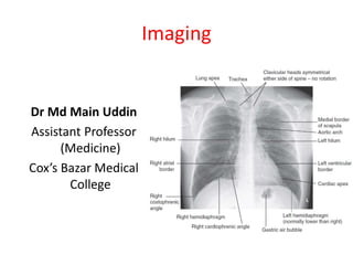
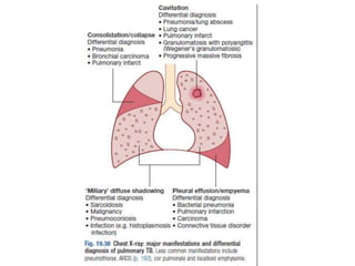
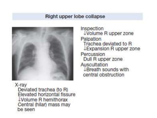
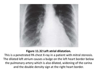
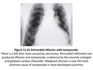
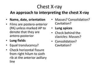
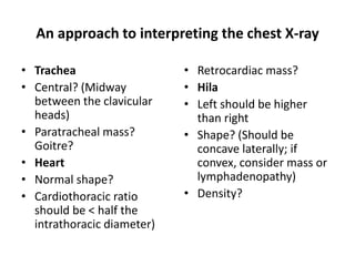
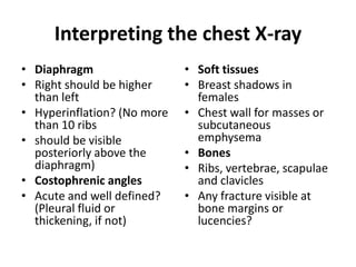
This document provides guidance on interpreting chest x-rays by assessing key anatomical structures and features including the lungs, heart, trachea, diaphragm, bones, and soft tissues. It describes the normal appearance of these structures and signs that may indicate abnormalities such as masses, consolidation, cavitation, enlarged organs, fluid, or fractures. A systematic approach is recommended to examine the chest x-ray starting from the lungs fields and working through other areas.


![Imaging in opaqe hemithorax [autosaved]](https://cdn.slidesharecdn.com/ss_thumbnails/imaginginopaqehemithoraxautosaved-161030071708-thumbnail.jpg?width=640&height=640&fit=bounds)


![PRINCIPLES OF PHARMOCODYNAMICS 2 [Autosaved].pptx](https://cdn.slidesharecdn.com/ss_thumbnails/principlesofpharmocodynamics2autosaved-230607181037-758ddb07-thumbnail.jpg?width=640&height=640&fit=bounds)


![Hypothalamus short ppt by Dr. Neha [PT].pptx](https://cdn.slidesharecdn.com/ss_thumbnails/hypothalamusbydr-260124145759-b9f94a93-thumbnail.jpg?width=640&height=640&fit=bounds)


