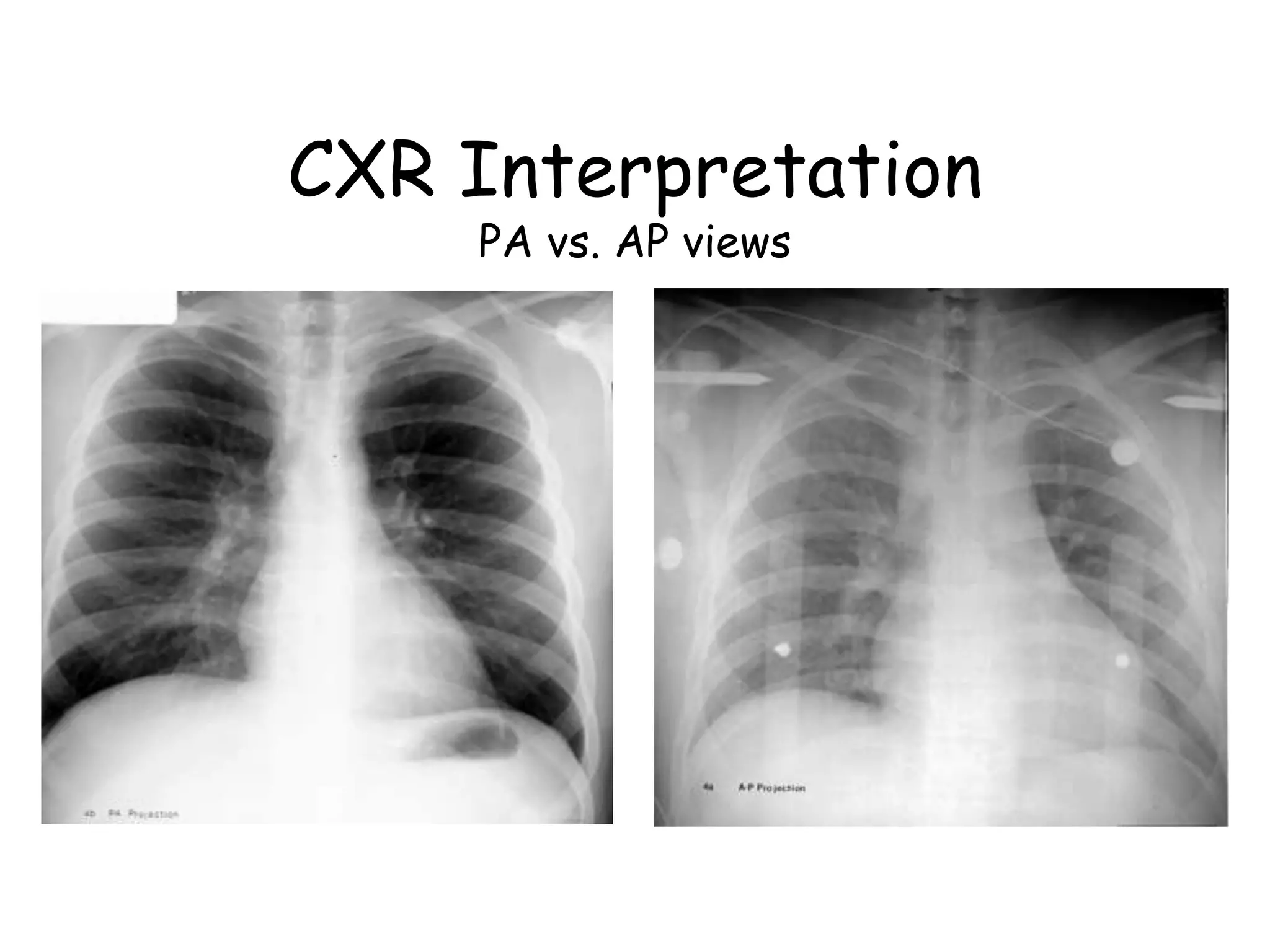
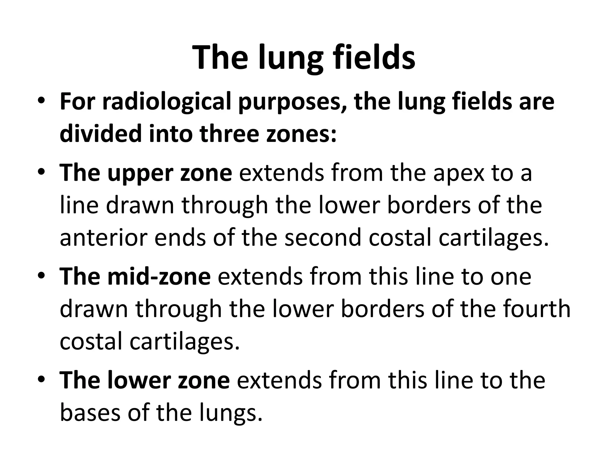
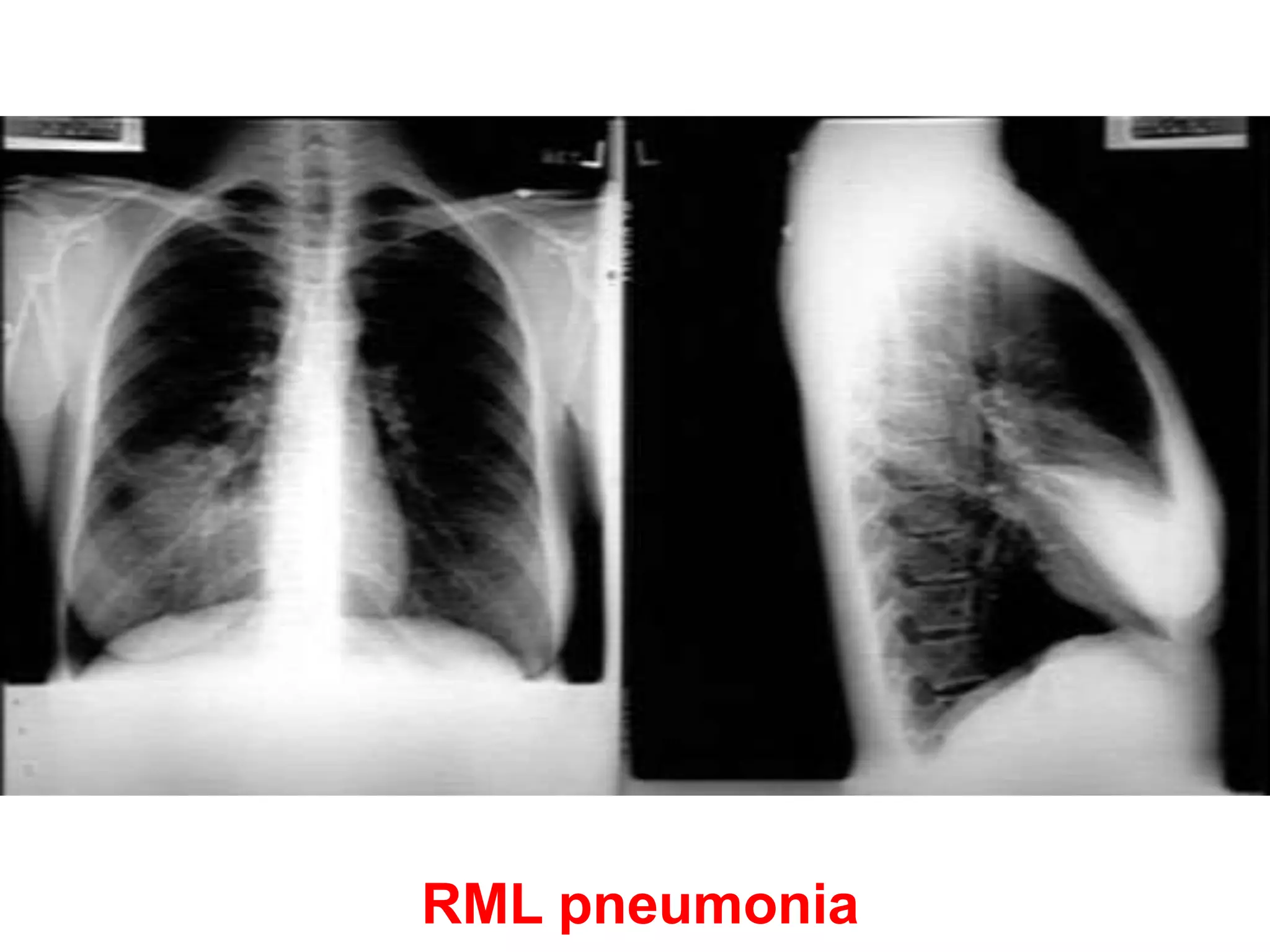
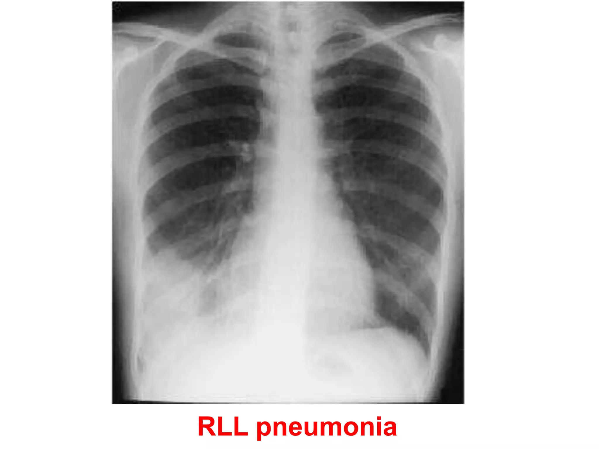
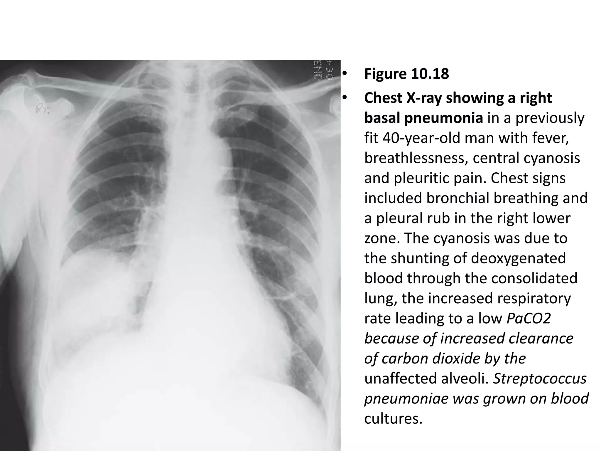
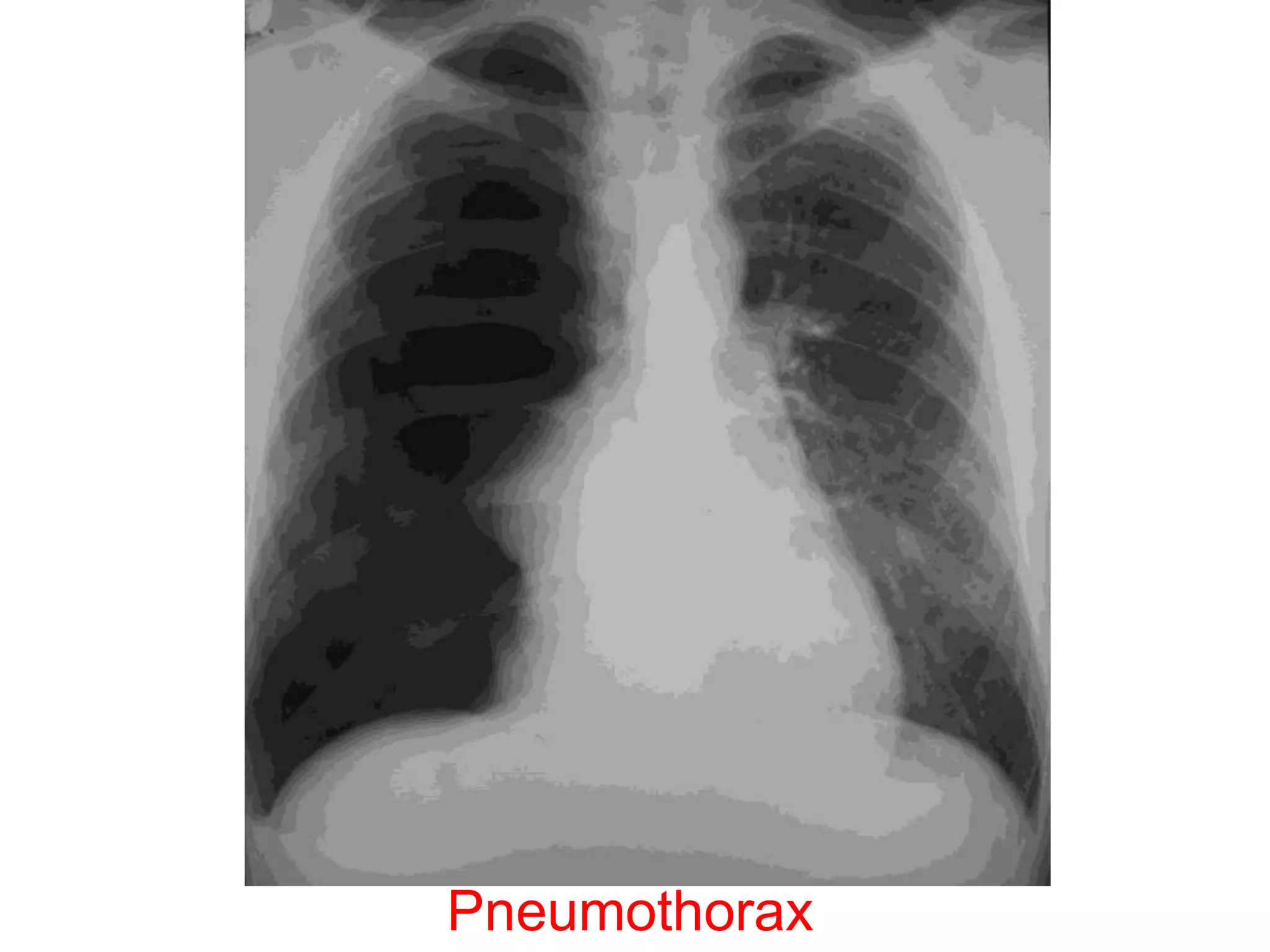
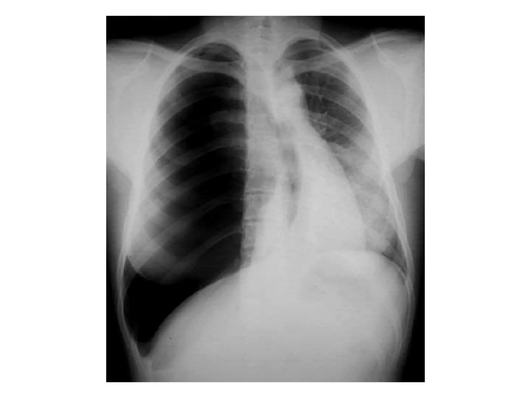
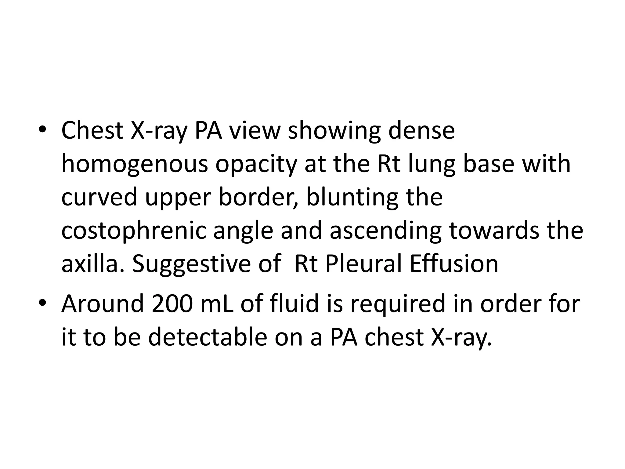
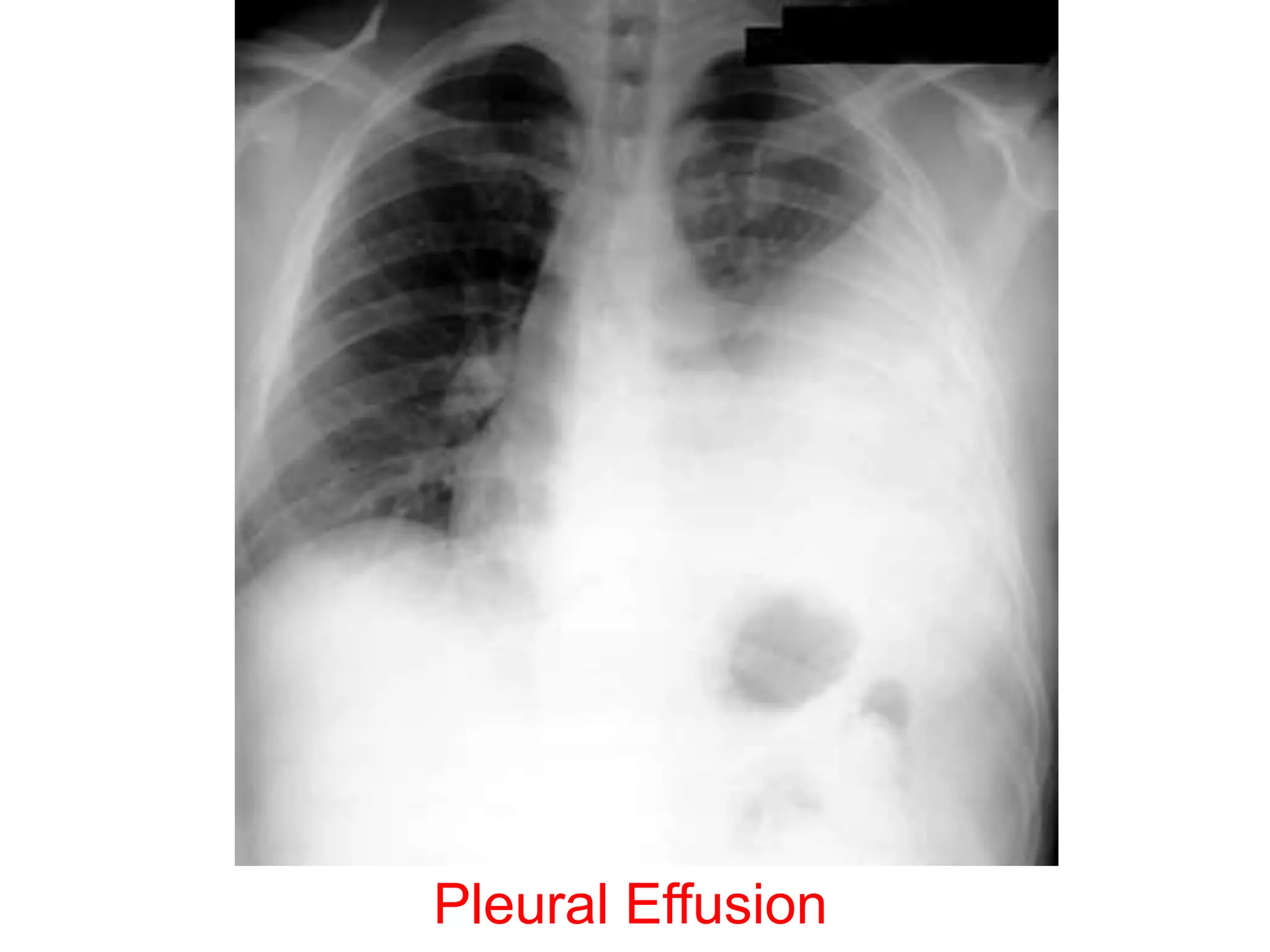
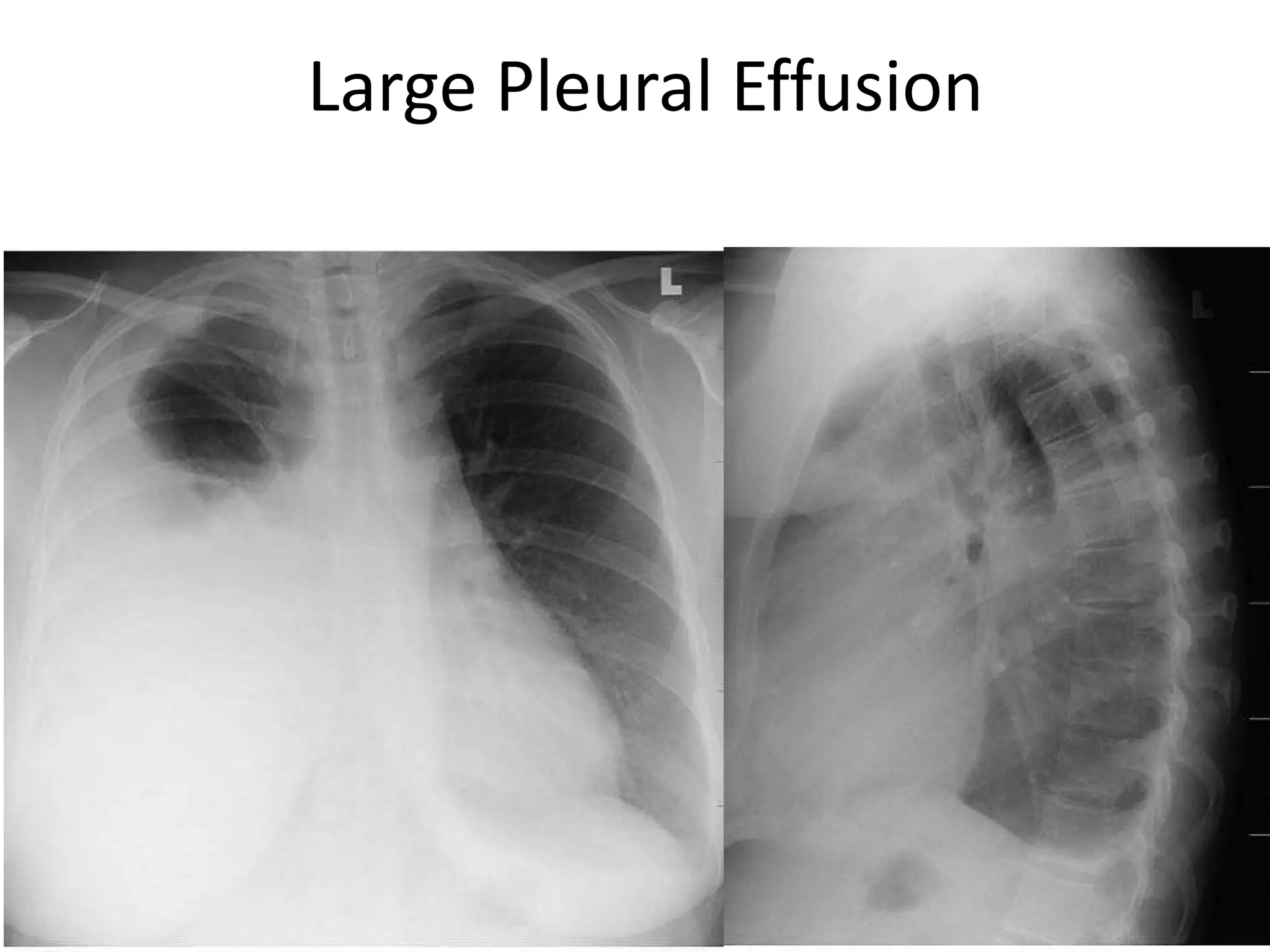
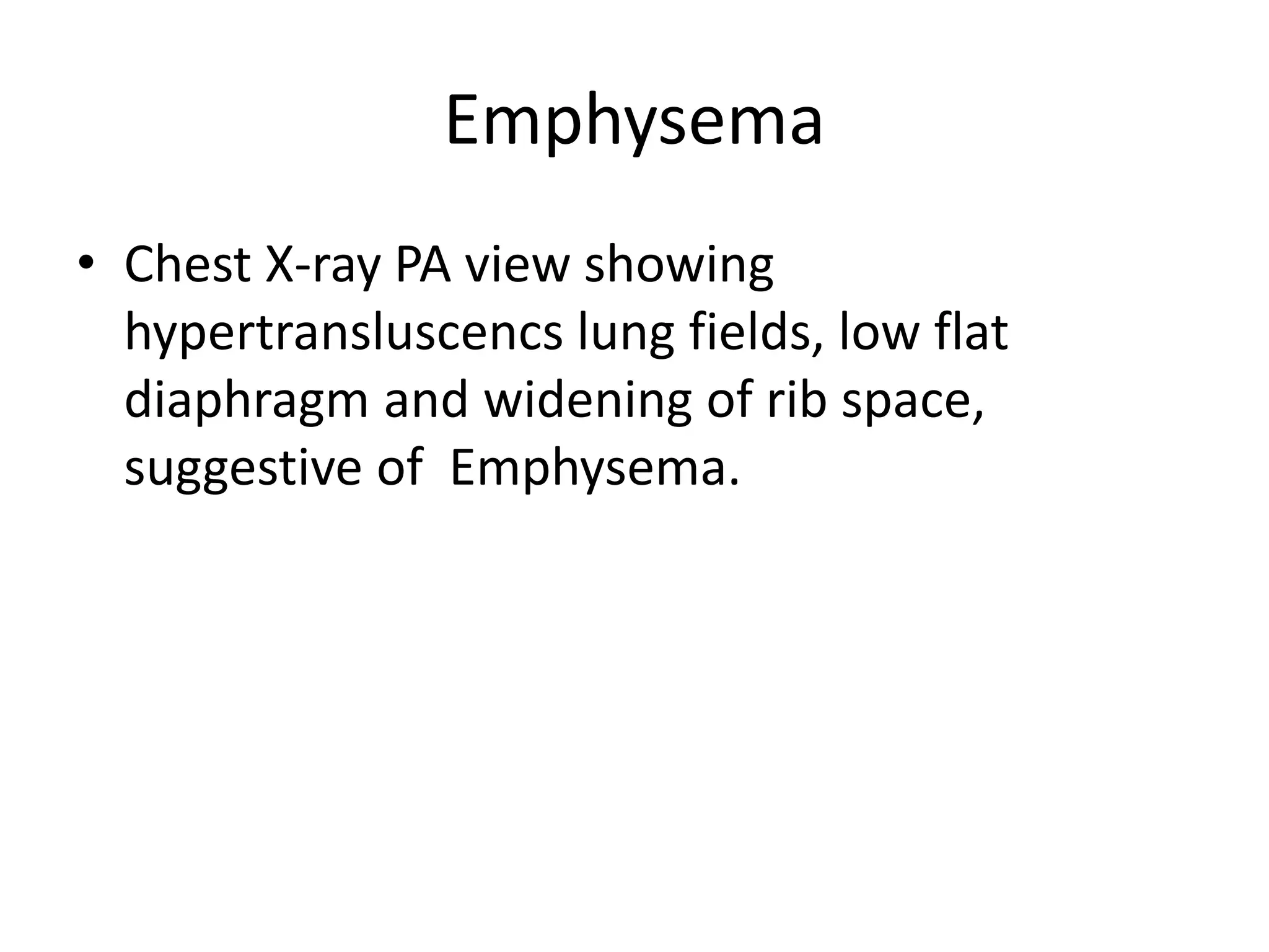
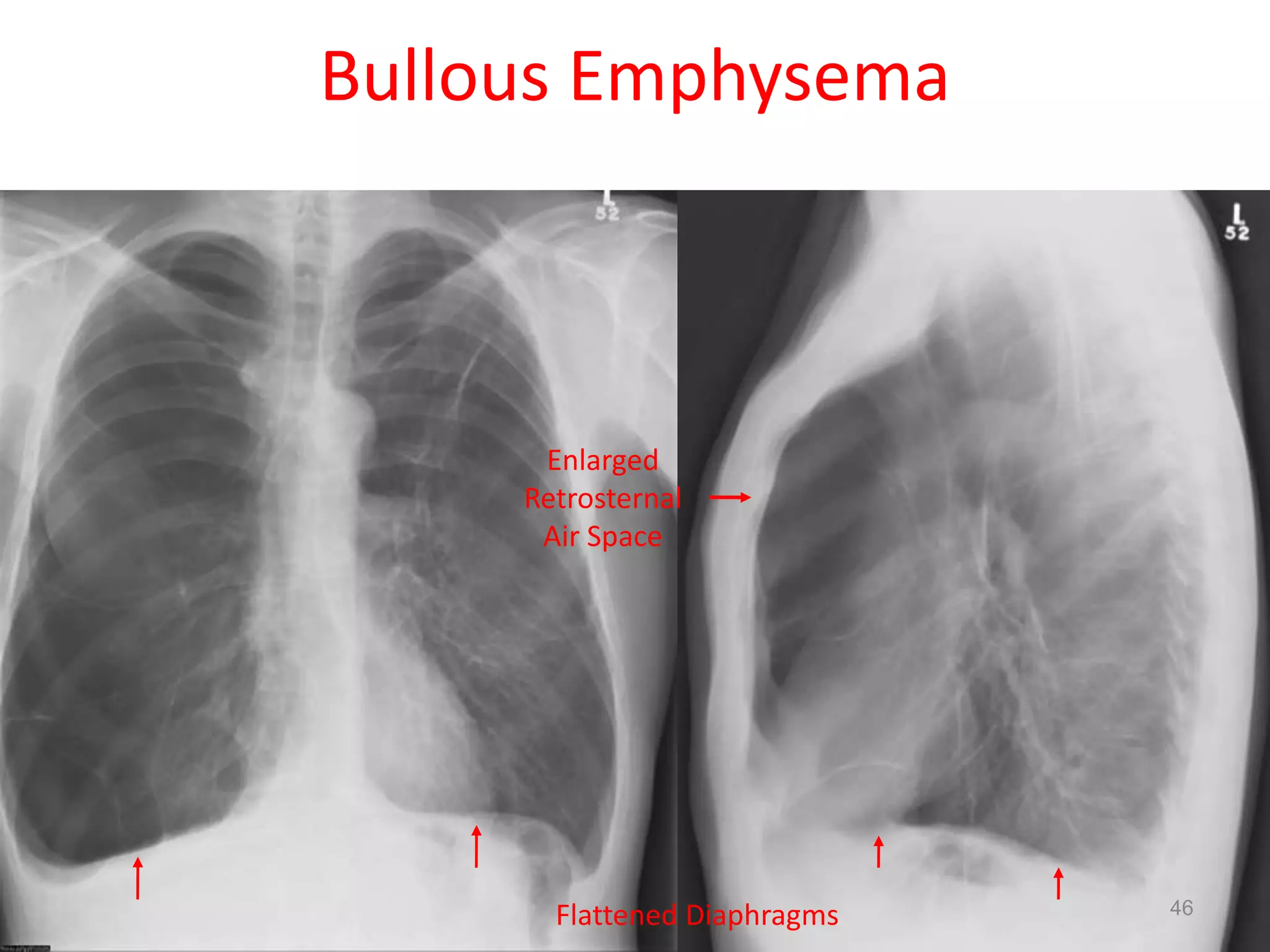
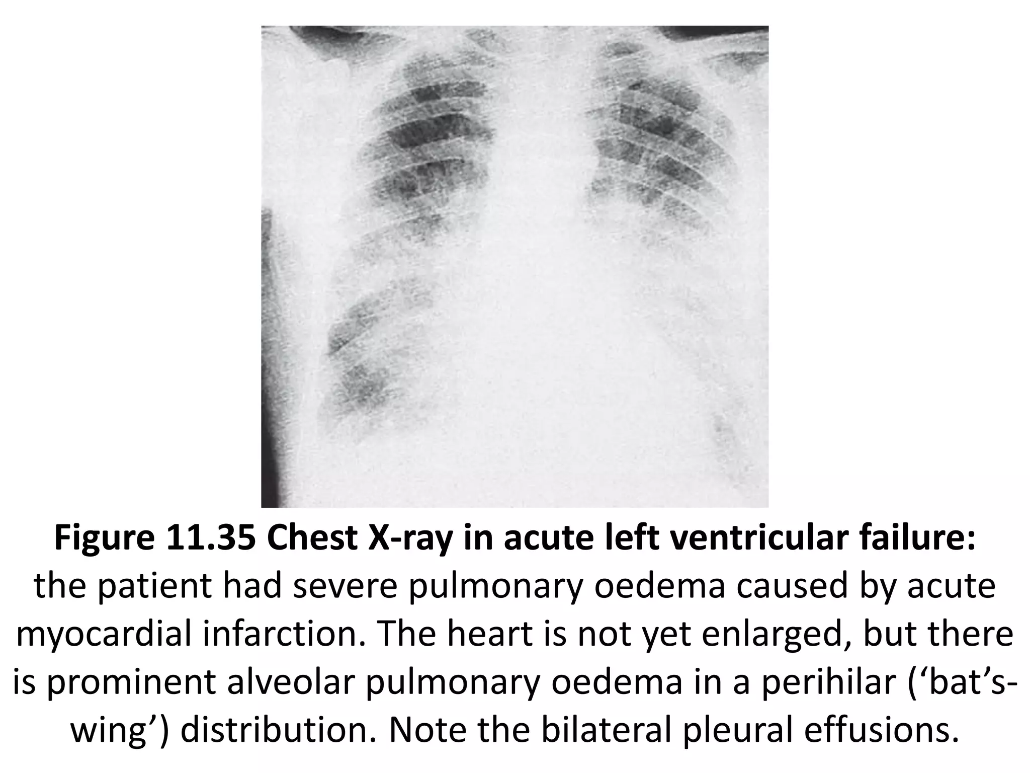
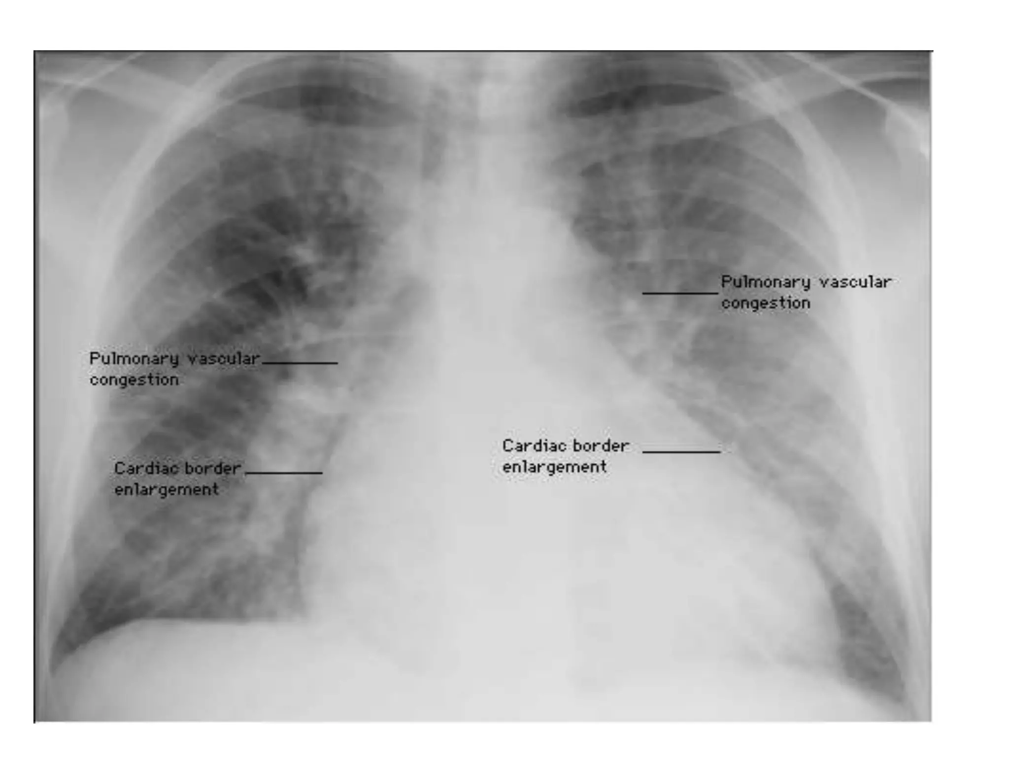
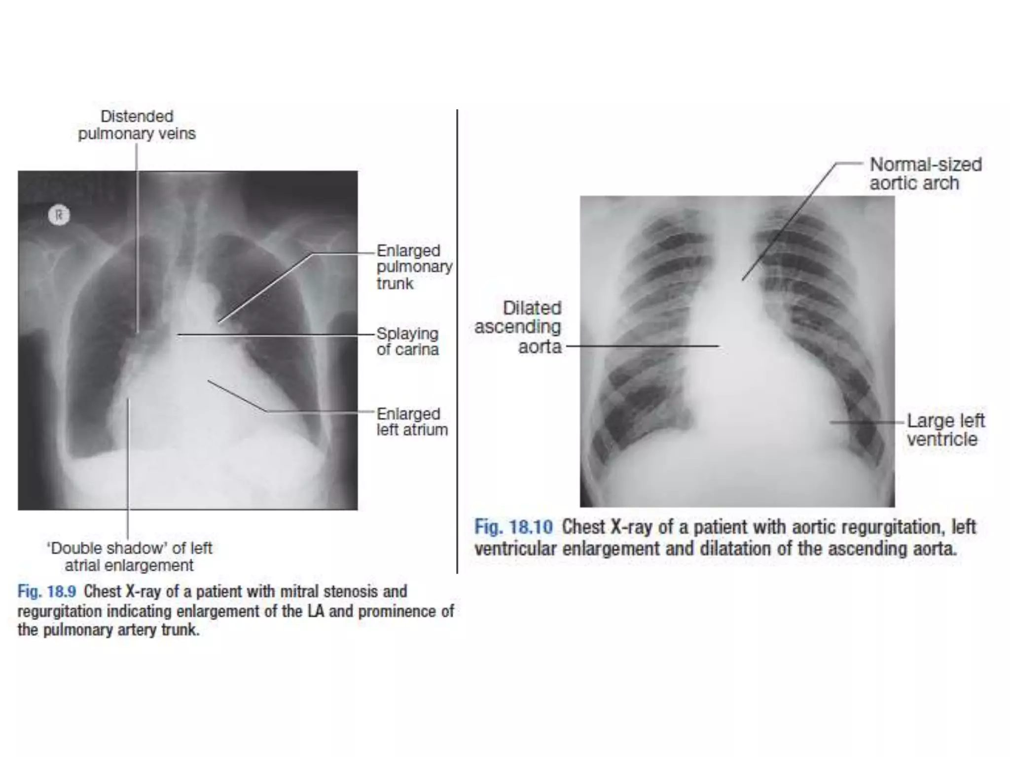
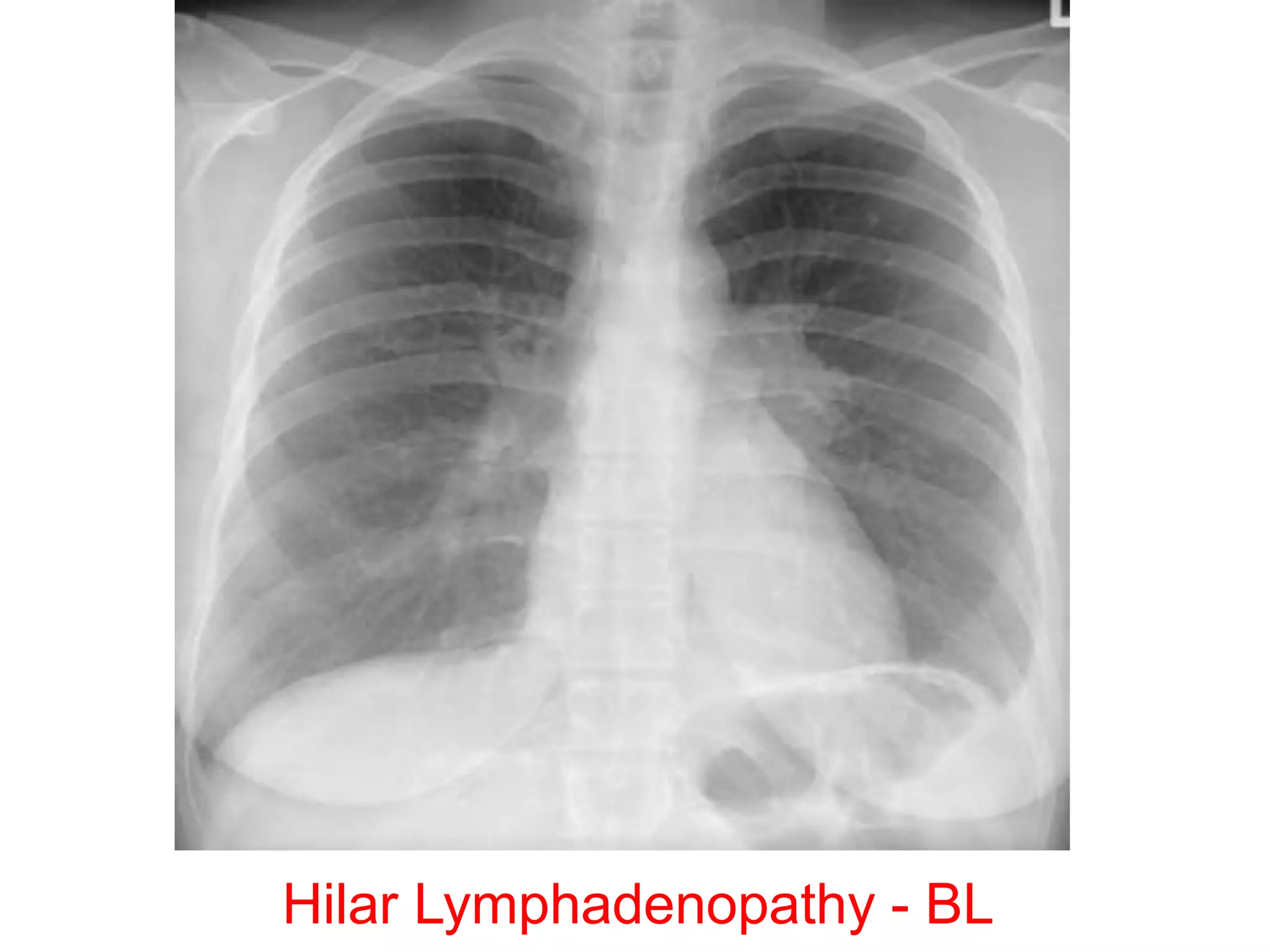
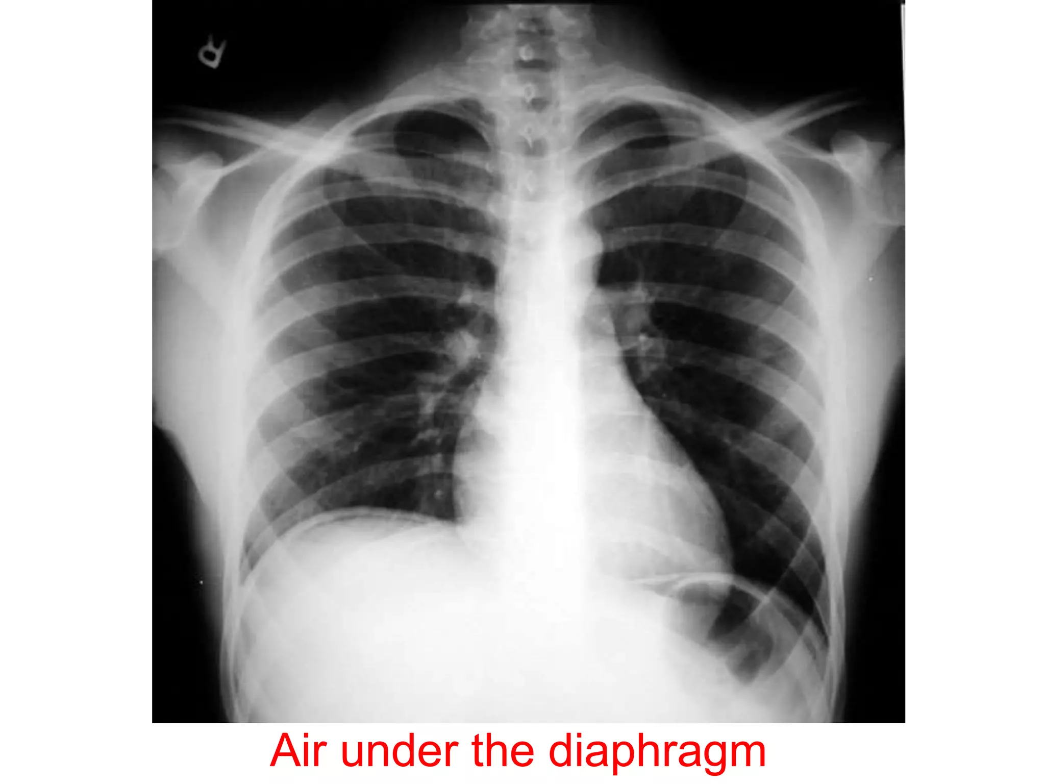
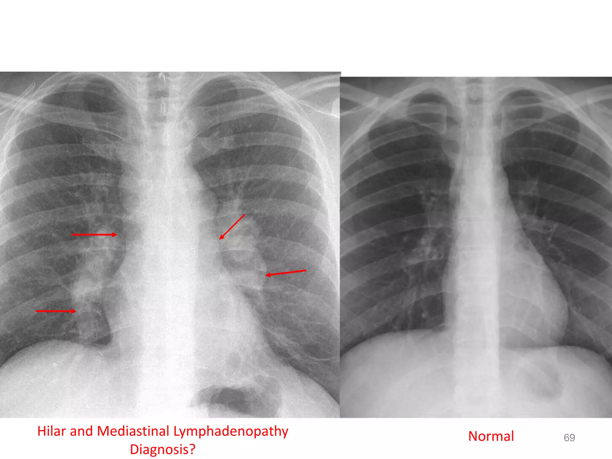
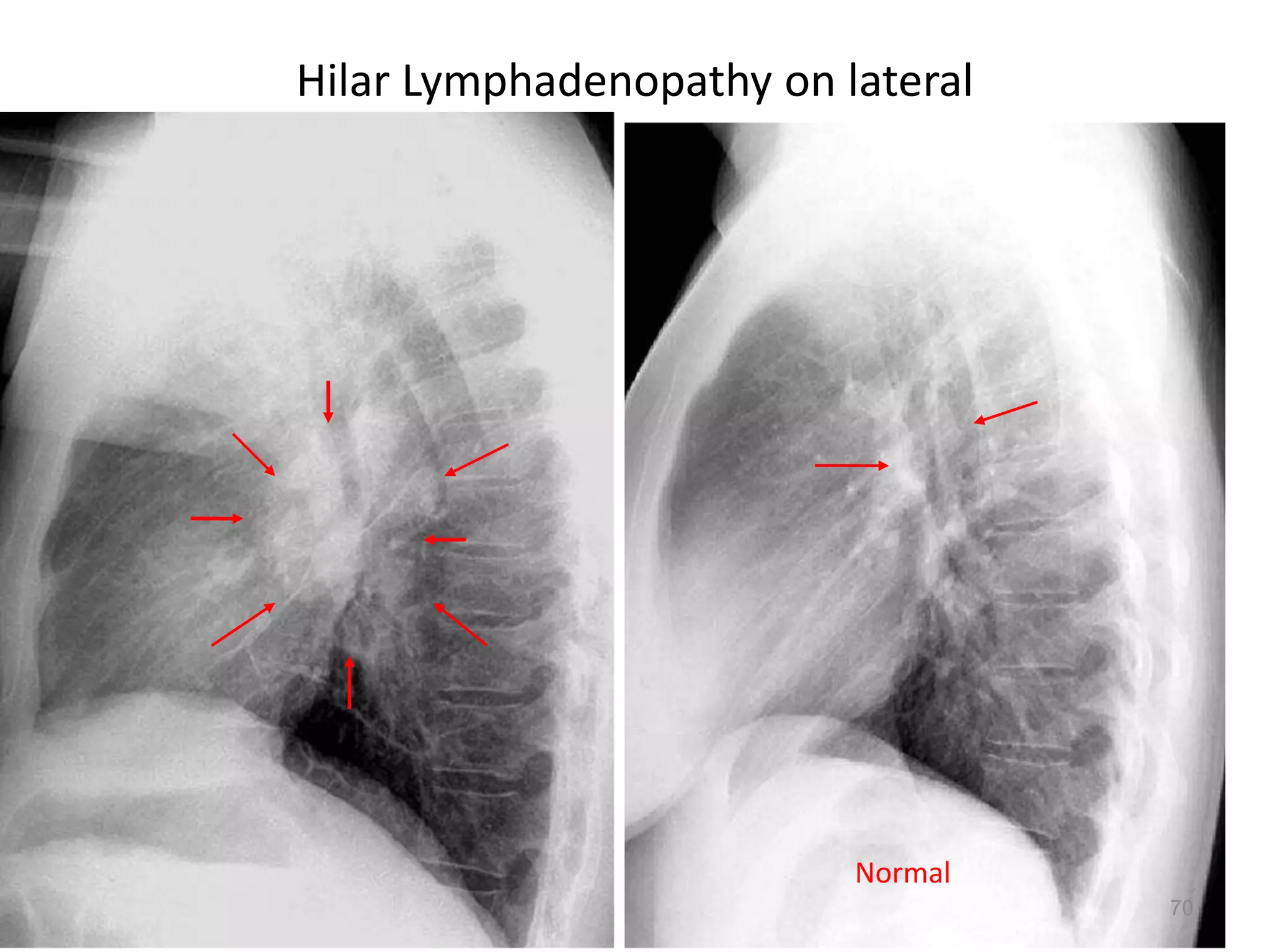
SlideShare a Scribd company logo
1 of 79
Imaging
Dr Md Main Uddin
Assistant Professor
(Medicine)
Cox’s Bazar Medical
College
Modalities
• X-Ray
CXR
Plain abdominal x-ray
• Computed tomography
• Magnetic resonance
imaging
• Ultrasound
• Nuclear imaging
Points to note when assessing the
chest X-ray
• Name of patient and date (and time) of X-ray
• Position of the patient
• Position of the trachea
• Outline of heart
• Outline of mediastinum
• Diaphragm
• Lung fields
• Bony skeleton
Assessing the chest X-ray
• The position of the patient
• Is the patient straight or rotated? If straight, the inner
ends of the clavicles will be equidistant from the
midline of the vertebral body. This is important
because any rotation will tend particularly to alter the
appearance of the mediastinum and the hilar shadows.
• The outline of the heart and the mediastinum
• Is this normal in size, shape and position?
• The position of the trachea
• This is seen as a dark column representing the air
within the trachea. Is the trachea centrally placed or
deviated to either side?
The Chest X-Ray
Techniques – Projection
•P-A (relation of x-ray beam to patient)
CXR Interpretation
PA vs. AP views
Inspiration/Expiration
Cardiac Silhouette
1. R Atrium
2. R Ventricle
3. Apex of L Ventricle
4. SVC
5. IVC
6. Tricuspid Valve
7. Pulmonary Valve
8. Pulmonary Trunk
9. R PA 10. L PA
Assessing the chest X-ray
• The diaphragm
• Can the diaphragm be seen on each side? Is it
normal in shape and position? Normally, the
anterior end of the sixth or seventh rib crosses
the mid-part of the diaphragm on each side,
although the diaphragm on the right is usually
a little higher than on the left. Are the
cardiophrenic angles clearly seen?
The lung fields
• For radiological purposes, the lung fields are
divided into three zones:
• The upper zone extends from the apex to a
line drawn through the lower borders of the
anterior ends of the second costal cartilages.
• The mid-zone extends from this line to one
drawn through the lower borders of the fourth
costal cartilages.
• The lower zone extends from this line to the
bases of the lungs.
• Each zone is systematically examined on both
sides, and any area that appears abnormal is
carefully compared with the corresponding
area on the opposite side. The horizontal
fissure, which separates the right upper and
middle lobes, may sometimes be seen running
horizontally in the third and fourth interspaces
on the right side.
The bony skeleton
• Is the chest symmetrical?
• Is scoliosis present?
• Are the ribs unduly crowded or widely spaced in
any area?
• Are cervical ribs present?
• Are any ribs eroded or absent?
• As well as the standard AP view, lateral views are
sometimes carried out to help localize any lesion
that is seen.
In examining a lateral view
follow this plan:
• Identify the sternum anteriorly and the vertebral bodies
posteriorly. The cardiac shadow lies anteriorly and
inferiorly.
• There should be a lucent (dark) area retrosternally which
has approximately the same density as the area posterior
to the heart and anterior to the vertebral bodies. Check for
any difference between the two, or for any discrete lesion
in either area.
• Check for any collapsed vertebrae.
• The lowest vertebrae should appear darkest, becoming
whiter as they progress superiorly.
• Interruption of this smooth gradation suggests an
abnormality overlying the vertebral bodies involved.
Figure 10.8 A lateral chest X-ray
The normal chest X-ray
• Chest X-ray PA view showing trachea is
central, lung fields are clear, both hilum are
normal, mediastinum is normal, cardiac
shadow is normal in size and contour, both
costophrenic and cardiophrenic angles are
clear, diaphragm is normal in shape and
position, bony skeleton and soft tissue
shadows are normal.
Pneumonia (consolidation) of the right
middle lobe
• CXR PA view showing opacification beneath
the horizontal fissure and loss of normal
contrast between the right heart border and
lung. Suggestive of Rt sided consolidation.
RML pneumonia
RLL pneumonia
LLL pneumonia
• Figure 10.18
• Chest X-ray showing a right
basal pneumonia in a previously
fit 40-year-old man with fever,
breathlessness, central cyanosis
and pleuritic pain. Chest signs
included bronchial breathing and
a pleural rub in the right lower
zone. The cyanosis was due to
the shunting of deoxygenated
blood through the consolidated
lung, the increased respiratory
rate leading to a low PaCO2
because of increased clearance
of carbon dioxide by the
unaffected alveoli. Streptococcus
pneumoniae was grown on blood
cultures.
25154 slides
Right Middle Lobe Pneumonia
25
26154 slides
Left Lower Lobe Pneumonia
26
• Chest X-ray PA view showing the sharply
defined edge of the deflated lung with
complete translucency (no lung markings, no
vascular markings) between this and the chest
wall in the Rt side with shifting of
mediastinum to left.
• Suggestive of Rt Tension Pneumothorax
Pneumothorax
32154 slides
Collapsed Right Lung
Tension Pneumothorax: Requires chest tube
Tracheal Deviation
What would you do with this patient?
32
34
35154 slides
Inspiration Expiration
35
• Chest X-ray PA view showing dense
homogenous opacity at the Rt lung base with
curved upper border, blunting the
costophrenic angle and ascending towards the
axilla. Suggestive of Rt Pleural Effusion
• Around 200 mL of fluid is required in order for
it to be detectable on a PA chest X-ray.
Pleural Effusion
• Figure 10.17
• Chest X-ray showing a
large left pleural effusion
in a young man with a 4-
month history of malaise,
fever, night sweats and
weight loss. The diagnosis
of tuberculosis was
confirmed on histology of
a pleural biopsy and
culture of the pleural
fluid.
42154 slides
Large Pleural Effusion
42
Emphysema
Emphysema
• Chest X-ray PA view showing
hypertransluscencs lung fields, low flat
diaphragm and widening of rib space,
suggestive of Emphysema.
46154 slides
Enlarged
Retrosternal
Air Space
Flattened Diaphragms
Bullous Emphysema
46
Figure 11.32 Left atrial dilatation.
This is a penetrated PA chest X-ray in a patient with mitral stenosis.
The dilated left atrium causes a bulge on the left heart border below
the pulmonary artery which is also dilated, widening of the carina
and the double density sign at the right heart border.
Figure 11.31 Pericardial effusion with tamponade:
There is a left hilar mass caused by carcinoma. Pericardial infiltration has
produced effusion and tamponade, evidenced by the severely enlarged
and globular cardiac silhouette. Malignant disease is now the most
common cause of tamponade in most developed countries.
Figure 11.35 Chest X-ray in acute left ventricular failure:
the patient had severe pulmonary oedema caused by acute
myocardial infarction. The heart is not yet enlarged, but there
is prominent alveolar pulmonary oedema in a perihilar (‘bat’s-
wing’) distribution. Note the bilateral pleural effusions.
Hilar mass
Multiple Masses
Hilar Lymphadenopathy - BL
Pulmonary Fibrosis
Air under the diaphragm
Cavitating lesion
68154 slides
Multiple Nodules: Diagnosis? Metastases
68
69154 slides NormalHilar and Mediastinal Lymphadenopathy
Diagnosis?
69
70154 slides
Hilar Lymphadenopathy on lateral
70
Normal
71154 slides
Sarcoidosis
71
Chest X-ray
An approach to interpreting the chest X-ray
• Name, date, orientation
• Films are postero-anterior
(PA) unless marked AP to
denote that they are
antero-posterior
• Lung fields
• Equal translucency?
• Check horizontal fissure
from right hilum to sixth
rib at the anterior axillary
line
• Masses? Consolidation?
Cavitation?
• Lung apices
• Check behind the
clavicles: Masses?
• Consolidation?
Cavitation?
An approach to interpreting the chest X-ray
• Trachea
• Central? (Midway
between the clavicular
heads)
• Paratracheal mass?
Goitre?
• Heart
• Normal shape?
• Cardiothoracic ratio
should be < half the
intrathoracic diameter)
• Retrocardiac mass?
• Hila
• Left should be higher
than right
• Shape? (Should be
concave laterally; if
convex, consider mass or
lymphadenopathy)
• Density?
Interpreting the chest X-ray
• Diaphragm
• Right should be higher
than left
• Hyperinflation? (No more
than 10 ribs
• should be visible
posteriorly above the
diaphragm)
• Costophrenic angles
• Acute and well defined?
(Pleural fluid or
thickening, if not)
• Soft tissues
• Breast shadows in
females
• Chest wall for masses or
subcutaneous
emphysema
• Bones
• Ribs, vertebrae, scapulae
and clavicles
• Any fracture visible at
bone margins or
lucencies?
Interpreting the chest X-ray
• This is useful for determining the size and
shape of the heart, and the state of the
pulmonary blood vessels and lung fields. Most
information is given by a posteroanterior (PA)
projection taken in full inspiration.
• Anteroposterior (AP) projections are
convenient when patient movement is
restricted but result in magnification of the
cardiac shadow.
Interpreting the chest X-ray
• ‘Cardiomegaly’ is the term used to describe an
enlarged cardiac silhouette where the
‘cardiothoracic ratio’ is greater than 0.5.
• It can be caused by chamber dilatation,
especially left ventricular dilatation, or by a
pericardial effusion.

More Related Content

What's hot

Learn Chest X-Ray With Its Normal Positioning & Radio-Anatomy
Learn Chest X-Ray With Its Normal Positioning & Radio-AnatomyLearn Chest X-Ray With Its Normal Positioning & Radio-Anatomy
Learn Chest X-Ray With Its Normal Positioning & Radio-AnatomyDr.Santosh Atreya
 
Chest x ray basic interpretation
Chest x ray basic interpretationChest x ray basic interpretation
Chest x ray basic interpretationVikram Patil
 
Presentation1.pptx. interpretation of x ray chest.
Presentation1.pptx. interpretation of x ray chest.Presentation1.pptx. interpretation of x ray chest.
Presentation1.pptx. interpretation of x ray chest.Abdellah Nazeer
 
basics of chest X- ray interpretation
basics of chest X- ray interpretationbasics of chest X- ray interpretation
basics of chest X- ray interpretationMaha Yousif
 
Heart imaging RADIOLOGY DEPT
Heart imaging RADIOLOGY DEPTHeart imaging RADIOLOGY DEPT
Heart imaging RADIOLOGY DEPTfarranajwa
 
Normal chest x ray- Radiology Basics
Normal chest x  ray- Radiology BasicsNormal chest x  ray- Radiology Basics
Normal chest x ray- Radiology BasicsSandeep Awal
 
An approach to cardiac xray Dr. Muhammad Bin Zulfiqar
An approach to cardiac xray Dr. Muhammad Bin ZulfiqarAn approach to cardiac xray Dr. Muhammad Bin Zulfiqar
An approach to cardiac xray Dr. Muhammad Bin ZulfiqarDr. Muhammad Bin Zulfiqar
 
Chest x ray positioning
Chest x ray  positioningChest x ray  positioning
Chest x ray positioningairwave12
 
Cxr revised 24 11-91
Cxr revised 24 11-91Cxr revised 24 11-91
Cxr revised 24 11-91aalmasi1970
 
Chest x ray-fundamentals
Chest x ray-fundamentalsChest x ray-fundamentals
Chest x ray-fundamentalsDr Emad efat
 

What's hot (20)

Learn Chest X-Ray With Its Normal Positioning & Radio-Anatomy
Learn Chest X-Ray With Its Normal Positioning & Radio-AnatomyLearn Chest X-Ray With Its Normal Positioning & Radio-Anatomy
Learn Chest X-Ray With Its Normal Positioning & Radio-Anatomy
 
Chest x ray basic interpretation
Chest x ray basic interpretationChest x ray basic interpretation
Chest x ray basic interpretation
 
Chest imaging
Chest imagingChest imaging
Chest imaging
 
BASIC CHEST X-RAY INTERPRETATION
BASIC CHEST X-RAY INTERPRETATIONBASIC CHEST X-RAY INTERPRETATION
BASIC CHEST X-RAY INTERPRETATION
 
Presentation1.pptx. interpretation of x ray chest.
Presentation1.pptx. interpretation of x ray chest.Presentation1.pptx. interpretation of x ray chest.
Presentation1.pptx. interpretation of x ray chest.
 
Chest Radiograph for Interns
Chest Radiograph for InternsChest Radiograph for Interns
Chest Radiograph for Interns
 
Normal Chest X-ray
Normal Chest X-rayNormal Chest X-ray
Normal Chest X-ray
 
basics of chest X- ray interpretation
basics of chest X- ray interpretationbasics of chest X- ray interpretation
basics of chest X- ray interpretation
 
Normal chest xray
Normal chest xrayNormal chest xray
Normal chest xray
 
Basics of CT chest
Basics of CT chestBasics of CT chest
Basics of CT chest
 
Abnormal Chest xray
Abnormal Chest xray Abnormal Chest xray
Abnormal Chest xray
 
Heart imaging RADIOLOGY DEPT
Heart imaging RADIOLOGY DEPTHeart imaging RADIOLOGY DEPT
Heart imaging RADIOLOGY DEPT
 
Chest xray
Chest xrayChest xray
Chest xray
 
Normal chest x ray- Radiology Basics
Normal chest x  ray- Radiology BasicsNormal chest x  ray- Radiology Basics
Normal chest x ray- Radiology Basics
 
An approach to cardiac xray Dr. Muhammad Bin Zulfiqar
An approach to cardiac xray Dr. Muhammad Bin ZulfiqarAn approach to cardiac xray Dr. Muhammad Bin Zulfiqar
An approach to cardiac xray Dr. Muhammad Bin Zulfiqar
 
Chest x ray positioning
Chest x ray  positioningChest x ray  positioning
Chest x ray positioning
 
Chest Xray
Chest XrayChest Xray
Chest Xray
 
Cxr revised 24 11-91
Cxr revised 24 11-91Cxr revised 24 11-91
Cxr revised 24 11-91
 
Chest x ray pathology
Chest x ray pathologyChest x ray pathology
Chest x ray pathology
 
Chest x ray-fundamentals
Chest x ray-fundamentalsChest x ray-fundamentals
Chest x ray-fundamentals
 

Similar to Interpretation of X-Ray and other imaging

xrayandotherimaging-180902063930.pdf
xrayandotherimaging-180902063930.pdfxrayandotherimaging-180902063930.pdf
xrayandotherimaging-180902063930.pdfEmmanuelOluseyi1
 
Normal chest x ray and collapse
Normal chest x ray and collapseNormal chest x ray and collapse
Normal chest x ray and collapseAabid Rahiman
 
Chest radiology
Chest radiologyChest radiology
Chest radiologyDr Vaziri
 
X RAY DETERMINATION AND EVALUATION.pptx
X RAY DETERMINATION AND EVALUATION.pptxX RAY DETERMINATION AND EVALUATION.pptx
X RAY DETERMINATION AND EVALUATION.pptxShoaibKhatik3
 
Chest basics + usg dr patil 21818
Chest basics + usg dr patil 21818Chest basics + usg dr patil 21818
Chest basics + usg dr patil 21818dypradio
 
Interpretation of chest xray ppt
Interpretation of chest xray pptInterpretation of chest xray ppt
Interpretation of chest xray pptRithwik Karumuri
 
Basic chest x ray interpretation
Basic chest x ray interpretationBasic chest x ray interpretation
Basic chest x ray interpretationHiba Ashibany
 
Radiology respiratory new.ppt
Radiology respiratory new.pptRadiology respiratory new.ppt
Radiology respiratory new.pptDarshuBoricha
 
Presentation2.pptx technique chest 2
Presentation2.pptx technique chest 2Presentation2.pptx technique chest 2
Presentation2.pptx technique chest 2Yashawant Yadav
 
Interpreting a chest x ray film
Interpreting a chest x ray filmInterpreting a chest x ray film
Interpreting a chest x ray filmPritom Das
 
chestx-ray-180804183634.pdf
chestx-ray-180804183634.pdfchestx-ray-180804183634.pdf
chestx-ray-180804183634.pdfrooqashali1
 

Similar to Interpretation of X-Ray and other imaging (20)

xrayandotherimaging-180902063930.pdf
xrayandotherimaging-180902063930.pdfxrayandotherimaging-180902063930.pdf
xrayandotherimaging-180902063930.pdf
 
Normal chest x ray and collapse
Normal chest x ray and collapseNormal chest x ray and collapse
Normal chest x ray and collapse
 
Chest x rays
Chest x raysChest x rays
Chest x rays
 
Chest radiology
Chest radiologyChest radiology
Chest radiology
 
X RAY DETERMINATION AND EVALUATION.pptx
X RAY DETERMINATION AND EVALUATION.pptxX RAY DETERMINATION AND EVALUATION.pptx
X RAY DETERMINATION AND EVALUATION.pptx
 
Normal chest x ray
Normal chest x rayNormal chest x ray
Normal chest x ray
 
Chest basics + usg dr patil 21818
Chest basics + usg dr patil 21818Chest basics + usg dr patil 21818
Chest basics + usg dr patil 21818
 
Chest X rays.pptx
Chest X rays.pptxChest X rays.pptx
Chest X rays.pptx
 
Interpretation of chest xray ppt
Interpretation of chest xray pptInterpretation of chest xray ppt
Interpretation of chest xray ppt
 
Chest imaging
Chest imagingChest imaging
Chest imaging
 
Basic chest x ray interpretation
Basic chest x ray interpretationBasic chest x ray interpretation
Basic chest x ray interpretation
 
Lung y3 2018 19 tl
Lung y3 2018 19 tlLung y3 2018 19 tl
Lung y3 2018 19 tl
 
Radiology respiratory new.ppt
Radiology respiratory new.pptRadiology respiratory new.ppt
Radiology respiratory new.ppt
 
Presentation2.pptx technique chest 2
Presentation2.pptx technique chest 2Presentation2.pptx technique chest 2
Presentation2.pptx technique chest 2
 
imaging.pptx
imaging.pptximaging.pptx
imaging.pptx
 
Interpreting a chest x ray film
Interpreting a chest x ray filmInterpreting a chest x ray film
Interpreting a chest x ray film
 
Chest imaging
Chest imagingChest imaging
Chest imaging
 
chest.pptx
chest.pptxchest.pptx
chest.pptx
 
chest-x-ray.pptx
chest-x-ray.pptxchest-x-ray.pptx
chest-x-ray.pptx
 
chestx-ray-180804183634.pdf
chestx-ray-180804183634.pdfchestx-ray-180804183634.pdf
chestx-ray-180804183634.pdf
 

More from drmainuddin

Rational use of Antibiotics
Rational use of AntibioticsRational use of Antibiotics
Rational use of Antibioticsdrmainuddin
 
Medical Negligence (ভুল চিকিৎসায় রোগীর মৃত্যু)
Medical Negligence (ভুল চিকিৎসায় রোগীর মৃত্যু)Medical Negligence (ভুল চিকিৎসায় রোগীর মৃত্যু)
Medical Negligence (ভুল চিকিৎসায় রোগীর মৃত্যু)drmainuddin
 
ECG Interpretation
ECG InterpretationECG Interpretation
ECG Interpretationdrmainuddin
 
6th Upload on DPP4 Inhibitors
6th Upload on DPP4 Inhibitors6th Upload on DPP4 Inhibitors
6th Upload on DPP4 Inhibitorsdrmainuddin
 
Diabetes Mellitus
Diabetes MellitusDiabetes Mellitus
Diabetes Mellitusdrmainuddin
 
Asthma COPD Overlap (ACO)
Asthma COPD Overlap (ACO)Asthma COPD Overlap (ACO)
Asthma COPD Overlap (ACO)drmainuddin
 
Interpretation of Chest X-Ray PPT
Interpretation of Chest X-Ray PPTInterpretation of Chest X-Ray PPT
Interpretation of Chest X-Ray PPTdrmainuddin
 

More from drmainuddin (7)

Rational use of Antibiotics
Rational use of AntibioticsRational use of Antibiotics
Rational use of Antibiotics
 
Medical Negligence (ভুল চিকিৎসায় রোগীর মৃত্যু)
Medical Negligence (ভুল চিকিৎসায় রোগীর মৃত্যু)Medical Negligence (ভুল চিকিৎসায় রোগীর মৃত্যু)
Medical Negligence (ভুল চিকিৎসায় রোগীর মৃত্যু)
 
ECG Interpretation
ECG InterpretationECG Interpretation
ECG Interpretation
 
6th Upload on DPP4 Inhibitors
6th Upload on DPP4 Inhibitors6th Upload on DPP4 Inhibitors
6th Upload on DPP4 Inhibitors
 
Diabetes Mellitus
Diabetes MellitusDiabetes Mellitus
Diabetes Mellitus
 
Asthma COPD Overlap (ACO)
Asthma COPD Overlap (ACO)Asthma COPD Overlap (ACO)
Asthma COPD Overlap (ACO)
 
Interpretation of Chest X-Ray PPT
Interpretation of Chest X-Ray PPTInterpretation of Chest X-Ray PPT
Interpretation of Chest X-Ray PPT
 

Recently uploaded

HMCS Max Bernays Pre-Deployment Brief (May 2024).pptx
HMCS Max Bernays Pre-Deployment Brief (May 2024).pptxHMCS Max Bernays Pre-Deployment Brief (May 2024).pptx
HMCS Max Bernays Pre-Deployment Brief (May 2024).pptxEsquimalt MFRC
 
Understanding Accommodations and Modifications
Understanding  Accommodations and ModificationsUnderstanding  Accommodations and Modifications
Understanding Accommodations and ModificationsMJDuyan
 
Basic Civil Engineering first year Notes- Chapter 4 Building.pptx
Basic Civil Engineering first year Notes- Chapter 4 Building.pptxBasic Civil Engineering first year Notes- Chapter 4 Building.pptx
Basic Civil Engineering first year Notes- Chapter 4 Building.pptxDenish Jangid
 
dusjagr & nano talk on open tools for agriculture research and learning
dusjagr & nano talk on open tools for agriculture research and learningdusjagr & nano talk on open tools for agriculture research and learning
dusjagr & nano talk on open tools for agriculture research and learningMarc Dusseiller Dusjagr
 
This PowerPoint helps students to consider the concept of infinity.
This PowerPoint helps students to consider the concept of infinity.This PowerPoint helps students to consider the concept of infinity.
This PowerPoint helps students to consider the concept of infinity.christianmathematics
 
How to Manage Call for Tendor in Odoo 17
How to Manage Call for Tendor in Odoo 17How to Manage Call for Tendor in Odoo 17
How to Manage Call for Tendor in Odoo 17Celine George
 
FSB Advising Checklist - Orientation 2024
FSB Advising Checklist - Orientation 2024FSB Advising Checklist - Orientation 2024
FSB Advising Checklist - Orientation 2024Elizabeth Walsh
 
How to setup Pycharm environment for Odoo 17.pptx
How to setup Pycharm environment for Odoo 17.pptxHow to setup Pycharm environment for Odoo 17.pptx
How to setup Pycharm environment for Odoo 17.pptxCeline George
 
TỔNG ÔN TẬP THI VÀO LỚP 10 MÔN TIẾNG ANH NĂM HỌC 2023 - 2024 CÓ ĐÁP ÁN (NGỮ Â...
TỔNG ÔN TẬP THI VÀO LỚP 10 MÔN TIẾNG ANH NĂM HỌC 2023 - 2024 CÓ ĐÁP ÁN (NGỮ Â...TỔNG ÔN TẬP THI VÀO LỚP 10 MÔN TIẾNG ANH NĂM HỌC 2023 - 2024 CÓ ĐÁP ÁN (NGỮ Â...
TỔNG ÔN TẬP THI VÀO LỚP 10 MÔN TIẾNG ANH NĂM HỌC 2023 - 2024 CÓ ĐÁP ÁN (NGỮ Â...Nguyen Thanh Tu Collection
 
AIM of Education-Teachers Training-2024.ppt
AIM of Education-Teachers Training-2024.pptAIM of Education-Teachers Training-2024.ppt
AIM of Education-Teachers Training-2024.pptNishitharanjan Rout
 
21st_Century_Skills_Framework_Final_Presentation_2.pptx
21st_Century_Skills_Framework_Final_Presentation_2.pptx21st_Century_Skills_Framework_Final_Presentation_2.pptx
21st_Century_Skills_Framework_Final_Presentation_2.pptxJoelynRubio1
 
Jamworks pilot and AI at Jisc (20/03/2024)
Jamworks pilot and AI at Jisc (20/03/2024)Jamworks pilot and AI at Jisc (20/03/2024)
Jamworks pilot and AI at Jisc (20/03/2024)Jisc
 
Interdisciplinary_Insights_Data_Collection_Methods.pptx
Interdisciplinary_Insights_Data_Collection_Methods.pptxInterdisciplinary_Insights_Data_Collection_Methods.pptx
Interdisciplinary_Insights_Data_Collection_Methods.pptxPooja Bhuva
 
Google Gemini An AI Revolution in Education.pptx
Google Gemini An AI Revolution in Education.pptxGoogle Gemini An AI Revolution in Education.pptx
Google Gemini An AI Revolution in Education.pptxDr. Sarita Anand
 
FICTIONAL SALESMAN/SALESMAN SNSW 2024.pdf
FICTIONAL SALESMAN/SALESMAN SNSW 2024.pdfFICTIONAL SALESMAN/SALESMAN SNSW 2024.pdf
FICTIONAL SALESMAN/SALESMAN SNSW 2024.pdfPondicherry University
 
Spellings Wk 4 and Wk 5 for Grade 4 at CAPS
Spellings Wk 4 and Wk 5 for Grade 4 at CAPSSpellings Wk 4 and Wk 5 for Grade 4 at CAPS
Spellings Wk 4 and Wk 5 for Grade 4 at CAPSAnaAcapella
 
Food safety_Challenges food safety laboratories_.pdf
Food safety_Challenges food safety laboratories_.pdfFood safety_Challenges food safety laboratories_.pdf
Food safety_Challenges food safety laboratories_.pdfSherif Taha
 
SOC 101 Demonstration of Learning Presentation
SOC 101 Demonstration of Learning PresentationSOC 101 Demonstration of Learning Presentation
SOC 101 Demonstration of Learning Presentationcamerronhm
 
latest AZ-104 Exam Questions and Answers
latest AZ-104 Exam Questions and Answerslatest AZ-104 Exam Questions and Answers
latest AZ-104 Exam Questions and Answersdalebeck957
 

Recently uploaded (20)

HMCS Max Bernays Pre-Deployment Brief (May 2024).pptx
HMCS Max Bernays Pre-Deployment Brief (May 2024).pptxHMCS Max Bernays Pre-Deployment Brief (May 2024).pptx
HMCS Max Bernays Pre-Deployment Brief (May 2024).pptx
 
Mehran University Newsletter Vol-X, Issue-I, 2024
Mehran University Newsletter Vol-X, Issue-I, 2024Mehran University Newsletter Vol-X, Issue-I, 2024
Mehran University Newsletter Vol-X, Issue-I, 2024
 
Understanding Accommodations and Modifications
Understanding  Accommodations and ModificationsUnderstanding  Accommodations and Modifications
Understanding Accommodations and Modifications
 
Basic Civil Engineering first year Notes- Chapter 4 Building.pptx
Basic Civil Engineering first year Notes- Chapter 4 Building.pptxBasic Civil Engineering first year Notes- Chapter 4 Building.pptx
Basic Civil Engineering first year Notes- Chapter 4 Building.pptx
 
dusjagr & nano talk on open tools for agriculture research and learning
dusjagr & nano talk on open tools for agriculture research and learningdusjagr & nano talk on open tools for agriculture research and learning
dusjagr & nano talk on open tools for agriculture research and learning
 
This PowerPoint helps students to consider the concept of infinity.
This PowerPoint helps students to consider the concept of infinity.This PowerPoint helps students to consider the concept of infinity.
This PowerPoint helps students to consider the concept of infinity.
 
How to Manage Call for Tendor in Odoo 17
How to Manage Call for Tendor in Odoo 17How to Manage Call for Tendor in Odoo 17
How to Manage Call for Tendor in Odoo 17
 
FSB Advising Checklist - Orientation 2024
FSB Advising Checklist - Orientation 2024FSB Advising Checklist - Orientation 2024
FSB Advising Checklist - Orientation 2024
 
How to setup Pycharm environment for Odoo 17.pptx
How to setup Pycharm environment for Odoo 17.pptxHow to setup Pycharm environment for Odoo 17.pptx
How to setup Pycharm environment for Odoo 17.pptx
 
TỔNG ÔN TẬP THI VÀO LỚP 10 MÔN TIẾNG ANH NĂM HỌC 2023 - 2024 CÓ ĐÁP ÁN (NGỮ Â...
TỔNG ÔN TẬP THI VÀO LỚP 10 MÔN TIẾNG ANH NĂM HỌC 2023 - 2024 CÓ ĐÁP ÁN (NGỮ Â...TỔNG ÔN TẬP THI VÀO LỚP 10 MÔN TIẾNG ANH NĂM HỌC 2023 - 2024 CÓ ĐÁP ÁN (NGỮ Â...
TỔNG ÔN TẬP THI VÀO LỚP 10 MÔN TIẾNG ANH NĂM HỌC 2023 - 2024 CÓ ĐÁP ÁN (NGỮ Â...
 
AIM of Education-Teachers Training-2024.ppt
AIM of Education-Teachers Training-2024.pptAIM of Education-Teachers Training-2024.ppt
AIM of Education-Teachers Training-2024.ppt
 
21st_Century_Skills_Framework_Final_Presentation_2.pptx
21st_Century_Skills_Framework_Final_Presentation_2.pptx21st_Century_Skills_Framework_Final_Presentation_2.pptx
21st_Century_Skills_Framework_Final_Presentation_2.pptx
 
Jamworks pilot and AI at Jisc (20/03/2024)
Jamworks pilot and AI at Jisc (20/03/2024)Jamworks pilot and AI at Jisc (20/03/2024)
Jamworks pilot and AI at Jisc (20/03/2024)
 
Interdisciplinary_Insights_Data_Collection_Methods.pptx
Interdisciplinary_Insights_Data_Collection_Methods.pptxInterdisciplinary_Insights_Data_Collection_Methods.pptx
Interdisciplinary_Insights_Data_Collection_Methods.pptx
 
Google Gemini An AI Revolution in Education.pptx
Google Gemini An AI Revolution in Education.pptxGoogle Gemini An AI Revolution in Education.pptx
Google Gemini An AI Revolution in Education.pptx
 
FICTIONAL SALESMAN/SALESMAN SNSW 2024.pdf
FICTIONAL SALESMAN/SALESMAN SNSW 2024.pdfFICTIONAL SALESMAN/SALESMAN SNSW 2024.pdf
FICTIONAL SALESMAN/SALESMAN SNSW 2024.pdf
 
Spellings Wk 4 and Wk 5 for Grade 4 at CAPS
Spellings Wk 4 and Wk 5 for Grade 4 at CAPSSpellings Wk 4 and Wk 5 for Grade 4 at CAPS
Spellings Wk 4 and Wk 5 for Grade 4 at CAPS
 
Food safety_Challenges food safety laboratories_.pdf
Food safety_Challenges food safety laboratories_.pdfFood safety_Challenges food safety laboratories_.pdf
Food safety_Challenges food safety laboratories_.pdf
 
SOC 101 Demonstration of Learning Presentation
SOC 101 Demonstration of Learning PresentationSOC 101 Demonstration of Learning Presentation
SOC 101 Demonstration of Learning Presentation
 
latest AZ-104 Exam Questions and Answers
latest AZ-104 Exam Questions and Answerslatest AZ-104 Exam Questions and Answers
latest AZ-104 Exam Questions and Answers
 

Interpretation of X-Ray and other imaging

  • 1. Imaging Dr Md Main Uddin Assistant Professor (Medicine) Cox’s Bazar Medical College
  • 2. Modalities • X-Ray CXR Plain abdominal x-ray • Computed tomography • Magnetic resonance imaging • Ultrasound • Nuclear imaging
  • 3.
  • 4. Points to note when assessing the chest X-ray • Name of patient and date (and time) of X-ray • Position of the patient • Position of the trachea • Outline of heart • Outline of mediastinum • Diaphragm • Lung fields • Bony skeleton
  • 5. Assessing the chest X-ray • The position of the patient • Is the patient straight or rotated? If straight, the inner ends of the clavicles will be equidistant from the midline of the vertebral body. This is important because any rotation will tend particularly to alter the appearance of the mediastinum and the hilar shadows. • The outline of the heart and the mediastinum • Is this normal in size, shape and position? • The position of the trachea • This is seen as a dark column representing the air within the trachea. Is the trachea centrally placed or deviated to either side?
  • 6. The Chest X-Ray Techniques – Projection •P-A (relation of x-ray beam to patient)
  • 9. Cardiac Silhouette 1. R Atrium 2. R Ventricle 3. Apex of L Ventricle 4. SVC 5. IVC 6. Tricuspid Valve 7. Pulmonary Valve 8. Pulmonary Trunk 9. R PA 10. L PA
  • 10. Assessing the chest X-ray • The diaphragm • Can the diaphragm be seen on each side? Is it normal in shape and position? Normally, the anterior end of the sixth or seventh rib crosses the mid-part of the diaphragm on each side, although the diaphragm on the right is usually a little higher than on the left. Are the cardiophrenic angles clearly seen?
  • 11. The lung fields • For radiological purposes, the lung fields are divided into three zones: • The upper zone extends from the apex to a line drawn through the lower borders of the anterior ends of the second costal cartilages. • The mid-zone extends from this line to one drawn through the lower borders of the fourth costal cartilages. • The lower zone extends from this line to the bases of the lungs.
  • 12. • Each zone is systematically examined on both sides, and any area that appears abnormal is carefully compared with the corresponding area on the opposite side. The horizontal fissure, which separates the right upper and middle lobes, may sometimes be seen running horizontally in the third and fourth interspaces on the right side.
  • 13. The bony skeleton • Is the chest symmetrical? • Is scoliosis present? • Are the ribs unduly crowded or widely spaced in any area? • Are cervical ribs present? • Are any ribs eroded or absent? • As well as the standard AP view, lateral views are sometimes carried out to help localize any lesion that is seen.
  • 14. In examining a lateral view follow this plan: • Identify the sternum anteriorly and the vertebral bodies posteriorly. The cardiac shadow lies anteriorly and inferiorly. • There should be a lucent (dark) area retrosternally which has approximately the same density as the area posterior to the heart and anterior to the vertebral bodies. Check for any difference between the two, or for any discrete lesion in either area. • Check for any collapsed vertebrae. • The lowest vertebrae should appear darkest, becoming whiter as they progress superiorly. • Interruption of this smooth gradation suggests an abnormality overlying the vertebral bodies involved.
  • 15. Figure 10.8 A lateral chest X-ray
  • 17. The normal chest X-ray • Chest X-ray PA view showing trachea is central, lung fields are clear, both hilum are normal, mediastinum is normal, cardiac shadow is normal in size and contour, both costophrenic and cardiophrenic angles are clear, diaphragm is normal in shape and position, bony skeleton and soft tissue shadows are normal.
  • 18.
  • 19. Pneumonia (consolidation) of the right middle lobe • CXR PA view showing opacification beneath the horizontal fissure and loss of normal contrast between the right heart border and lung. Suggestive of Rt sided consolidation.
  • 20.
  • 24. • Figure 10.18 • Chest X-ray showing a right basal pneumonia in a previously fit 40-year-old man with fever, breathlessness, central cyanosis and pleuritic pain. Chest signs included bronchial breathing and a pleural rub in the right lower zone. The cyanosis was due to the shunting of deoxygenated blood through the consolidated lung, the increased respiratory rate leading to a low PaCO2 because of increased clearance of carbon dioxide by the unaffected alveoli. Streptococcus pneumoniae was grown on blood cultures.
  • 25. 25154 slides Right Middle Lobe Pneumonia 25
  • 26. 26154 slides Left Lower Lobe Pneumonia 26
  • 27.
  • 28.
  • 29. • Chest X-ray PA view showing the sharply defined edge of the deflated lung with complete translucency (no lung markings, no vascular markings) between this and the chest wall in the Rt side with shifting of mediastinum to left. • Suggestive of Rt Tension Pneumothorax
  • 31.
  • 32. 32154 slides Collapsed Right Lung Tension Pneumothorax: Requires chest tube Tracheal Deviation What would you do with this patient? 32
  • 33. 34
  • 35.
  • 36.
  • 37. • Chest X-ray PA view showing dense homogenous opacity at the Rt lung base with curved upper border, blunting the costophrenic angle and ascending towards the axilla. Suggestive of Rt Pleural Effusion • Around 200 mL of fluid is required in order for it to be detectable on a PA chest X-ray.
  • 39. • Figure 10.17 • Chest X-ray showing a large left pleural effusion in a young man with a 4- month history of malaise, fever, night sweats and weight loss. The diagnosis of tuberculosis was confirmed on histology of a pleural biopsy and culture of the pleural fluid.
  • 40.
  • 42.
  • 44. Emphysema • Chest X-ray PA view showing hypertransluscencs lung fields, low flat diaphragm and widening of rib space, suggestive of Emphysema.
  • 46.
  • 47.
  • 48. Figure 11.32 Left atrial dilatation. This is a penetrated PA chest X-ray in a patient with mitral stenosis. The dilated left atrium causes a bulge on the left heart border below the pulmonary artery which is also dilated, widening of the carina and the double density sign at the right heart border.
  • 49. Figure 11.31 Pericardial effusion with tamponade: There is a left hilar mass caused by carcinoma. Pericardial infiltration has produced effusion and tamponade, evidenced by the severely enlarged and globular cardiac silhouette. Malignant disease is now the most common cause of tamponade in most developed countries.
  • 50. Figure 11.35 Chest X-ray in acute left ventricular failure: the patient had severe pulmonary oedema caused by acute myocardial infarction. The heart is not yet enlarged, but there is prominent alveolar pulmonary oedema in a perihilar (‘bat’s- wing’) distribution. Note the bilateral pleural effusions.
  • 51.
  • 52.
  • 53.
  • 54.
  • 55.
  • 56.
  • 57.
  • 58.
  • 59.
  • 60.
  • 65. Air under the diaphragm
  • 67. 68154 slides Multiple Nodules: Diagnosis? Metastases 68
  • 68. 69154 slides NormalHilar and Mediastinal Lymphadenopathy Diagnosis? 69
  • 69. 70154 slides Hilar Lymphadenopathy on lateral 70 Normal
  • 71.
  • 72.
  • 73.
  • 74.
  • 75. Chest X-ray An approach to interpreting the chest X-ray • Name, date, orientation • Films are postero-anterior (PA) unless marked AP to denote that they are antero-posterior • Lung fields • Equal translucency? • Check horizontal fissure from right hilum to sixth rib at the anterior axillary line • Masses? Consolidation? Cavitation? • Lung apices • Check behind the clavicles: Masses? • Consolidation? Cavitation?
  • 76. An approach to interpreting the chest X-ray • Trachea • Central? (Midway between the clavicular heads) • Paratracheal mass? Goitre? • Heart • Normal shape? • Cardiothoracic ratio should be < half the intrathoracic diameter) • Retrocardiac mass? • Hila • Left should be higher than right • Shape? (Should be concave laterally; if convex, consider mass or lymphadenopathy) • Density?
  • 77. Interpreting the chest X-ray • Diaphragm • Right should be higher than left • Hyperinflation? (No more than 10 ribs • should be visible posteriorly above the diaphragm) • Costophrenic angles • Acute and well defined? (Pleural fluid or thickening, if not) • Soft tissues • Breast shadows in females • Chest wall for masses or subcutaneous emphysema • Bones • Ribs, vertebrae, scapulae and clavicles • Any fracture visible at bone margins or lucencies?
  • 78. Interpreting the chest X-ray • This is useful for determining the size and shape of the heart, and the state of the pulmonary blood vessels and lung fields. Most information is given by a posteroanterior (PA) projection taken in full inspiration. • Anteroposterior (AP) projections are convenient when patient movement is restricted but result in magnification of the cardiac shadow.
  • 79. Interpreting the chest X-ray • ‘Cardiomegaly’ is the term used to describe an enlarged cardiac silhouette where the ‘cardiothoracic ratio’ is greater than 0.5. • It can be caused by chamber dilatation, especially left ventricular dilatation, or by a pericardial effusion.