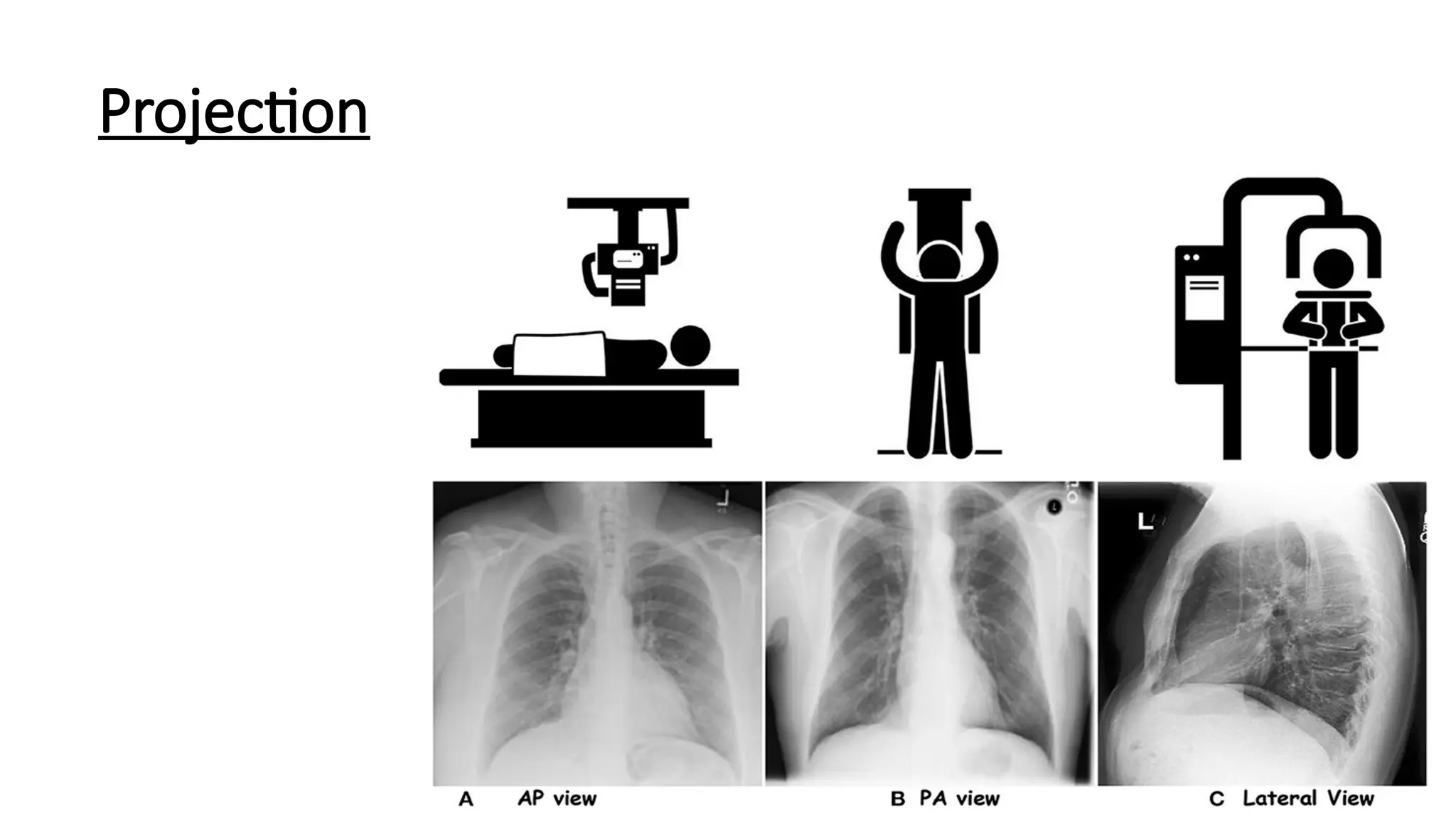
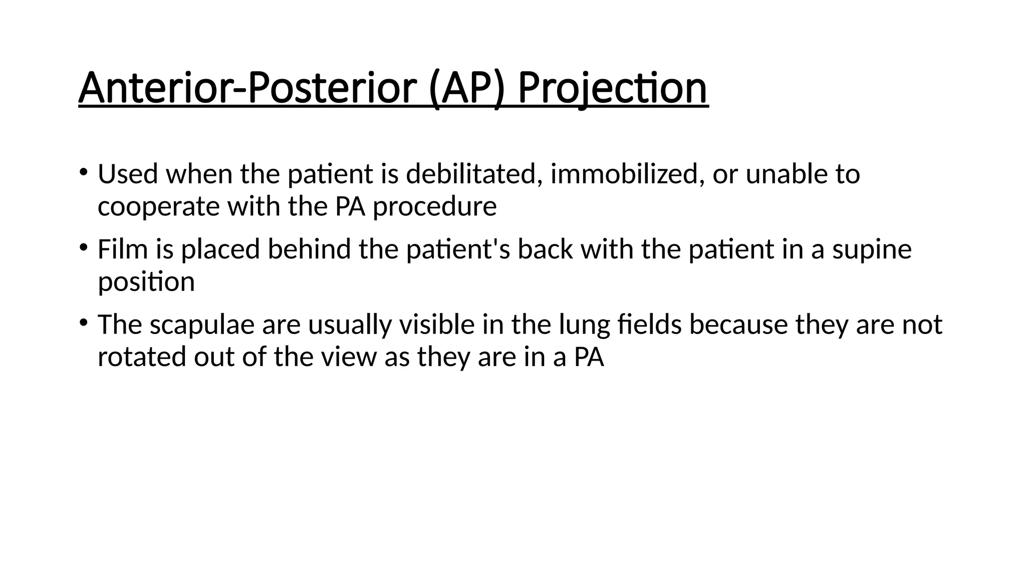
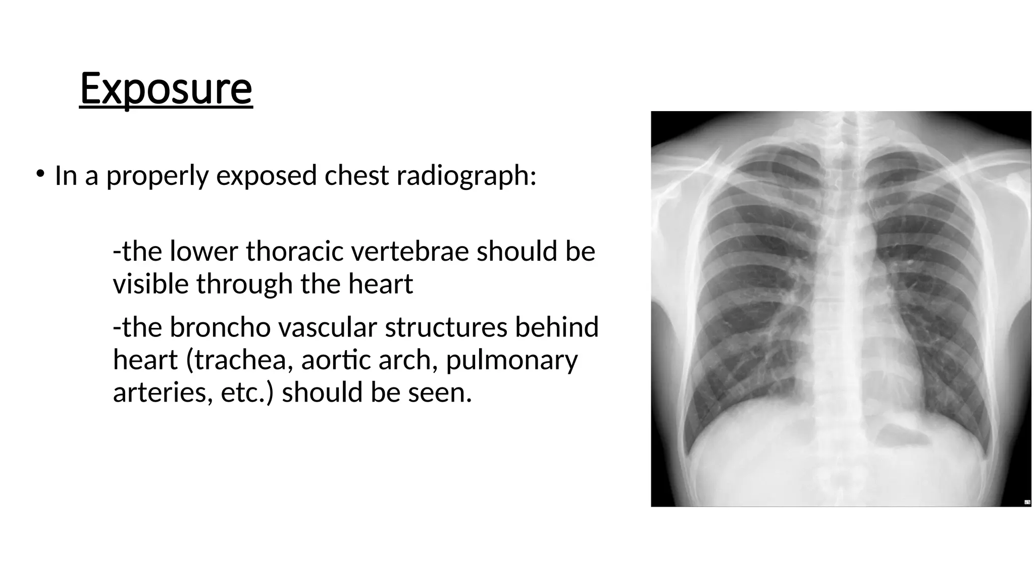
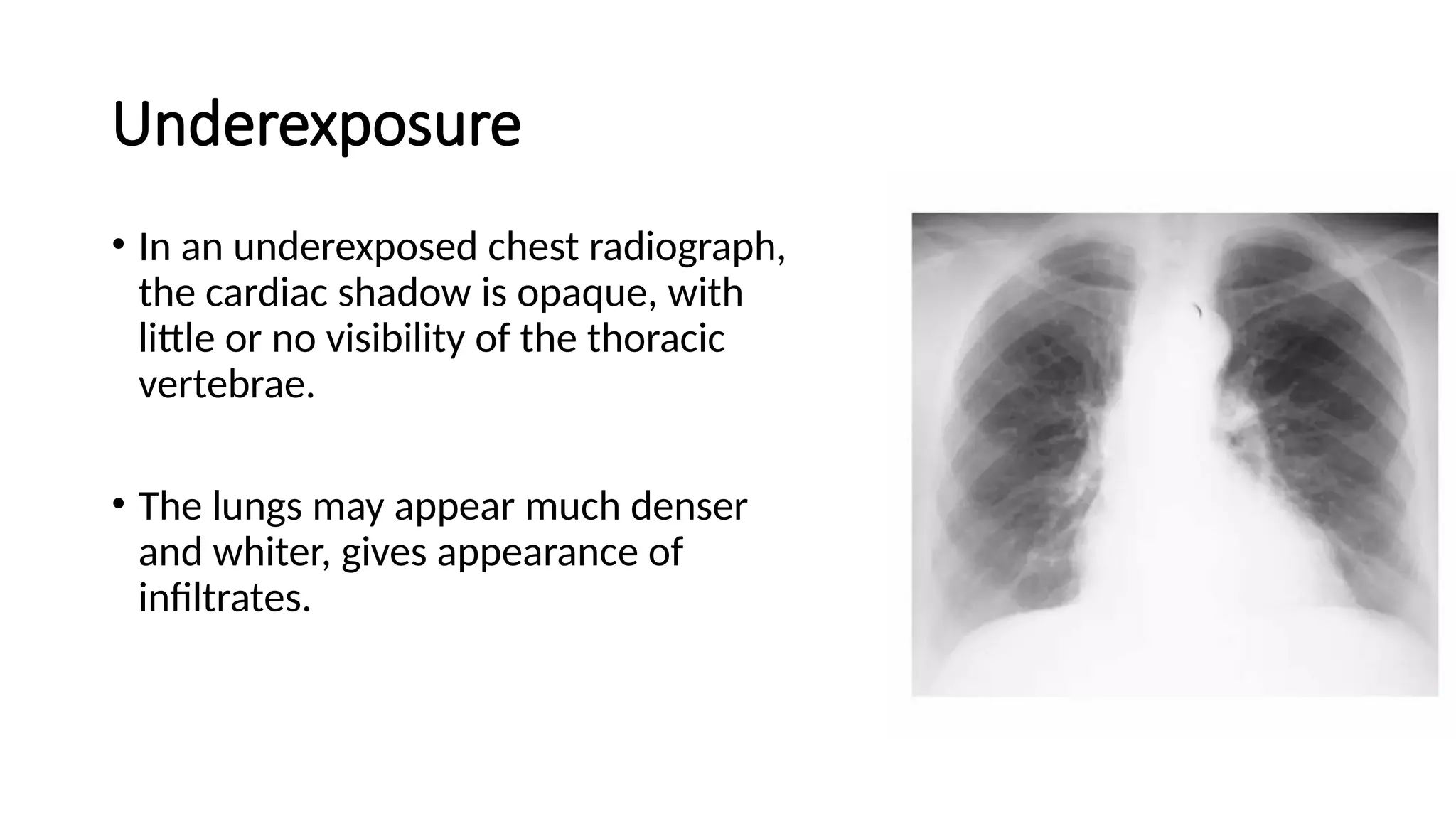
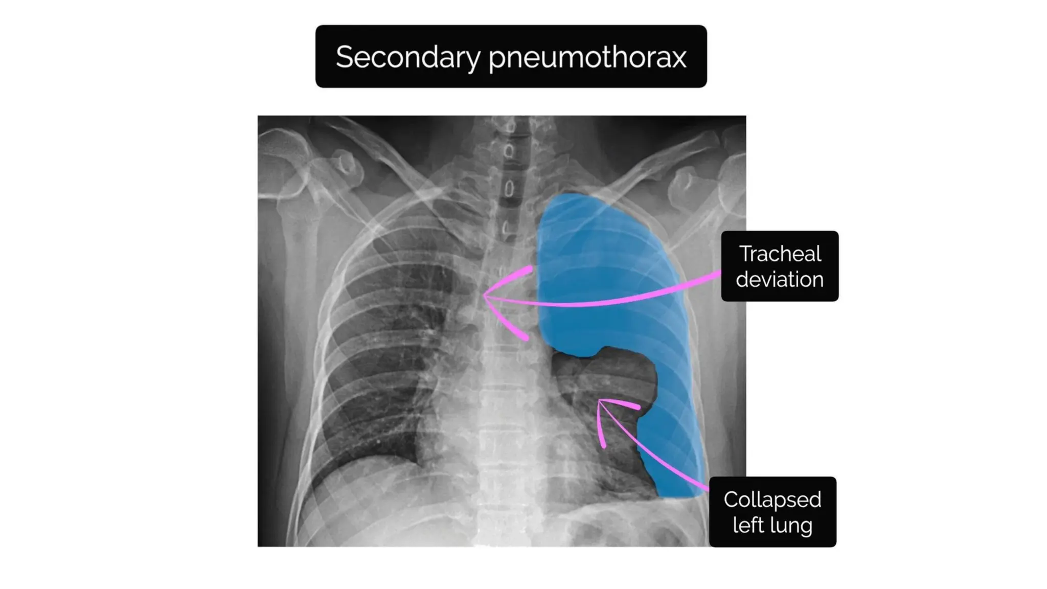
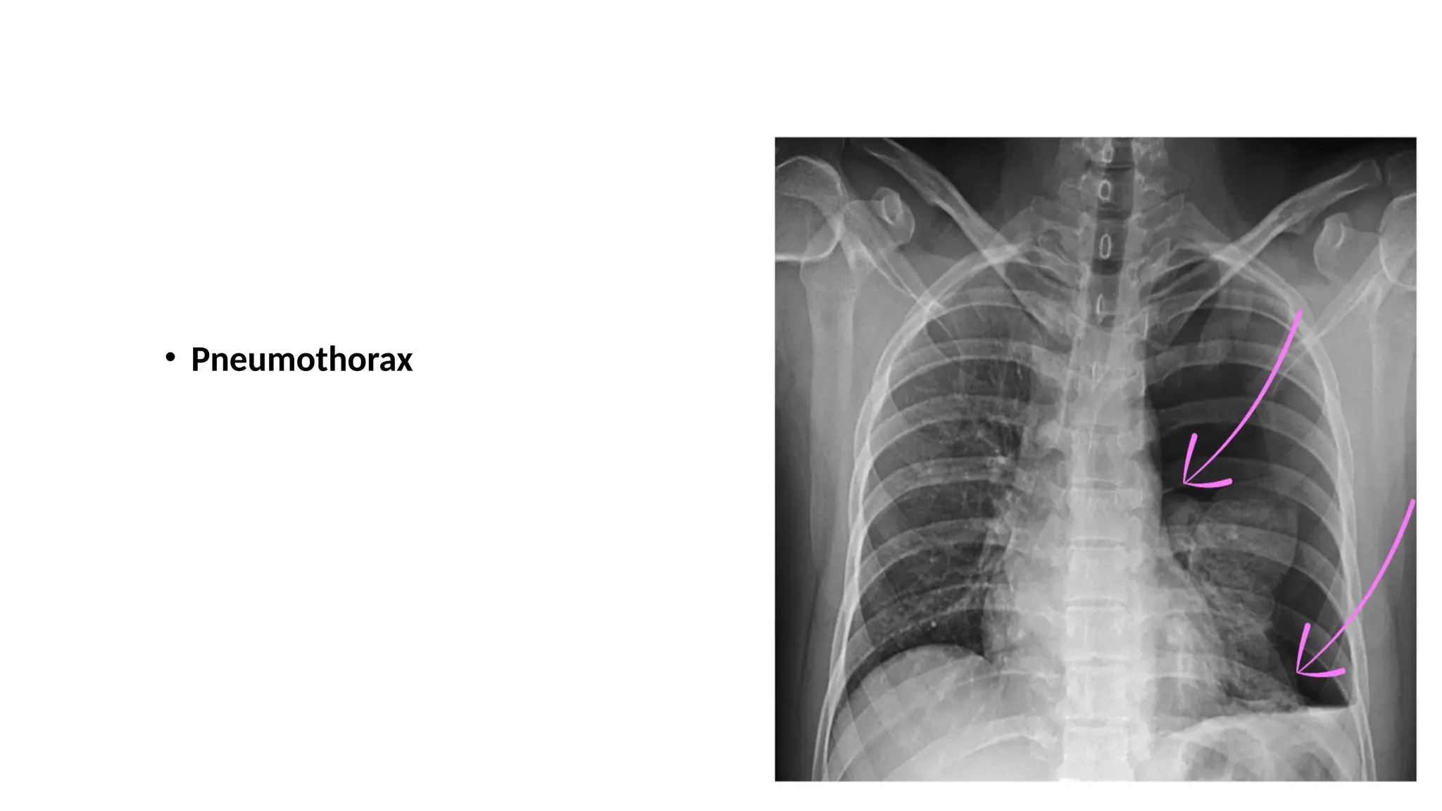
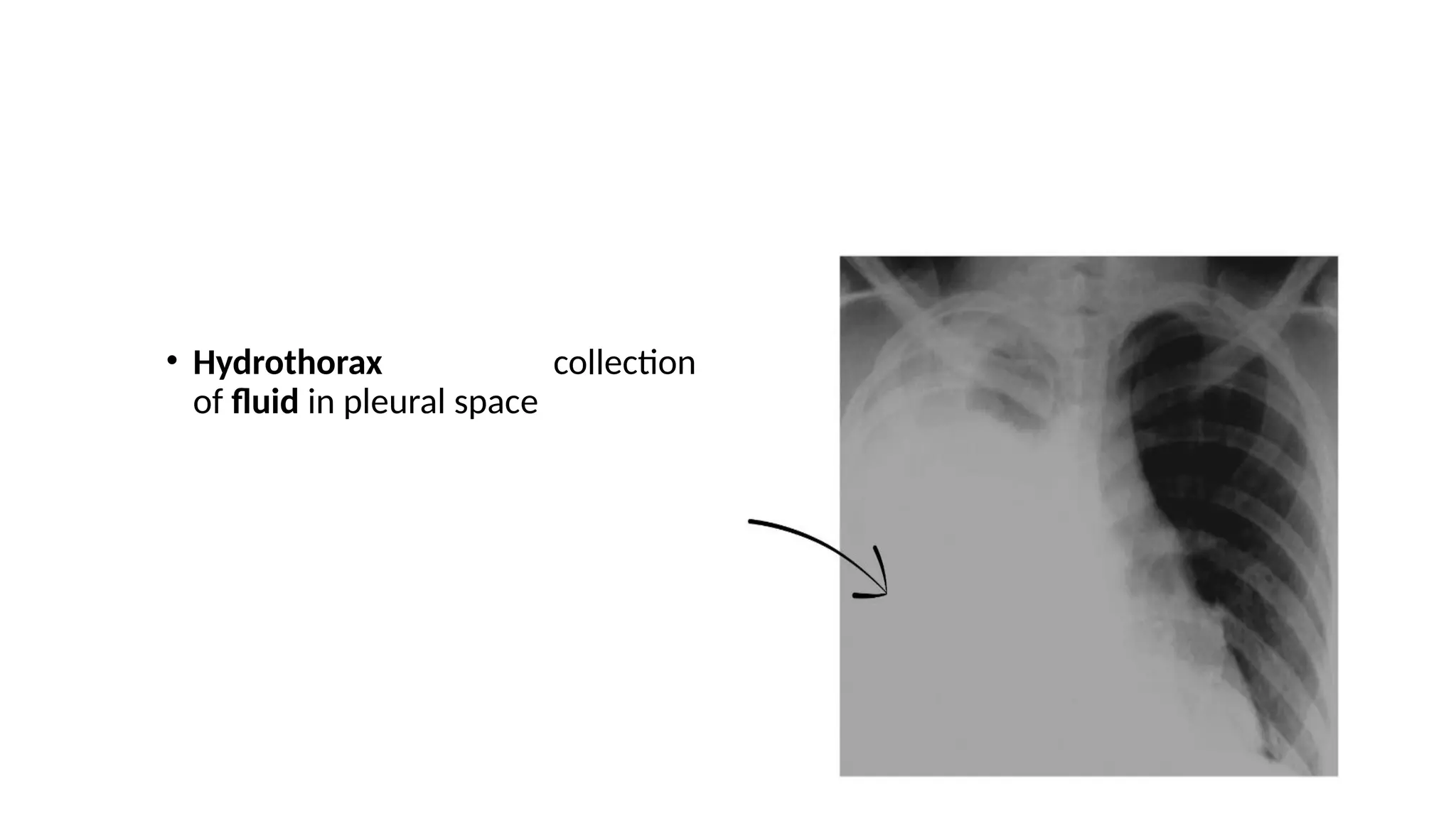
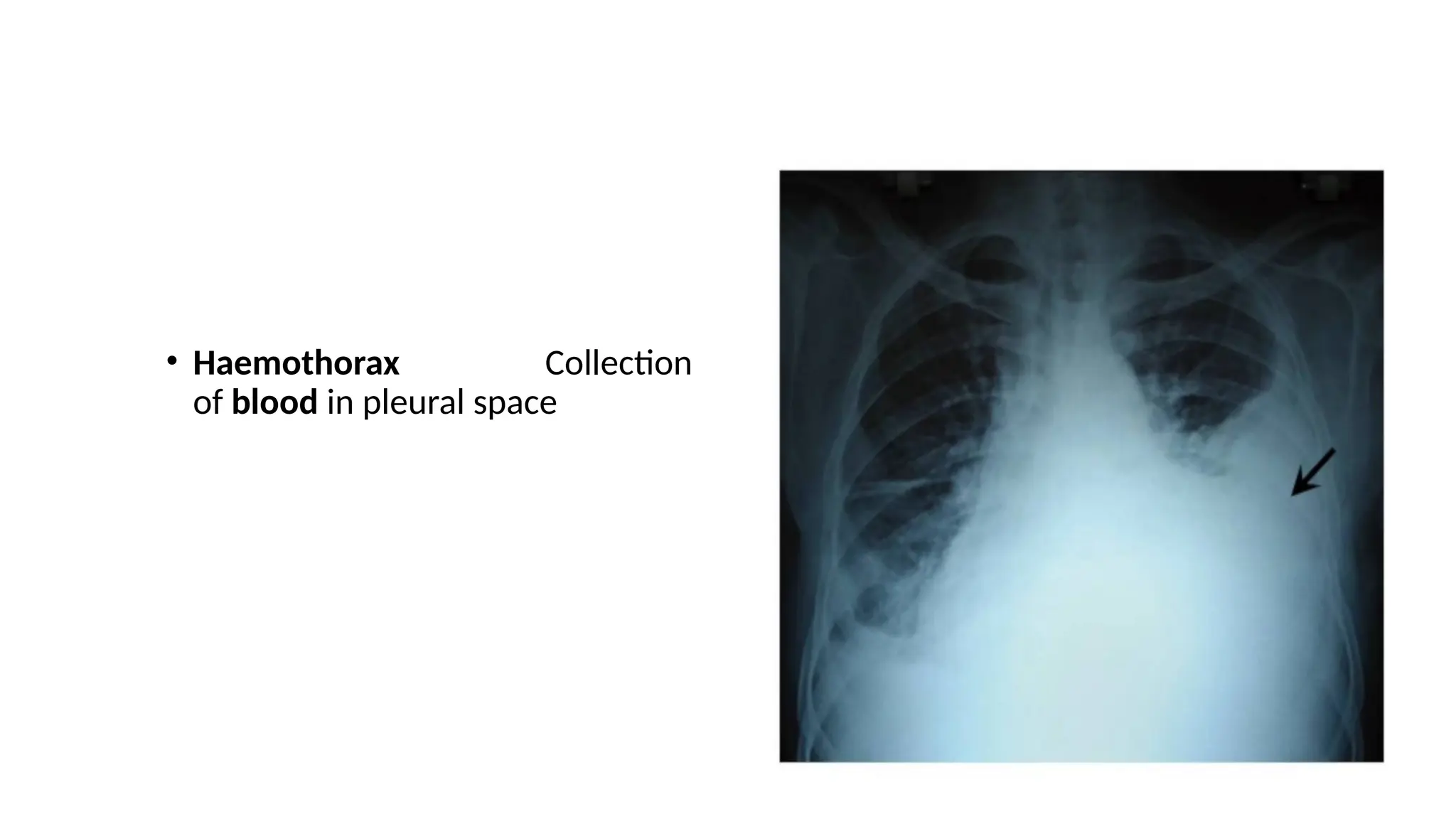
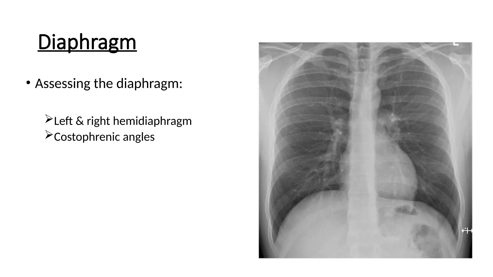
CHEST X-RAY
ART OF DIAGNOSIS
Presenter: Dr. Ravi Rokaya
Moderator: Dr. Bidesh Bista
Contents
• Basics of X-rays
• Assessing image quality
• Approach to interpreting X-ray
• X-ray review
• Documenting details
• Take home message
Basics of an X-ray
• X-rays use radiation (photons) to create 2D images
• Structure that are less dense appear dark/black (eg lungs)
• Structure that are more dense appear white (eg bones, tumor,
metals)
• Structure that are medium dense appear grey (eg soft tissues)
Before you begin
• Patient details (name, DOB, hospital number)
• Date & time x-ray was performed
• Previous imaging - for comparison
Assess image quality
RIPE
• R – Rotation
• I – Inspiration
• P – Projection
• E – Exposure
Rotation
• Vertebral bodies & spinous
processes should be vertically
aligned.
• The medial aspect of each
clavicle should be equidistant
from the spinous processes.
Rotation
Inspiration
• If inspiration is adequate, the
following should be visible:
5-6 anterior ribs
Both lung apices
Both costophrenic angles
Lateral rib edges
Projection
Posterior-Anterior (PA) Position
• The standard position for obtaining a routine chest radiograph.
• Patient stands upright with the anterior wall of chest placed against
the front of the film.
• Usually taken with the patient in full inspiration
• The PA film is viewed as if the patient is standing in front of you
Anterior-Posterior (AP) Projection
• Used when the patient is debilitated, immobilized, or unable to
cooperate with the PA procedure
• Film is placed behind the patient's back with the patient in a supine
position
• The scapulae are usually visible in the lung fields because they are not
rotated out of the view as they are in a PA
Lateral Projection
• Patient stands upright with the left side of the chest against the film
and the arms raised over the head
• Allows the viewer to see behind the heart and diaphragmatic dome
• Typically used in conjunction with a PA view of the same side of chest
to help determine the three-dimensional position of organs or
abnormal densities
Exposure
• In a properly exposed chest radiograph:
-the lower thoracic vertebrae should be
visible through the heart
-the broncho vascular structures behind
heart (trachea, aortic arch, pulmonary
arteries, etc.) should be seen.
Underexposure
• In an underexposed chest radiograph,
the cardiac shadow is opaque, with
little or no visibility of the thoracic
vertebrae.
• The lungs may appear much denser
and whiter, gives appearance of
infiltrates.
Overexposure
• With greater exposure of the chest
radiograph, the heart becomes more
radiolucent and the lungs become
proportionately darker.
• Often gives the appearance of lacking
lung tissue, as would be seen in a
condition such as emphysema.
ABCDE Approach
• Airway | trachea, carina, bronchi & hilar structures
• Breathing | lungs & pleura
• Cardiac | heart size & borders
• Diaphragm | assessment of diaphragm & costophrenic angles
• Everything else | mediastinal contours, bones, soft tissues, tubes,
valves, pacemakers & review areas
Airway
• Airway structures to
examine:
Trachea
Carina & bronchi
Hilar structures
Trachea
Inspect the trachea for signs of deviation
True deviation Apparent deviation
Large pleural effusion
Tension pneumothorax
Lobar collapse
Rotation of the patient
Carina and Bronchi
• The carina is located at the point
where trachea divides into the
Left & Right main bronchus.
Hilar structures
• The Hilar contain
 Pulmonary vessels
 Major bronchi
 Lymph nodes
• Hilar enlargement can be unilateral
or bilateral
• Bilateral enlargement –
1. Metastasis
2. Tuberculosis
3. Sarcoidosis
4. Lymphoma
5. Histoplasmosis
• Hilar enlargement can be unilateral
or bilateral
• Unilateral enlargement –
• Bronchogenic cyst
• Lung Cancer
• Large right pulmonary artery
• Aneurysm of descending aorta
Breathing
Breathing
• Divide each of the lungs into three zones & inspect each zone,
noting any asymmetry.
Lungs
• Abnormalities may include:
Pulmonary edema
Consolidation/Malignancy
Pneumothorax
Pulmonary Edema
• Common findings may include:
Air space opacification (bat wing distribution)
Kerley B lines
Pleural effusions (look for costophrenic blunting)
Fluid in interlobular fissures
Fig: Pulmonary edema
• Common causes of pulmonary edema includes:
Left heart failure
Mitral regurgitation
Fluid overload
Pneumothorax
Pleura
• Membrane surrounding the lungs
• Usually not visible in healthy individuals
• But can be visible when thickened
• Common causes of pleural
thickening:
• Mesothelioma
Pleural Thickening
• Pneumothorax
• Hydrothorax collection
of fluid in pleural space
• Haemothorax Collection
of blood in pleural space
• Empyema Collection
of pus in pleural space
Cardia
• Assess the following:
Cardiac size
Cardiac borders
Cardiac size
• Normal <50% of the thoracic
window
• >50% of thoracic window
Cardiomegaly
Accurate assessment requires PA chest x-ray
As AP films exaggerate the size of the heart
Causes of cardiomegaly
• There are many different causes of
cardiomegaly:
Valvular heart diseases
Coronary artery diseases
Pulmonary hypertension
Congenital heart diseases
Cardiac borders
• In healthy individuals the borders
of the heart should be clearly
visible & well-defined.
Diaphragm
• Assessing the diaphragm:
Left & right hemidiaphragm
Costophrenic angles
Hemidiaphragm
• Right hemidiaphragm higher than the left
due to presence of liver below
• Stomach sits under the left hemidiaphragm
and gastric bubble can be visualized
• Pneumoperitoneum= accumulation of air
under the diaphragm
• Results in the separation of the diaphragm
from liver.
• Often secondary to a bowel perforation
Costophrenic angles
• The costophrenic angles reflect the dome
shape of hemidiaphragm as it meets the
lateral chest wall.
• The loss of this angle is termed as
costophrenic blunting & causes
includes:
Pleural effusion
Consolidation
Hyperinflation (e.g. asthma copd)
Everything else
Review everything else:
Mediastinal contours
Aortic knuckle
Aortopulmonary window
Bones & soft tissues
Tubes, valves & pacemakers
Review areas
Mediastinal contours
Bones & soft tissues
• Inspect the bones for visible fractures (e.g. ribs & clavicles) or
lytic/sclerotic lesions (e.g. myeloma, metastases)
• Inspect the soft tissues for any abnormalities such as hematoma
Tubes, valves and pacemakers
We may see:
Nasogastric tubes
Central lines
ECG leads
Artificial heart valves
Pacemakers
Final Review areas
Lung apices
Retrocardiac region
Lung peripheries
Document your findings
Patient details (name, DOB and ID number)
Date and time chest x-ray was performed
Indication/clinical context
Document each finding using ABCDE format
Overall impression
Plan
Your name, grade, signature, registration
number and contact number
Take Home Message
• Patient details should always be checked before reading an x-ray
• Appropriate image quality is necessary for interpreting of an x- ray
• PA projection is standard projection
• It is important to know the clinical context before ordering an x-ray as
it will help in overall interpretation & reaching DD
References
• UpToDate
• Radipaedia.org
• Davidson’s Principles & Practice of Medicine
• Harrison’s Principle of Internal Medicine
• Washington Manual of Medical Therapeutics
CHEST XRAY INTERPRETATION . A SLIDE IN CSH

CHEST XRAY INTERPRETATION . A SLIDE IN CSH

  • 1.
    CHEST X-RAY ART OFDIAGNOSIS Presenter: Dr. Ravi Rokaya Moderator: Dr. Bidesh Bista
  • 2.
    Contents • Basics ofX-rays • Assessing image quality • Approach to interpreting X-ray • X-ray review • Documenting details • Take home message
  • 3.
    Basics of anX-ray • X-rays use radiation (photons) to create 2D images • Structure that are less dense appear dark/black (eg lungs) • Structure that are more dense appear white (eg bones, tumor, metals) • Structure that are medium dense appear grey (eg soft tissues)
  • 4.
    Before you begin •Patient details (name, DOB, hospital number) • Date & time x-ray was performed • Previous imaging - for comparison
  • 5.
    Assess image quality RIPE •R – Rotation • I – Inspiration • P – Projection • E – Exposure
  • 6.
    Rotation • Vertebral bodies& spinous processes should be vertically aligned. • The medial aspect of each clavicle should be equidistant from the spinous processes.
  • 7.
  • 8.
    Inspiration • If inspirationis adequate, the following should be visible: 5-6 anterior ribs Both lung apices Both costophrenic angles Lateral rib edges
  • 9.
  • 10.
    Posterior-Anterior (PA) Position •The standard position for obtaining a routine chest radiograph. • Patient stands upright with the anterior wall of chest placed against the front of the film. • Usually taken with the patient in full inspiration • The PA film is viewed as if the patient is standing in front of you
  • 11.
    Anterior-Posterior (AP) Projection •Used when the patient is debilitated, immobilized, or unable to cooperate with the PA procedure • Film is placed behind the patient's back with the patient in a supine position • The scapulae are usually visible in the lung fields because they are not rotated out of the view as they are in a PA
  • 12.
    Lateral Projection • Patientstands upright with the left side of the chest against the film and the arms raised over the head • Allows the viewer to see behind the heart and diaphragmatic dome • Typically used in conjunction with a PA view of the same side of chest to help determine the three-dimensional position of organs or abnormal densities
  • 13.
    Exposure • In aproperly exposed chest radiograph: -the lower thoracic vertebrae should be visible through the heart -the broncho vascular structures behind heart (trachea, aortic arch, pulmonary arteries, etc.) should be seen.
  • 14.
    Underexposure • In anunderexposed chest radiograph, the cardiac shadow is opaque, with little or no visibility of the thoracic vertebrae. • The lungs may appear much denser and whiter, gives appearance of infiltrates.
  • 15.
    Overexposure • With greaterexposure of the chest radiograph, the heart becomes more radiolucent and the lungs become proportionately darker. • Often gives the appearance of lacking lung tissue, as would be seen in a condition such as emphysema.
  • 16.
    ABCDE Approach • Airway| trachea, carina, bronchi & hilar structures • Breathing | lungs & pleura • Cardiac | heart size & borders • Diaphragm | assessment of diaphragm & costophrenic angles • Everything else | mediastinal contours, bones, soft tissues, tubes, valves, pacemakers & review areas
  • 17.
    Airway • Airway structuresto examine: Trachea Carina & bronchi Hilar structures
  • 18.
    Trachea Inspect the tracheafor signs of deviation True deviation Apparent deviation Large pleural effusion Tension pneumothorax Lobar collapse Rotation of the patient
  • 21.
    Carina and Bronchi •The carina is located at the point where trachea divides into the Left & Right main bronchus.
  • 22.
    Hilar structures • TheHilar contain  Pulmonary vessels  Major bronchi  Lymph nodes
  • 23.
    • Hilar enlargementcan be unilateral or bilateral • Bilateral enlargement – 1. Metastasis 2. Tuberculosis 3. Sarcoidosis 4. Lymphoma 5. Histoplasmosis
  • 24.
    • Hilar enlargementcan be unilateral or bilateral • Unilateral enlargement – • Bronchogenic cyst • Lung Cancer • Large right pulmonary artery • Aneurysm of descending aorta
  • 25.
  • 26.
    Breathing • Divide eachof the lungs into three zones & inspect each zone, noting any asymmetry.
  • 28.
    Lungs • Abnormalities mayinclude: Pulmonary edema Consolidation/Malignancy Pneumothorax
  • 29.
    Pulmonary Edema • Commonfindings may include: Air space opacification (bat wing distribution) Kerley B lines Pleural effusions (look for costophrenic blunting) Fluid in interlobular fissures
  • 30.
  • 31.
    • Common causesof pulmonary edema includes: Left heart failure Mitral regurgitation Fluid overload
  • 34.
  • 37.
    Pleura • Membrane surroundingthe lungs • Usually not visible in healthy individuals • But can be visible when thickened
  • 38.
    • Common causesof pleural thickening: • Mesothelioma Pleural Thickening
  • 39.
  • 40.
    • Hydrothorax collection offluid in pleural space
  • 41.
    • Haemothorax Collection ofblood in pleural space
  • 42.
    • Empyema Collection ofpus in pleural space
  • 43.
    Cardia • Assess thefollowing: Cardiac size Cardiac borders
  • 44.
    Cardiac size • Normal<50% of the thoracic window • >50% of thoracic window Cardiomegaly Accurate assessment requires PA chest x-ray As AP films exaggerate the size of the heart
  • 46.
    Causes of cardiomegaly •There are many different causes of cardiomegaly: Valvular heart diseases Coronary artery diseases Pulmonary hypertension Congenital heart diseases
  • 47.
    Cardiac borders • Inhealthy individuals the borders of the heart should be clearly visible & well-defined.
  • 52.
    Diaphragm • Assessing thediaphragm: Left & right hemidiaphragm Costophrenic angles
  • 53.
    Hemidiaphragm • Right hemidiaphragmhigher than the left due to presence of liver below • Stomach sits under the left hemidiaphragm and gastric bubble can be visualized
  • 54.
    • Pneumoperitoneum= accumulationof air under the diaphragm • Results in the separation of the diaphragm from liver. • Often secondary to a bowel perforation
  • 56.
    Costophrenic angles • Thecostophrenic angles reflect the dome shape of hemidiaphragm as it meets the lateral chest wall.
  • 57.
    • The lossof this angle is termed as costophrenic blunting & causes includes: Pleural effusion Consolidation Hyperinflation (e.g. asthma copd)
  • 58.
    Everything else Review everythingelse: Mediastinal contours Aortic knuckle Aortopulmonary window Bones & soft tissues Tubes, valves & pacemakers Review areas
  • 59.
  • 60.
    Bones & softtissues • Inspect the bones for visible fractures (e.g. ribs & clavicles) or lytic/sclerotic lesions (e.g. myeloma, metastases) • Inspect the soft tissues for any abnormalities such as hematoma
  • 61.
    Tubes, valves andpacemakers We may see: Nasogastric tubes Central lines ECG leads Artificial heart valves Pacemakers
  • 65.
    Final Review areas Lungapices Retrocardiac region Lung peripheries
  • 66.
    Document your findings Patientdetails (name, DOB and ID number) Date and time chest x-ray was performed Indication/clinical context Document each finding using ABCDE format Overall impression Plan Your name, grade, signature, registration number and contact number
  • 67.
    Take Home Message •Patient details should always be checked before reading an x-ray • Appropriate image quality is necessary for interpreting of an x- ray • PA projection is standard projection • It is important to know the clinical context before ordering an x-ray as it will help in overall interpretation & reaching DD
  • 68.
    References • UpToDate • Radipaedia.org •Davidson’s Principles & Practice of Medicine • Harrison’s Principle of Internal Medicine • Washington Manual of Medical Therapeutics