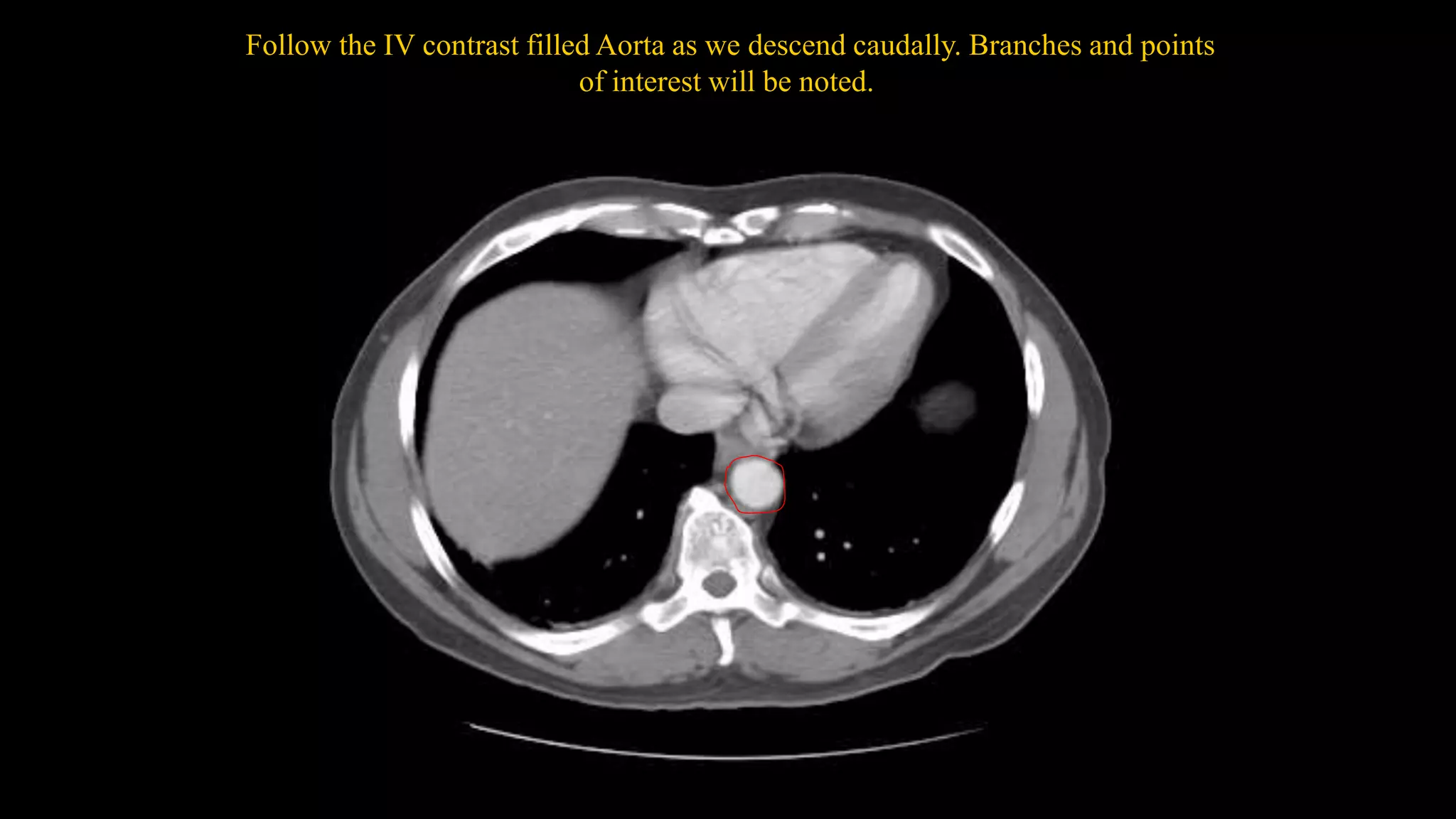
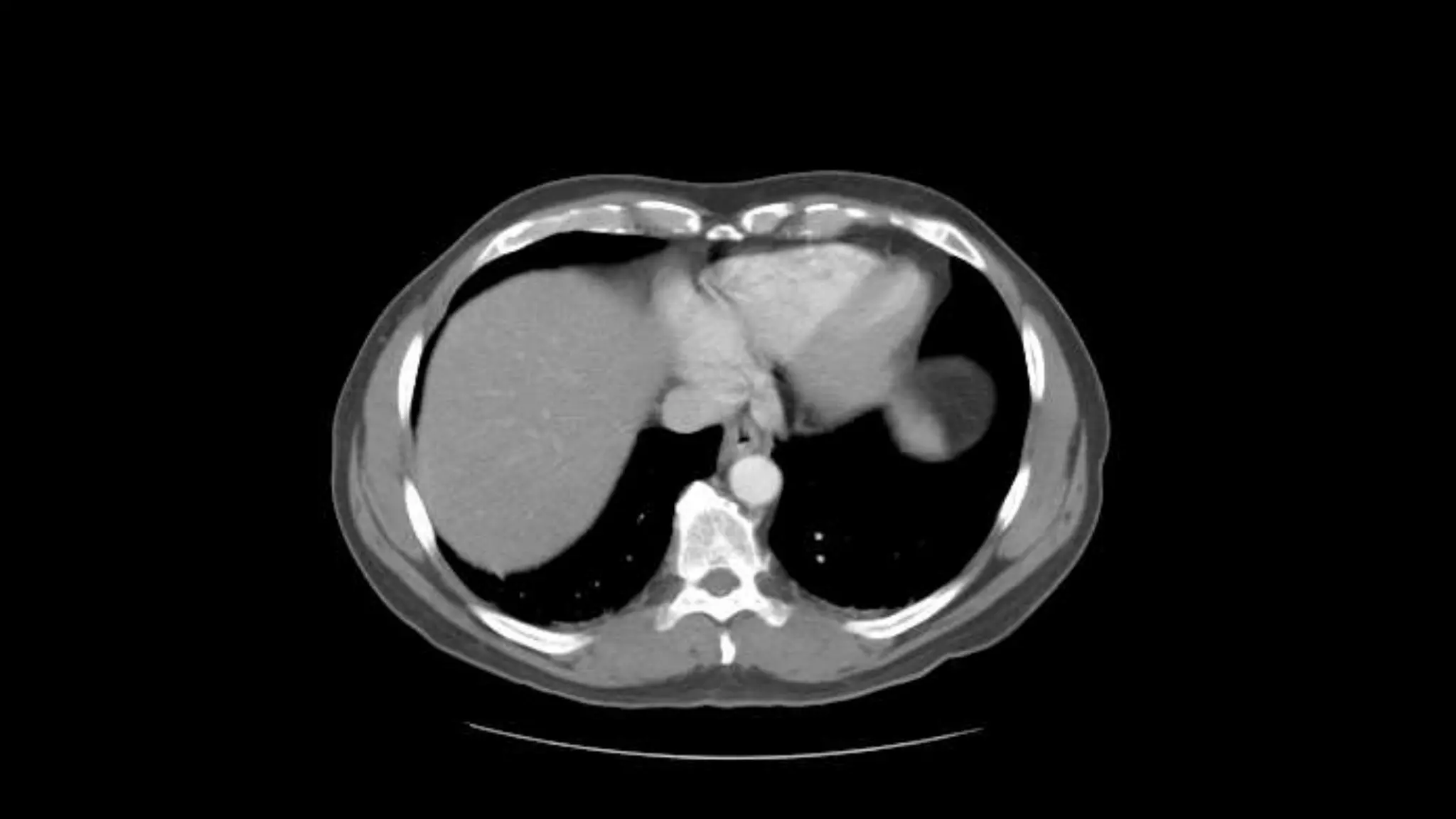
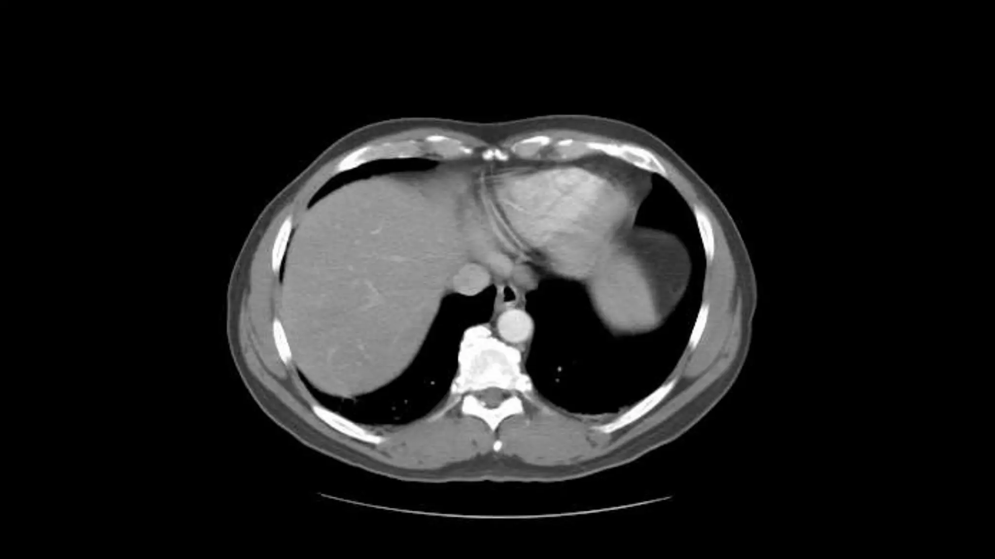
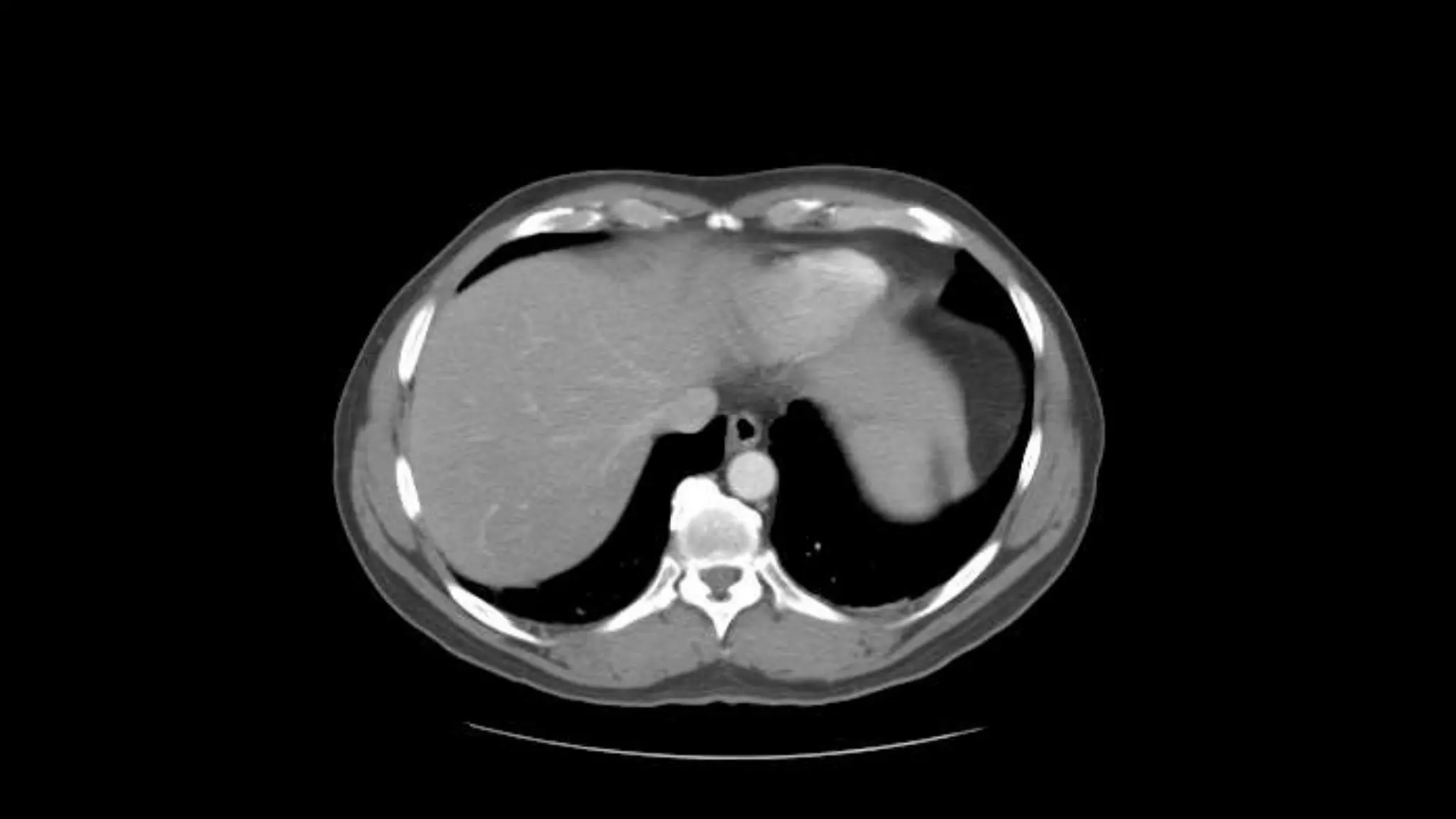
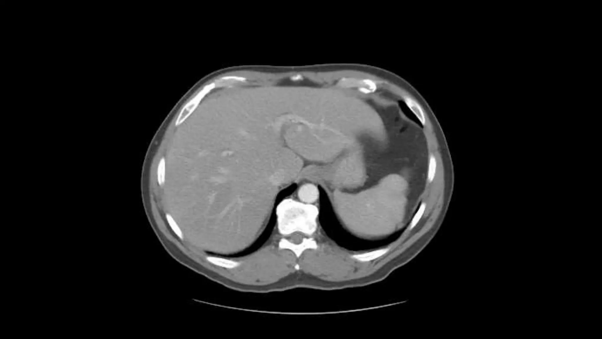
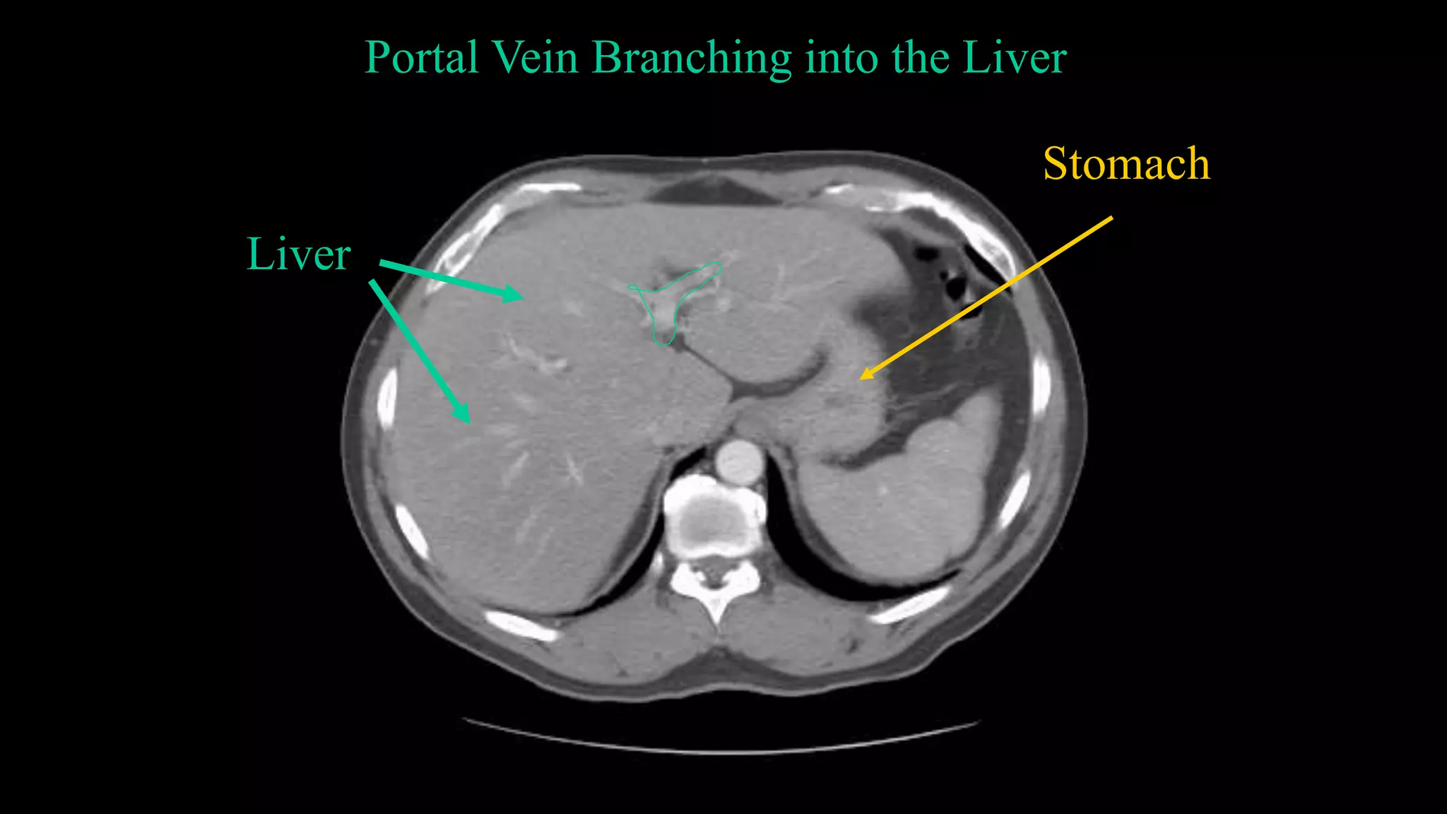
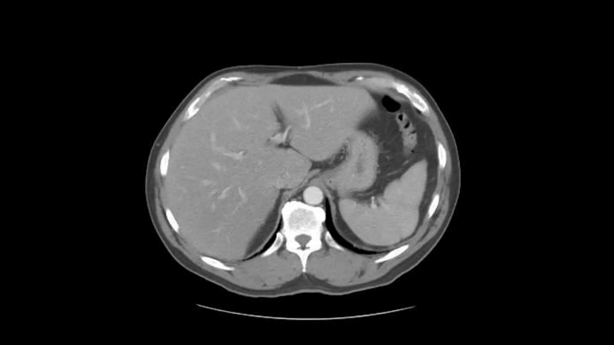
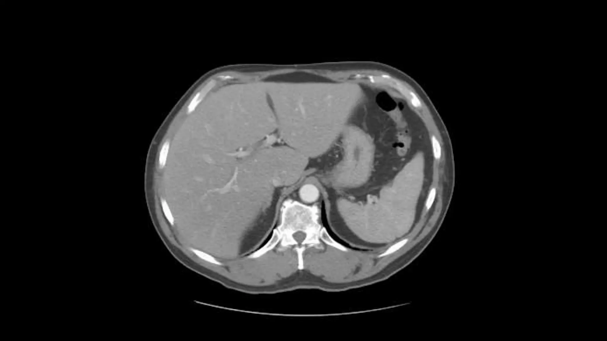
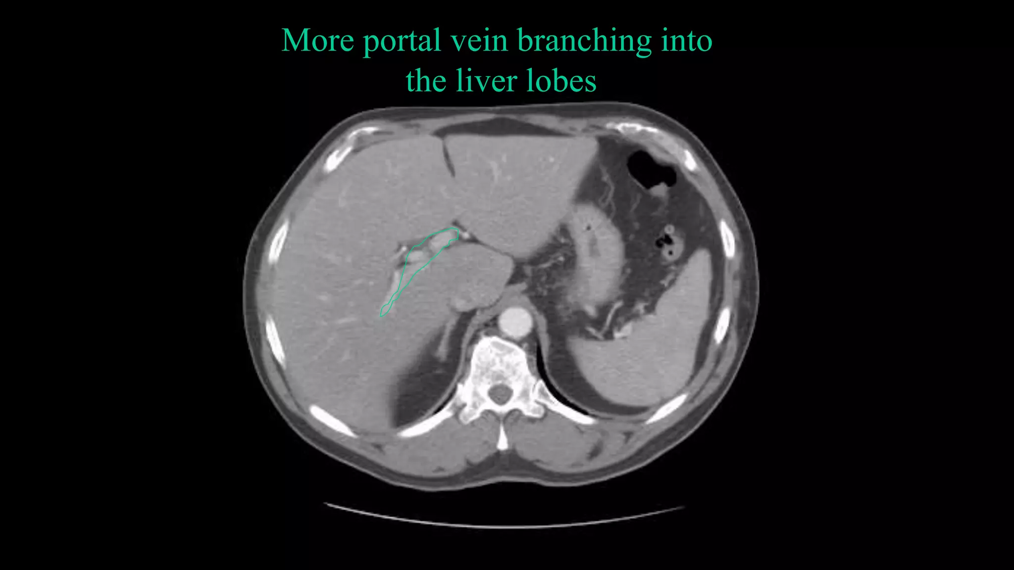
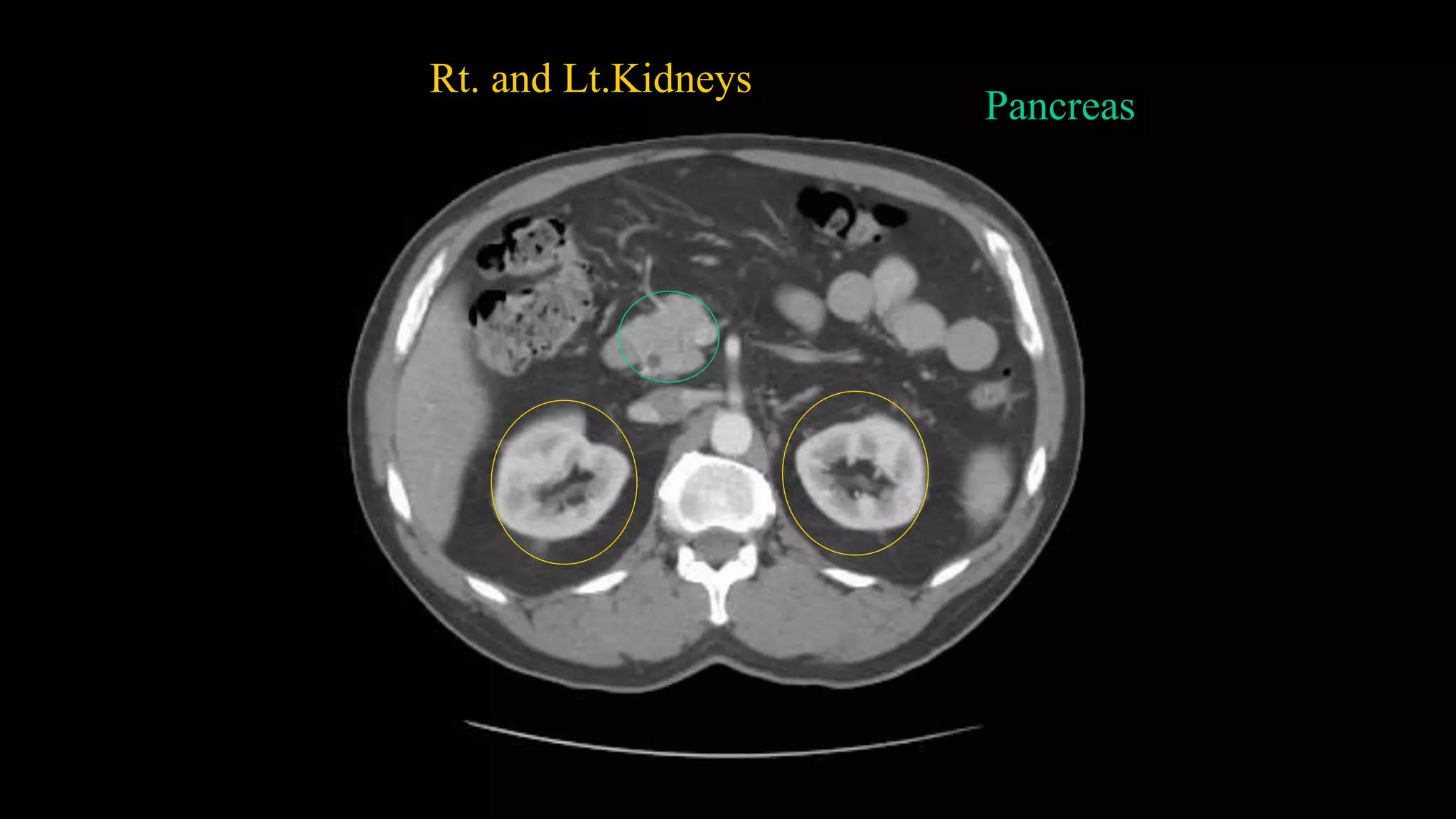
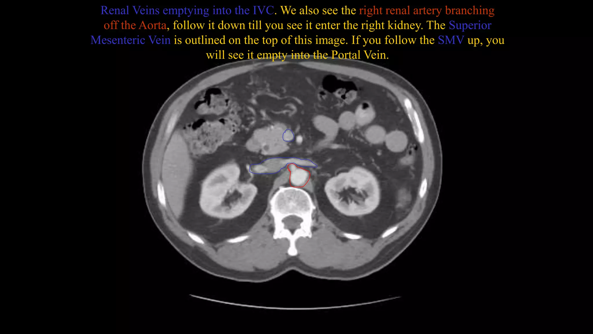
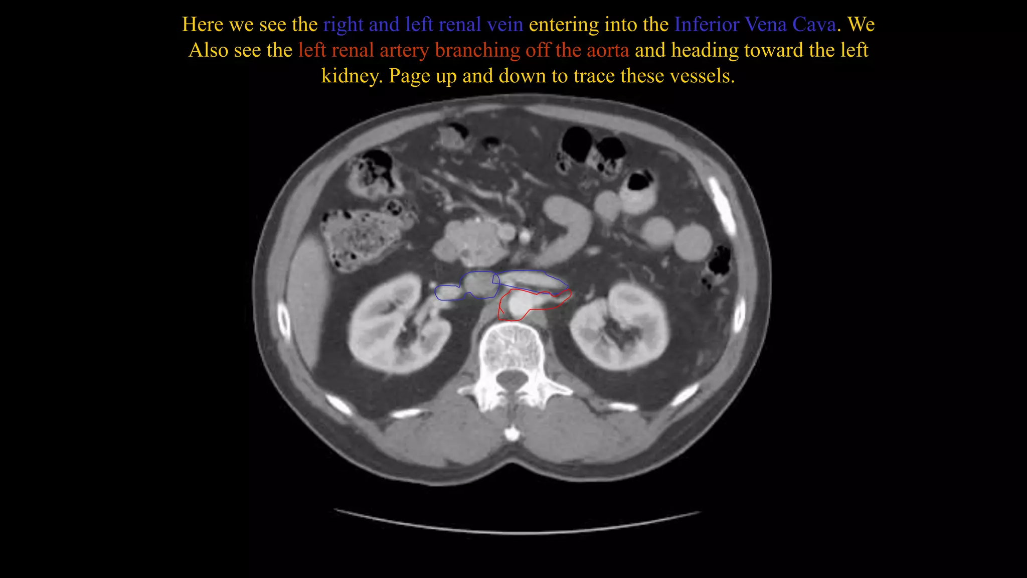
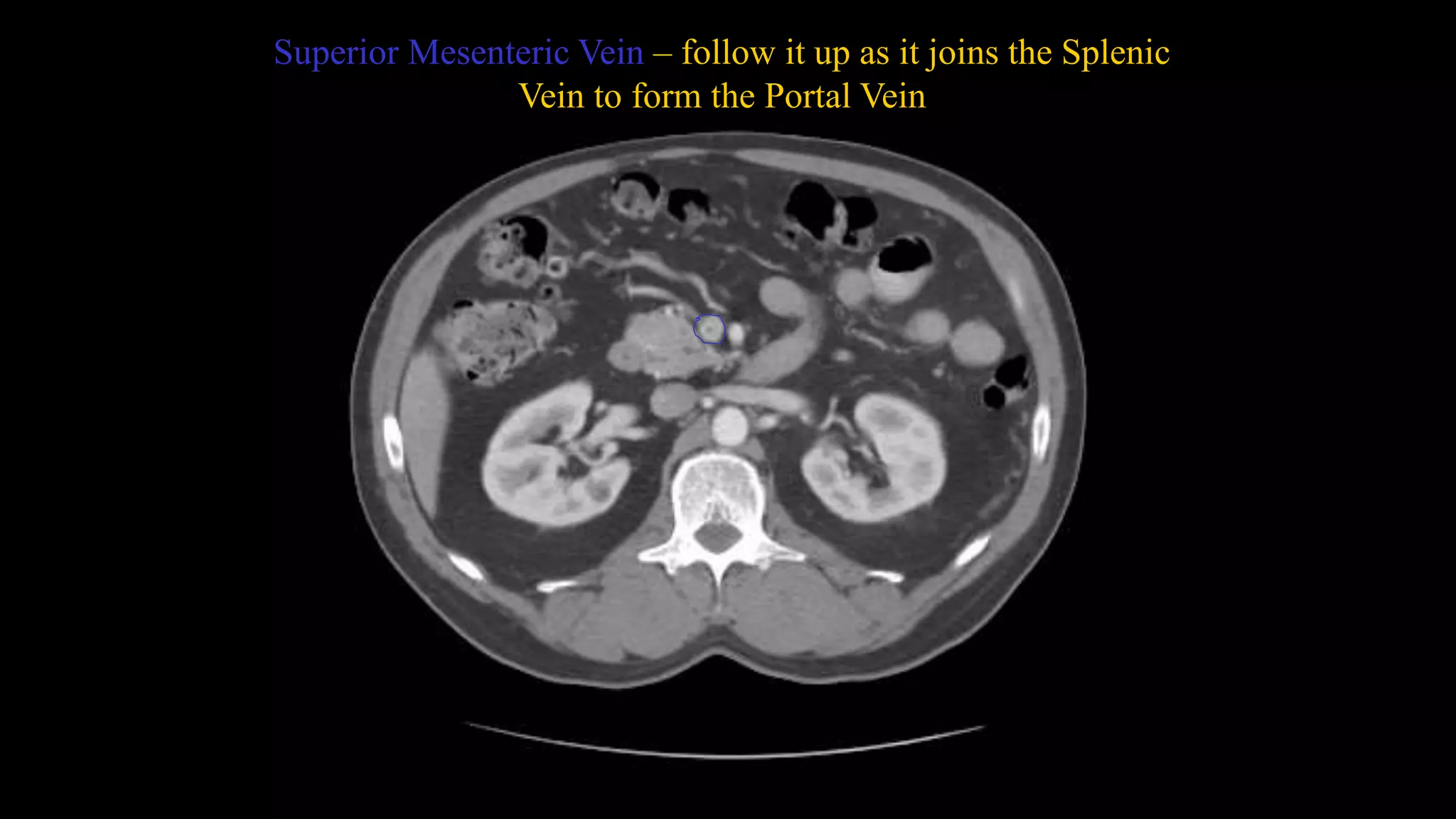
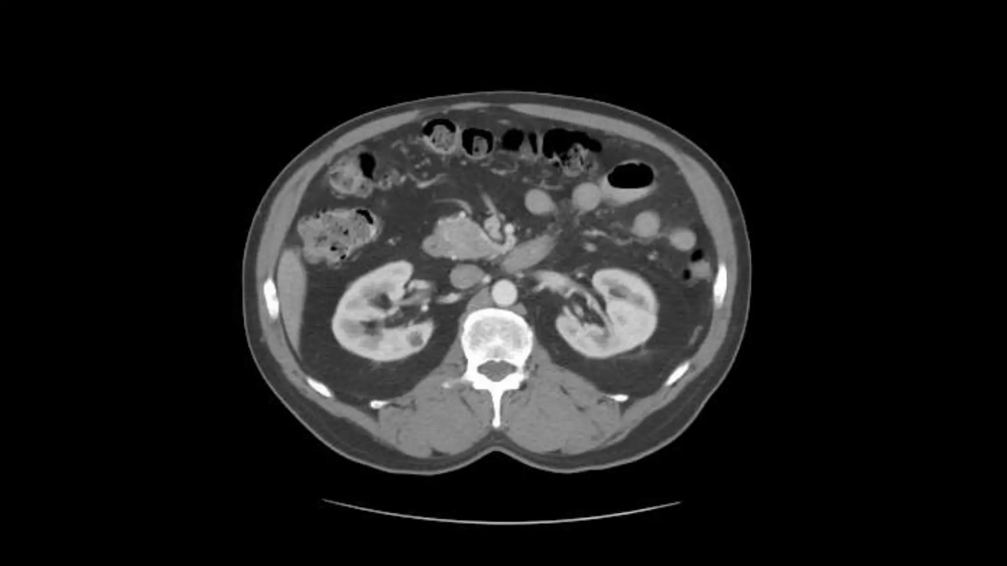
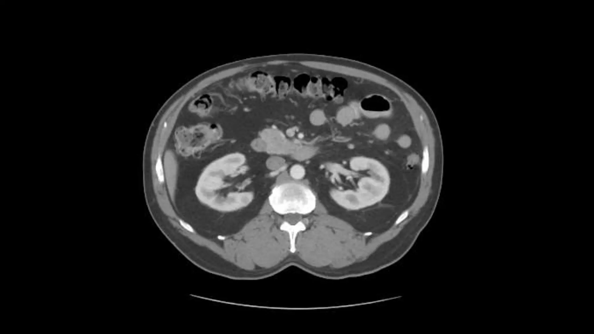
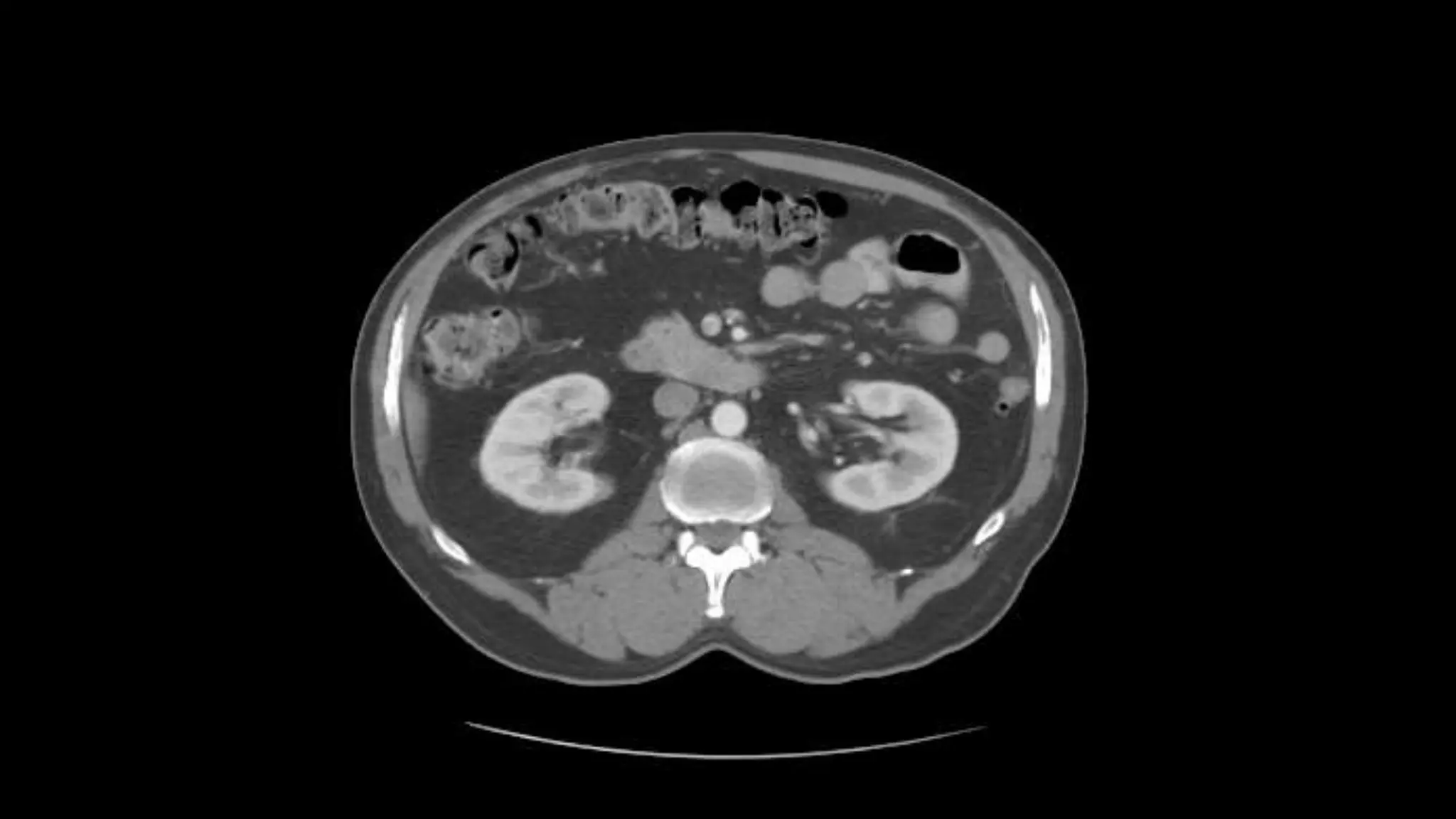
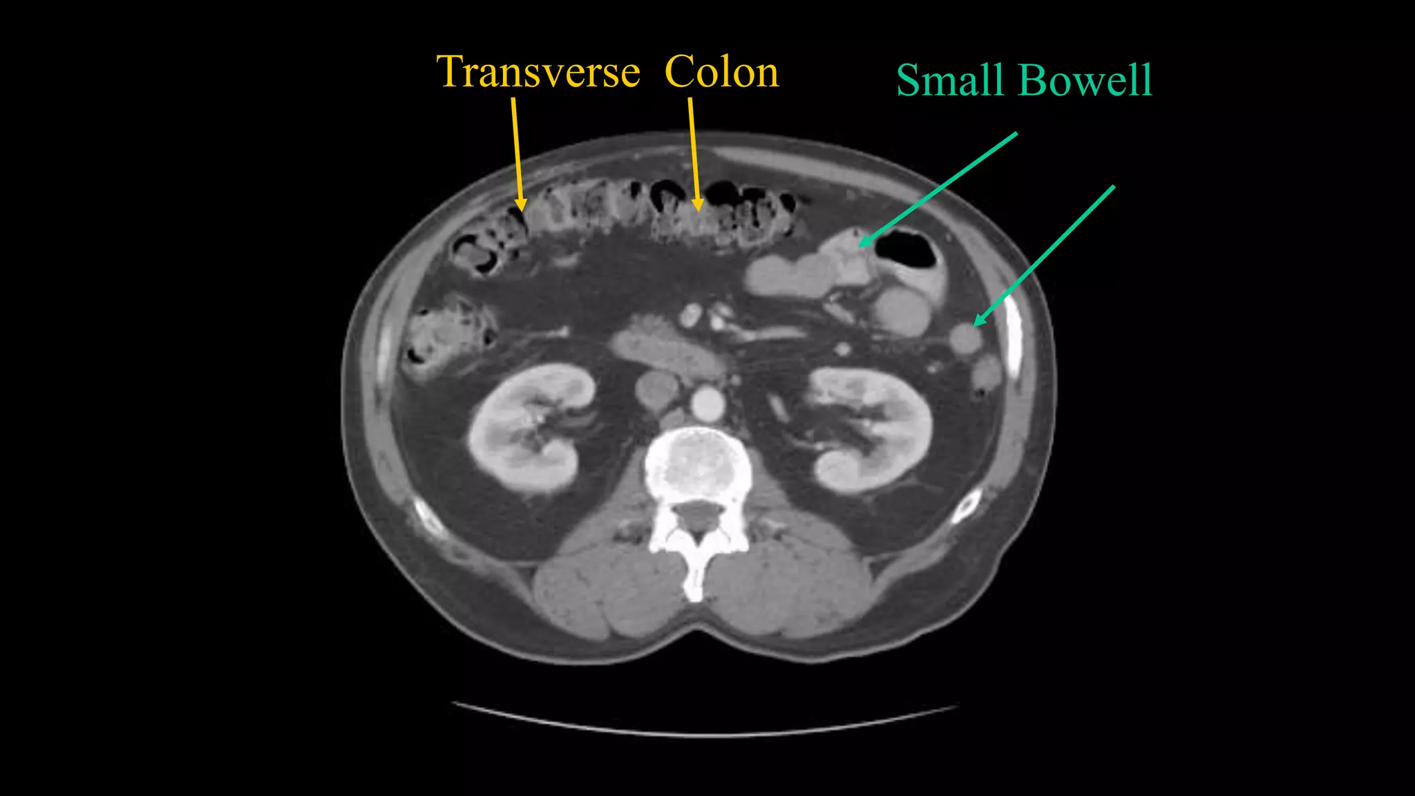
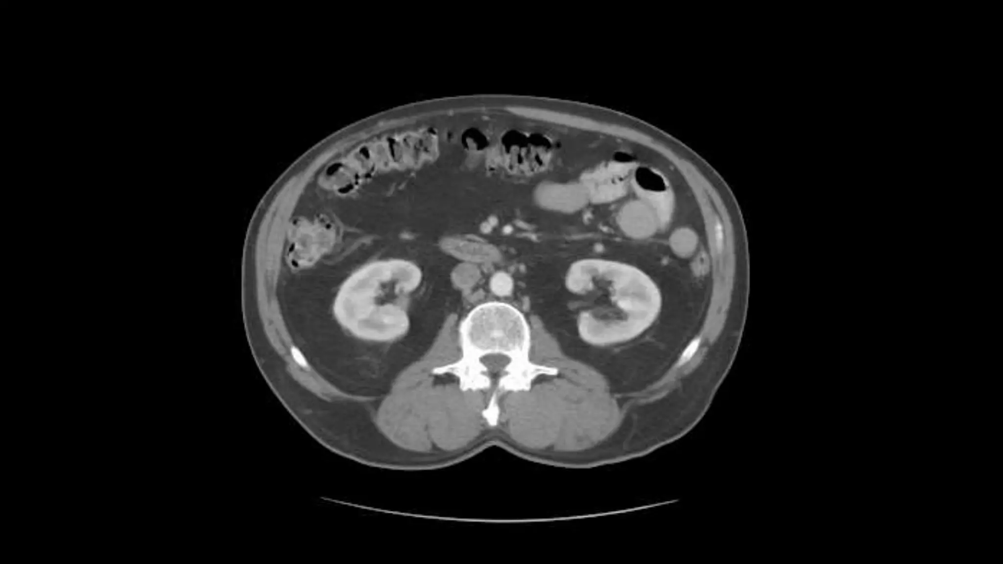
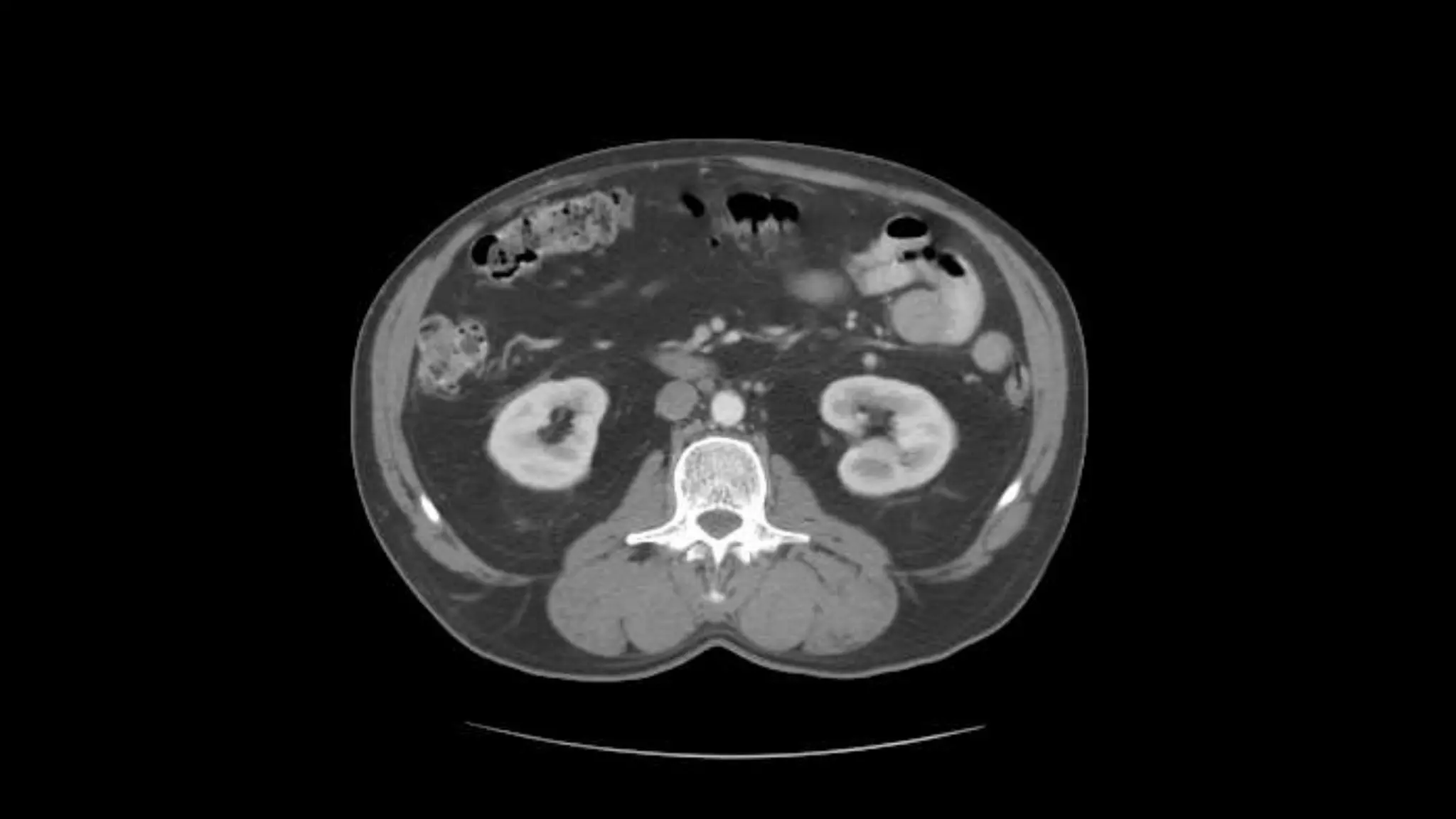
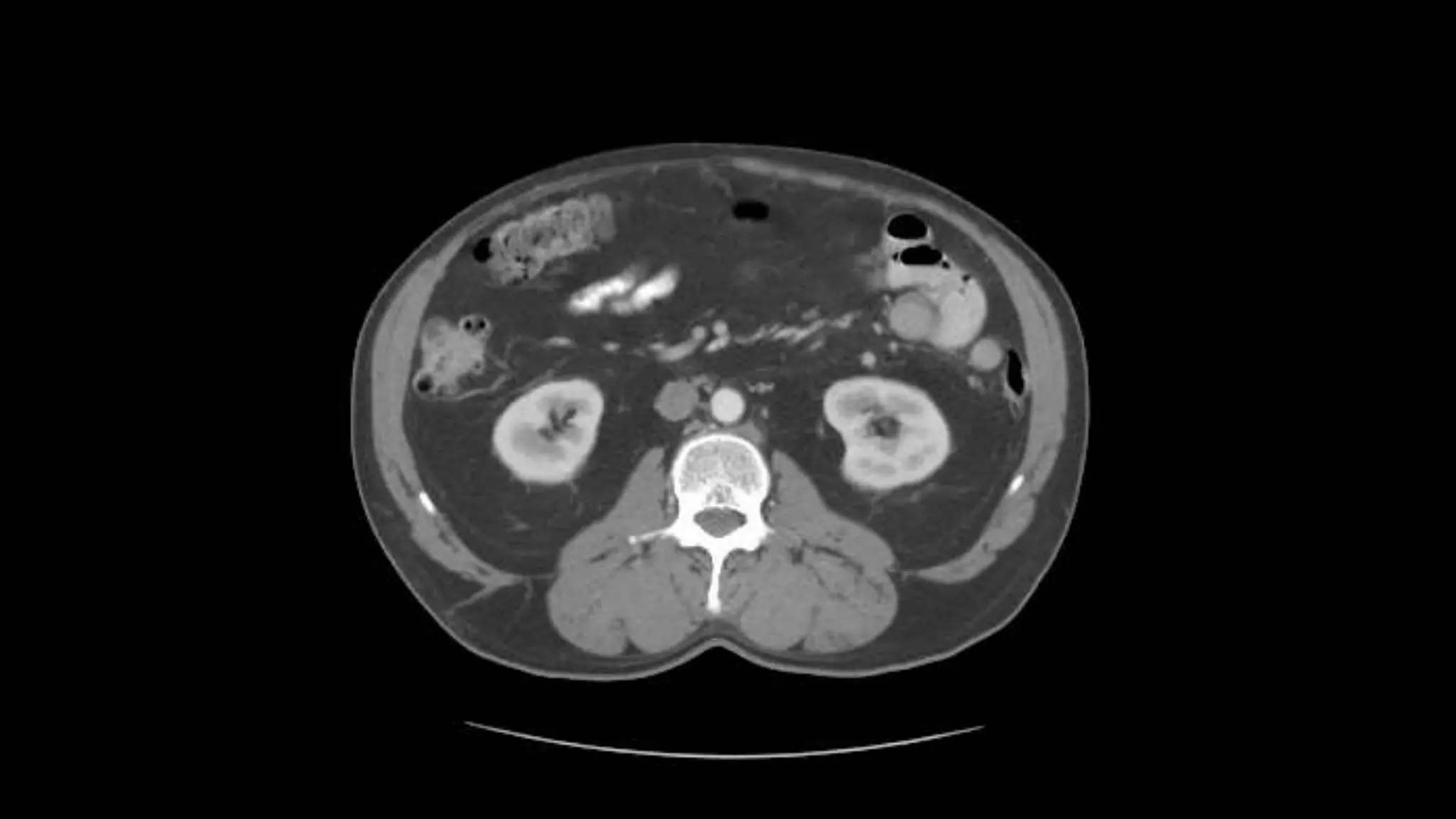
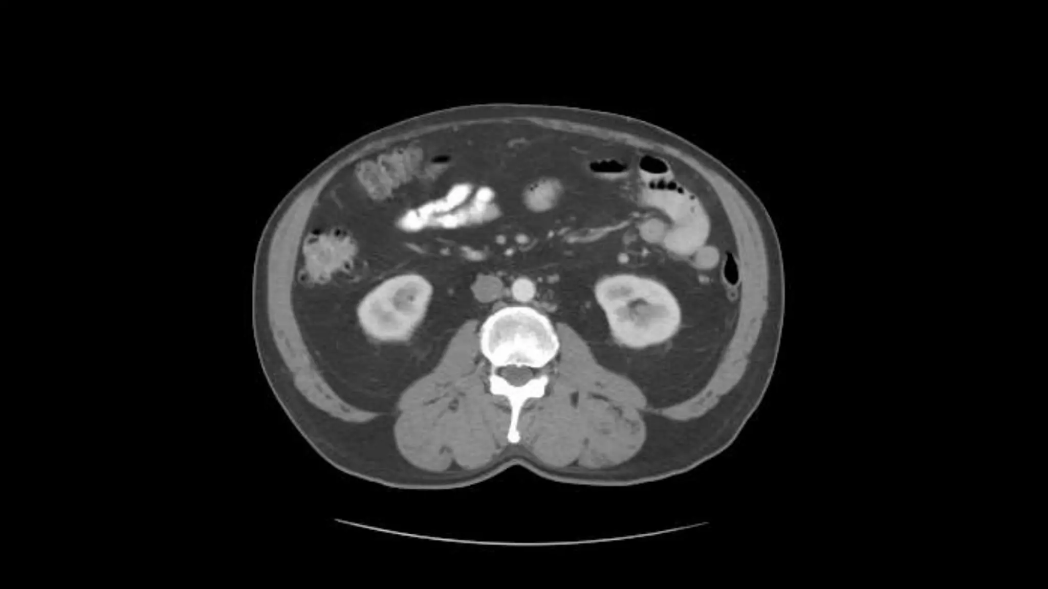
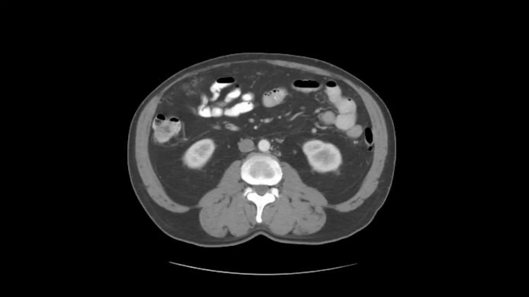
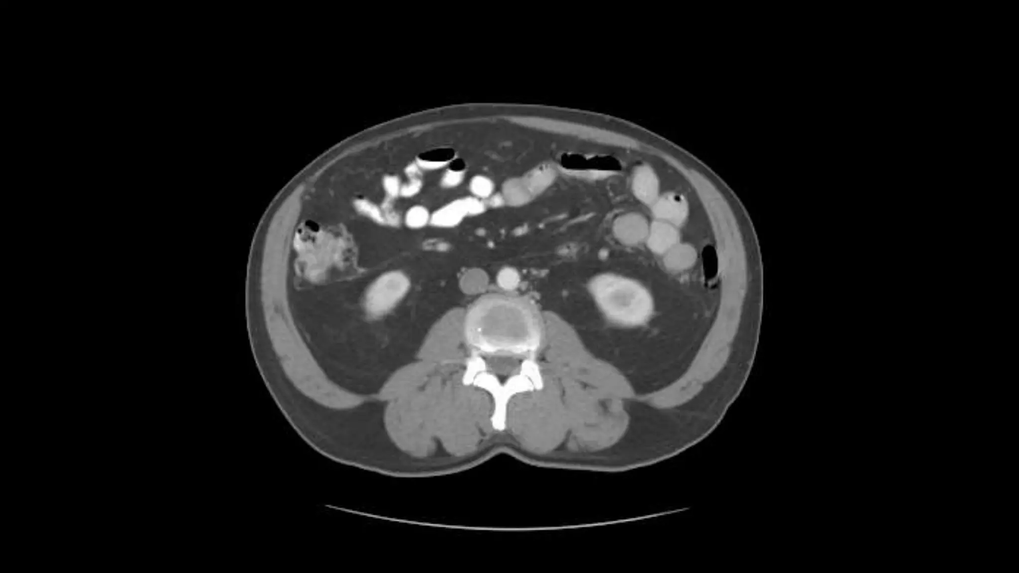
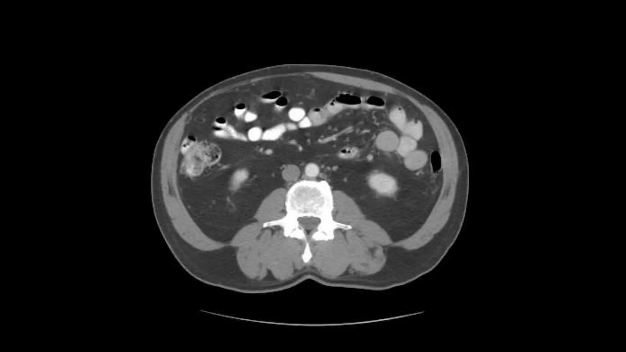
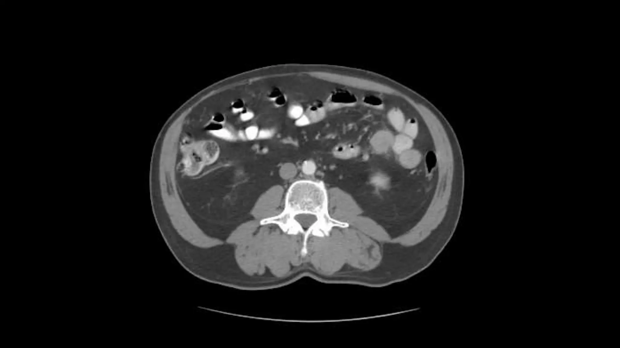
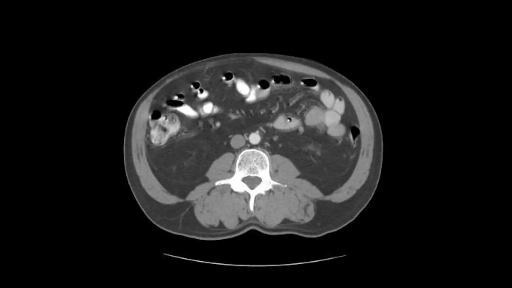
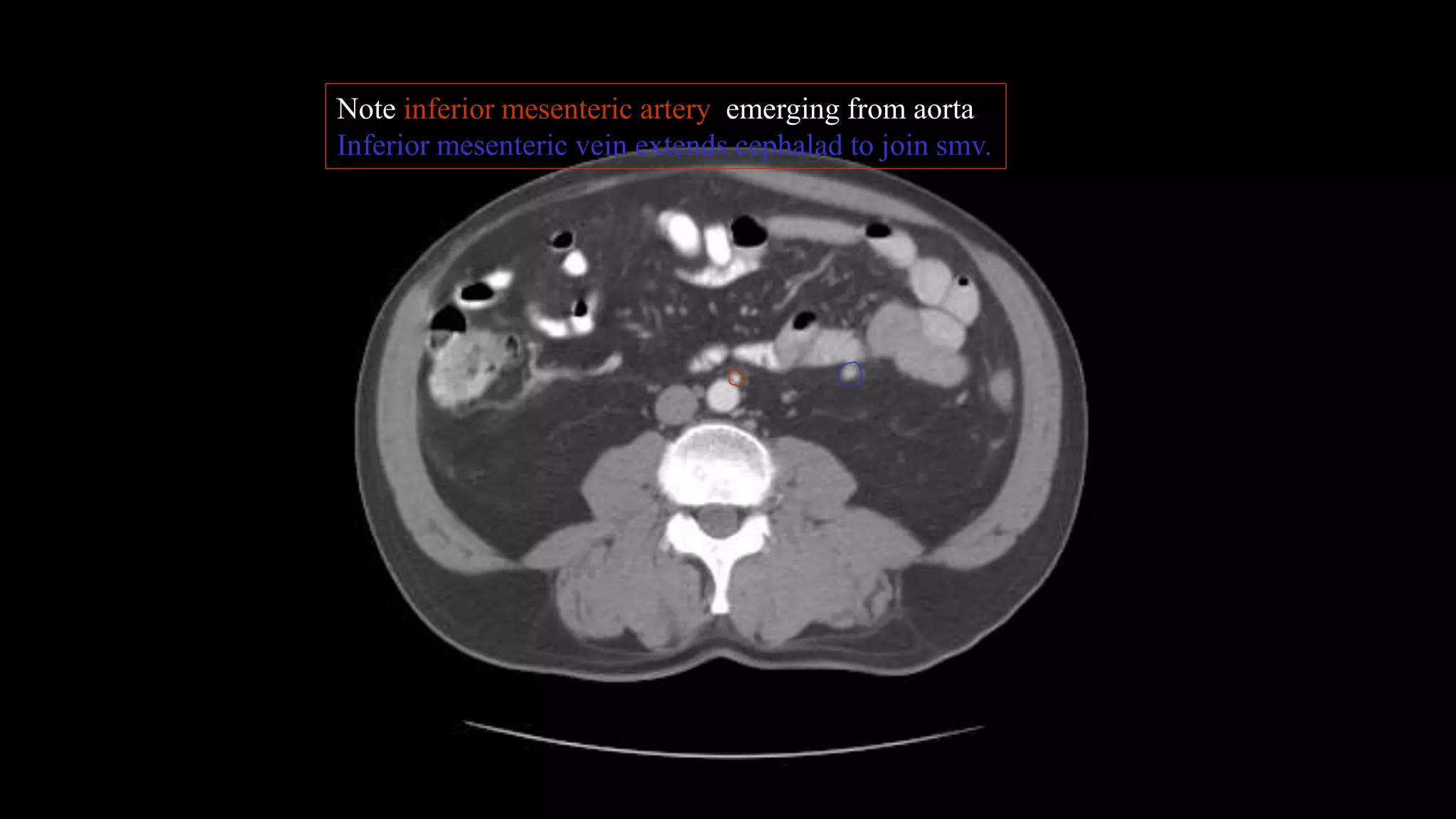
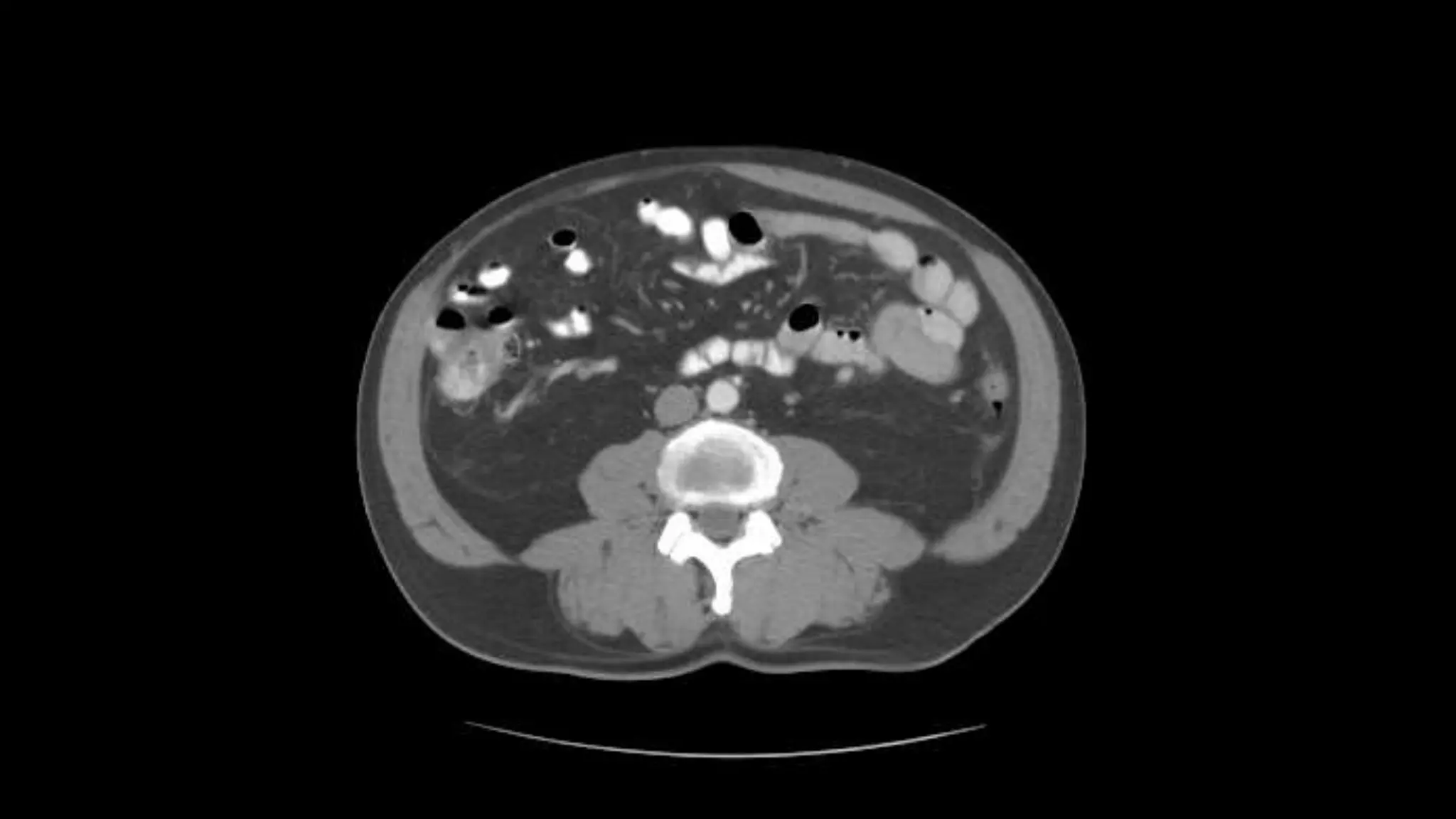
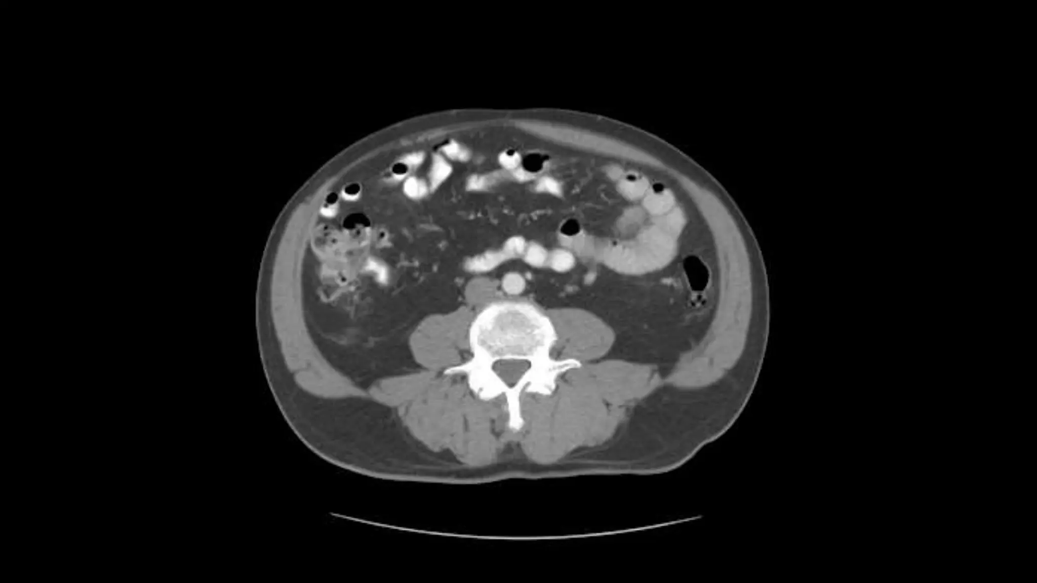
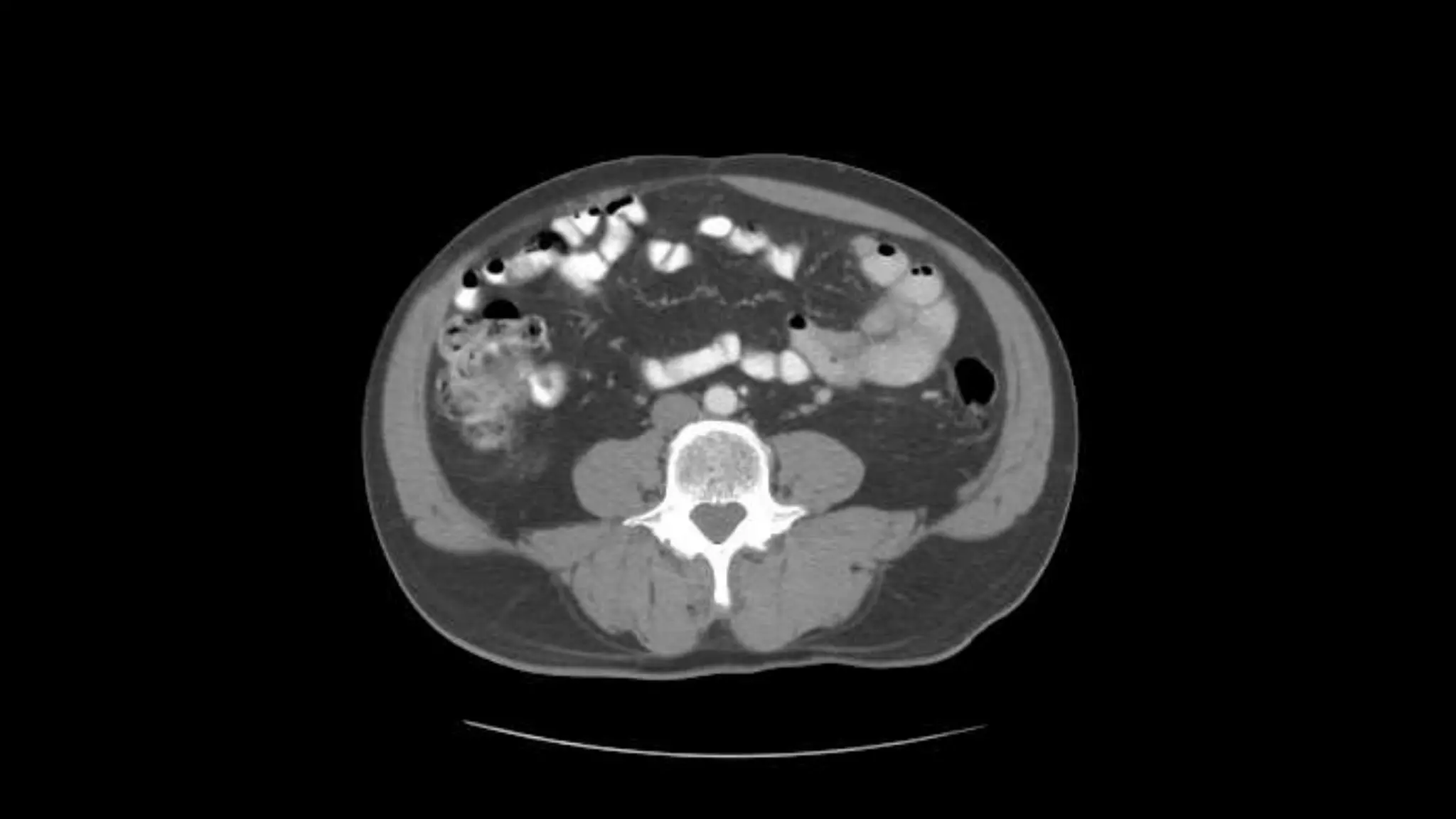
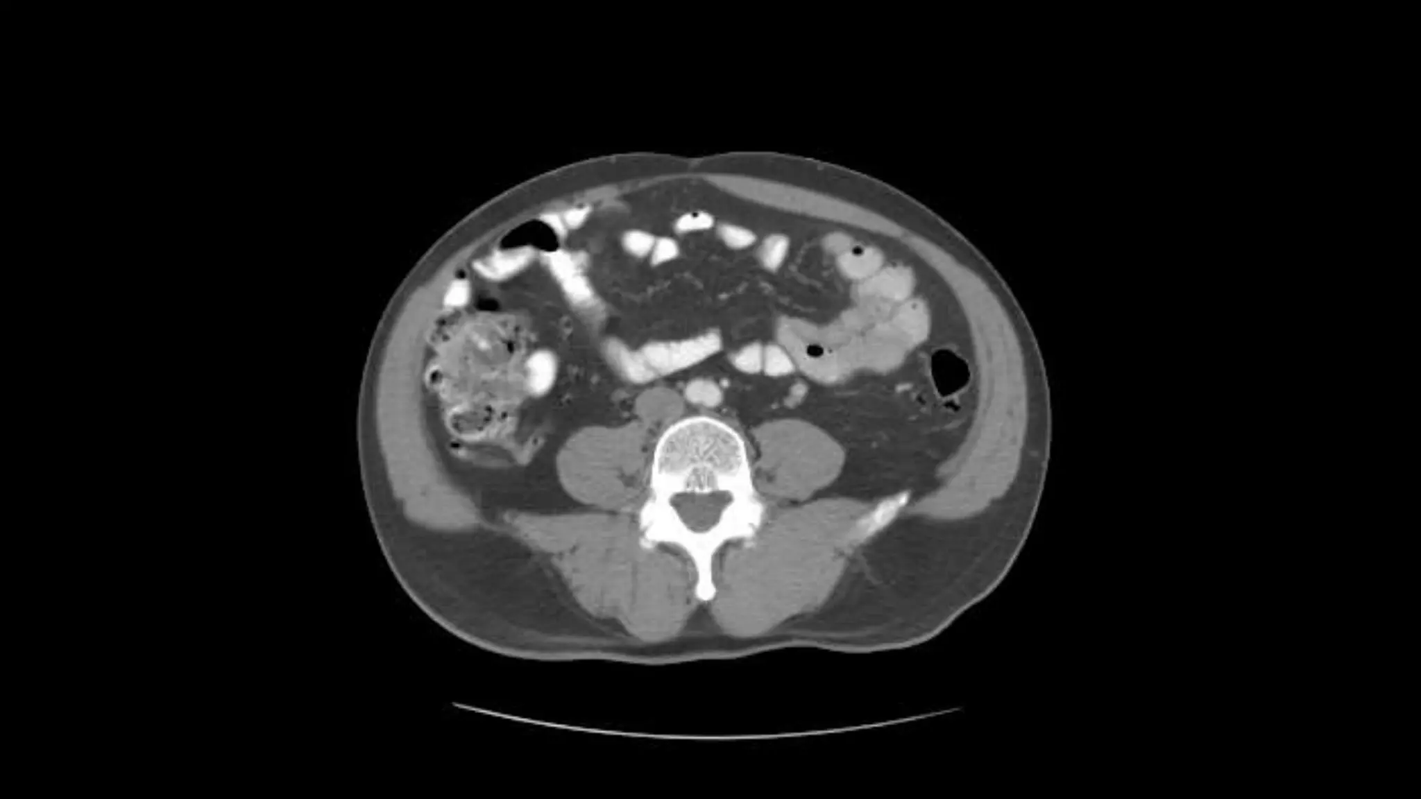
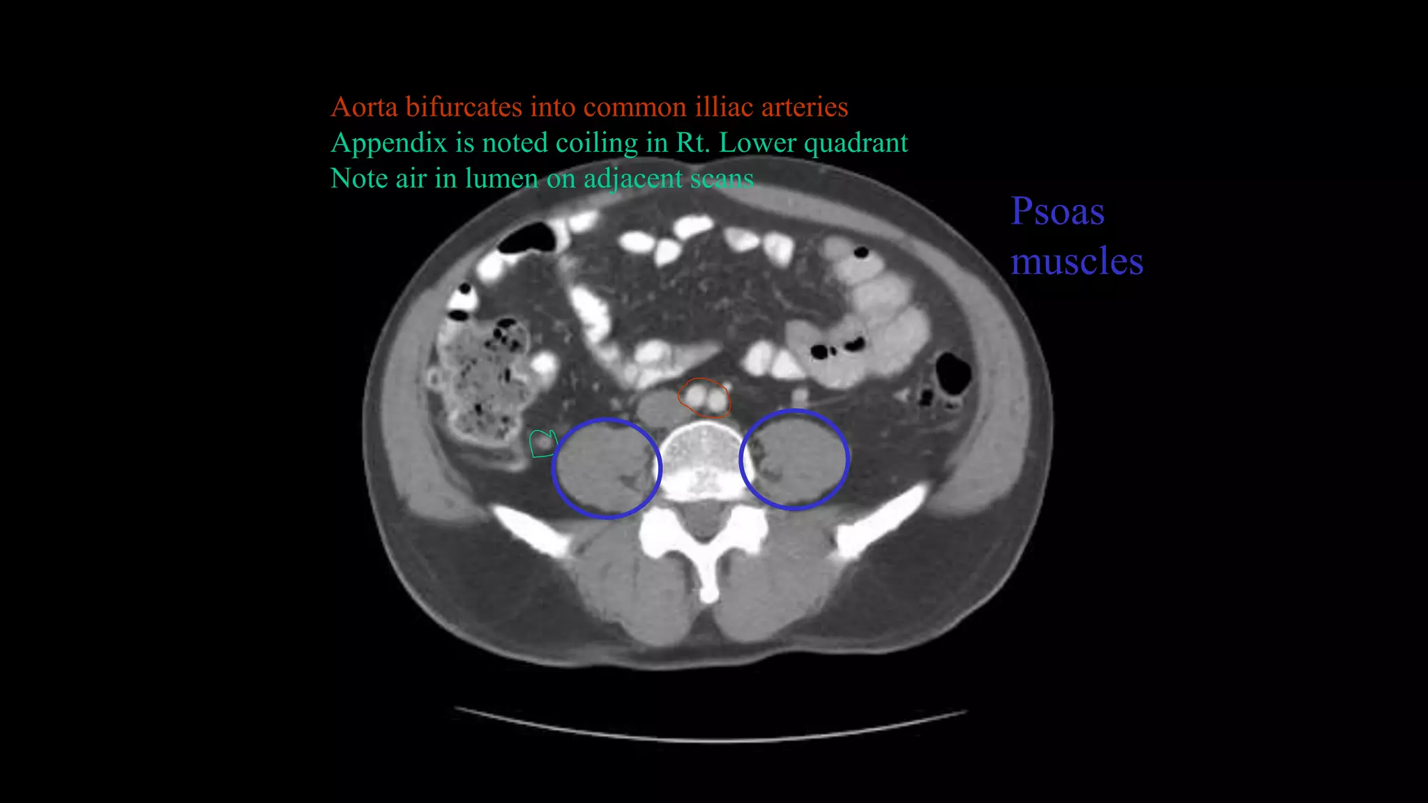
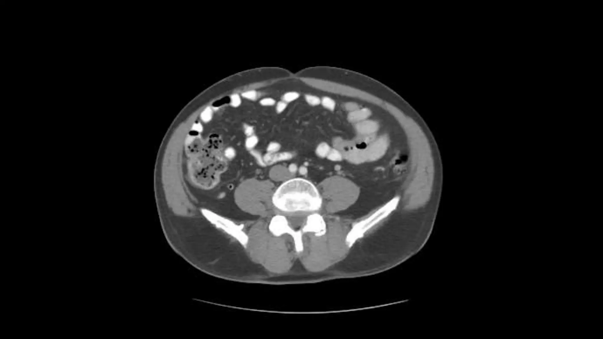
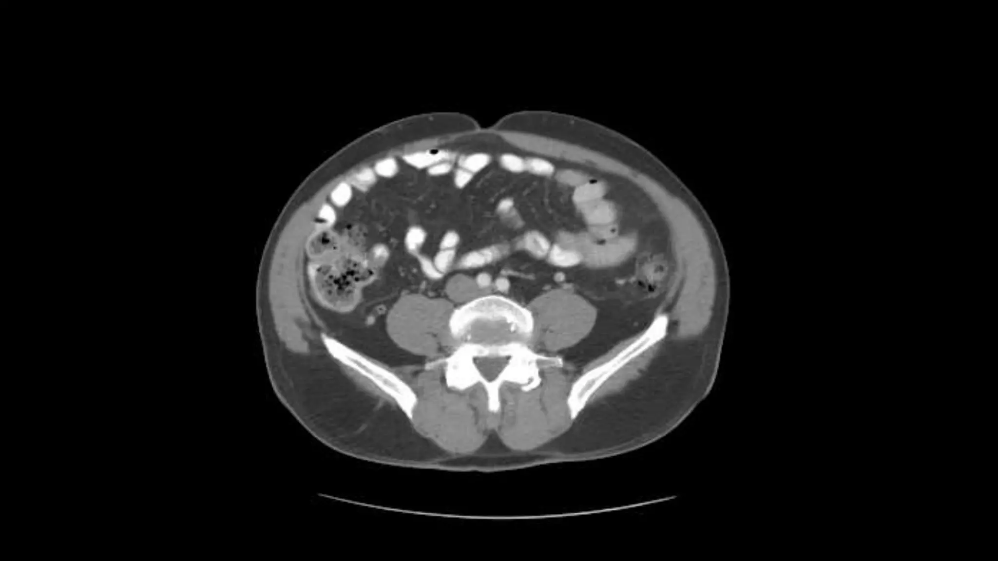
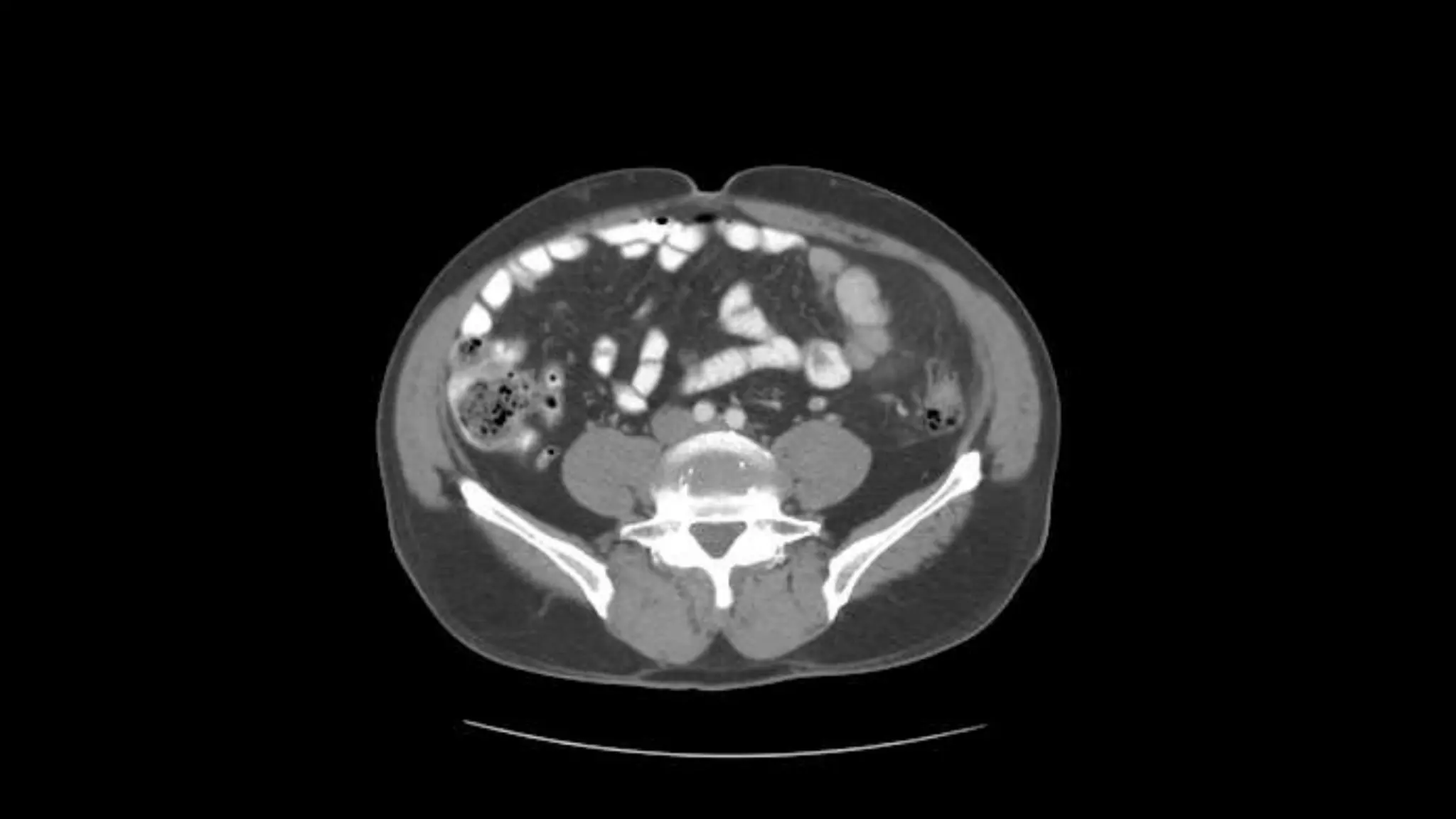
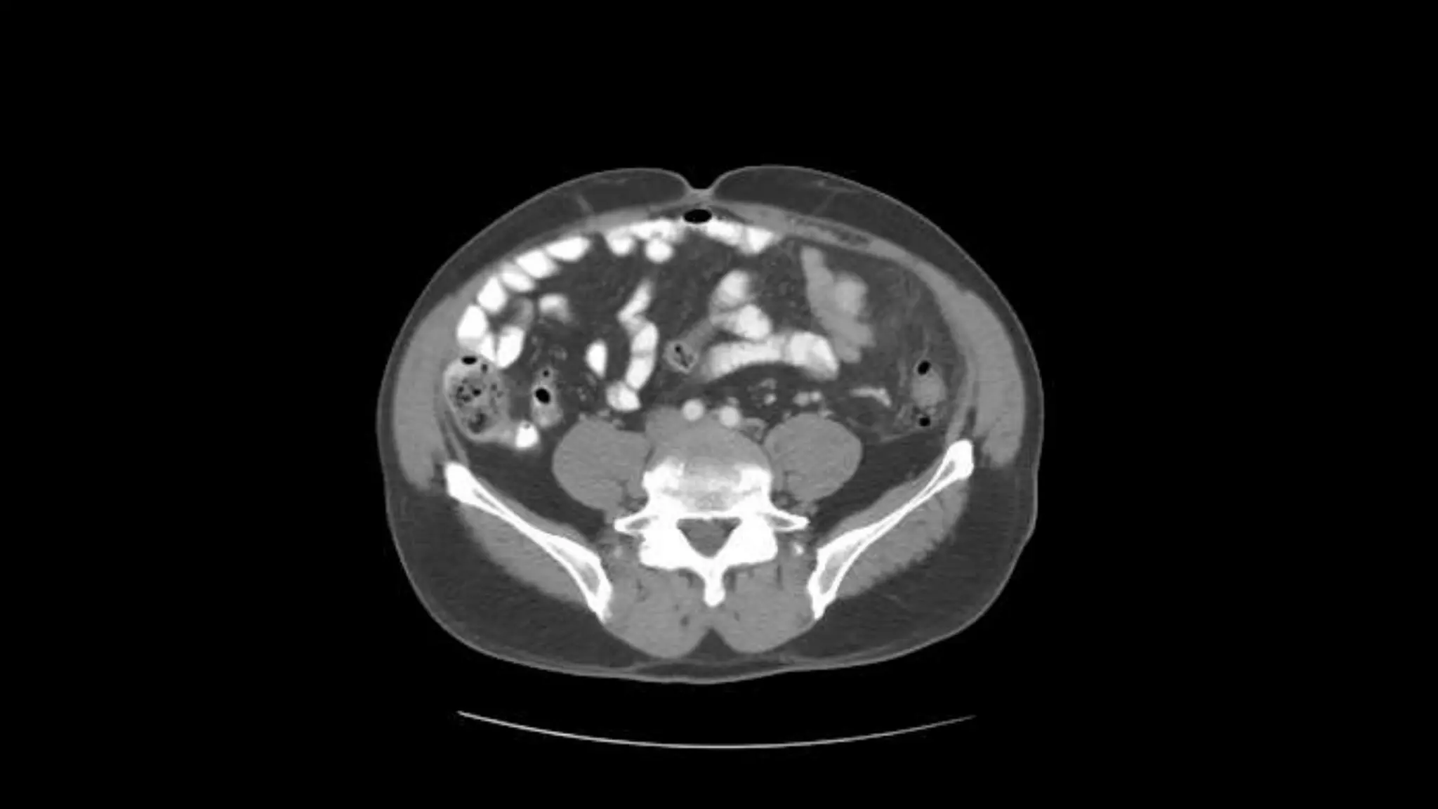
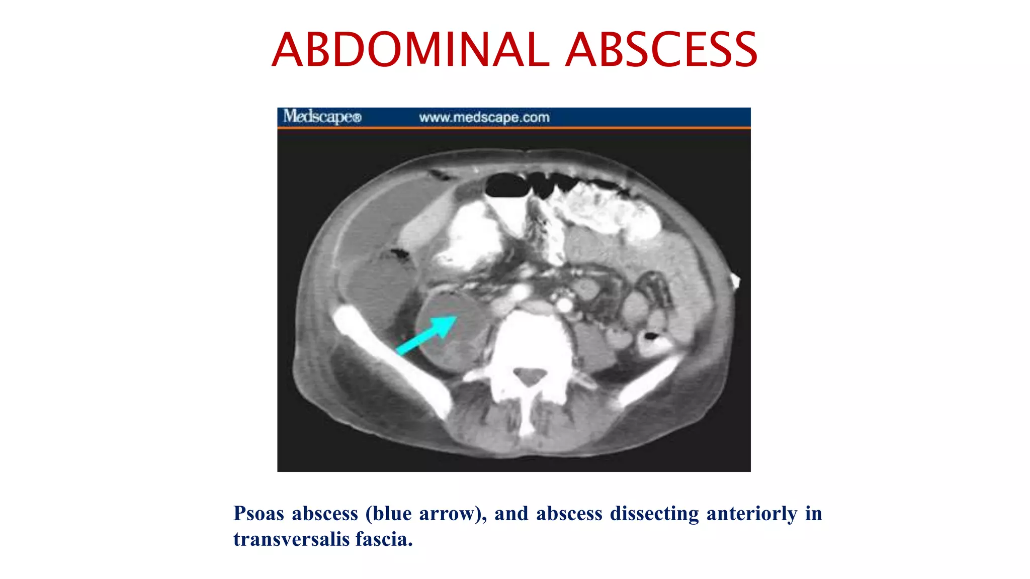
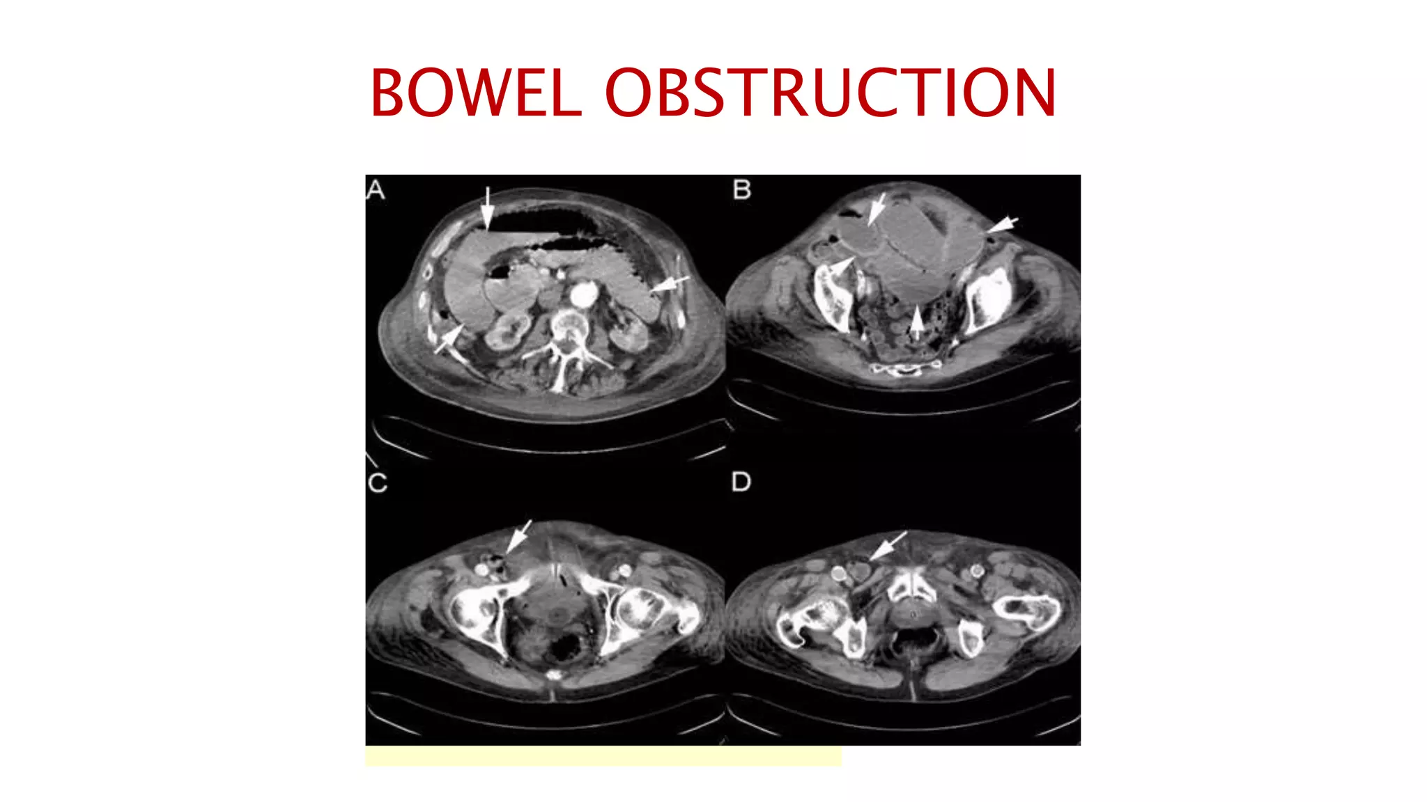
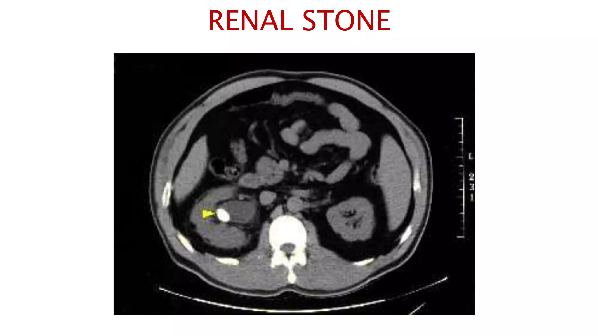
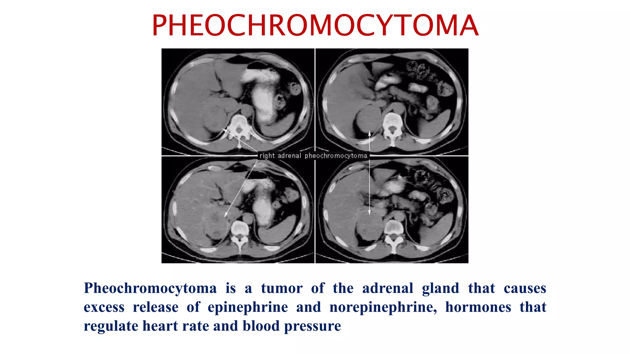
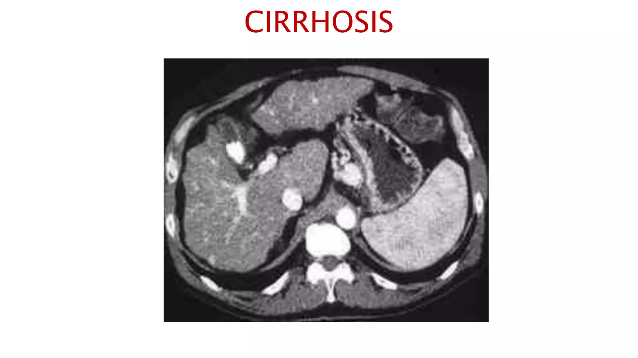
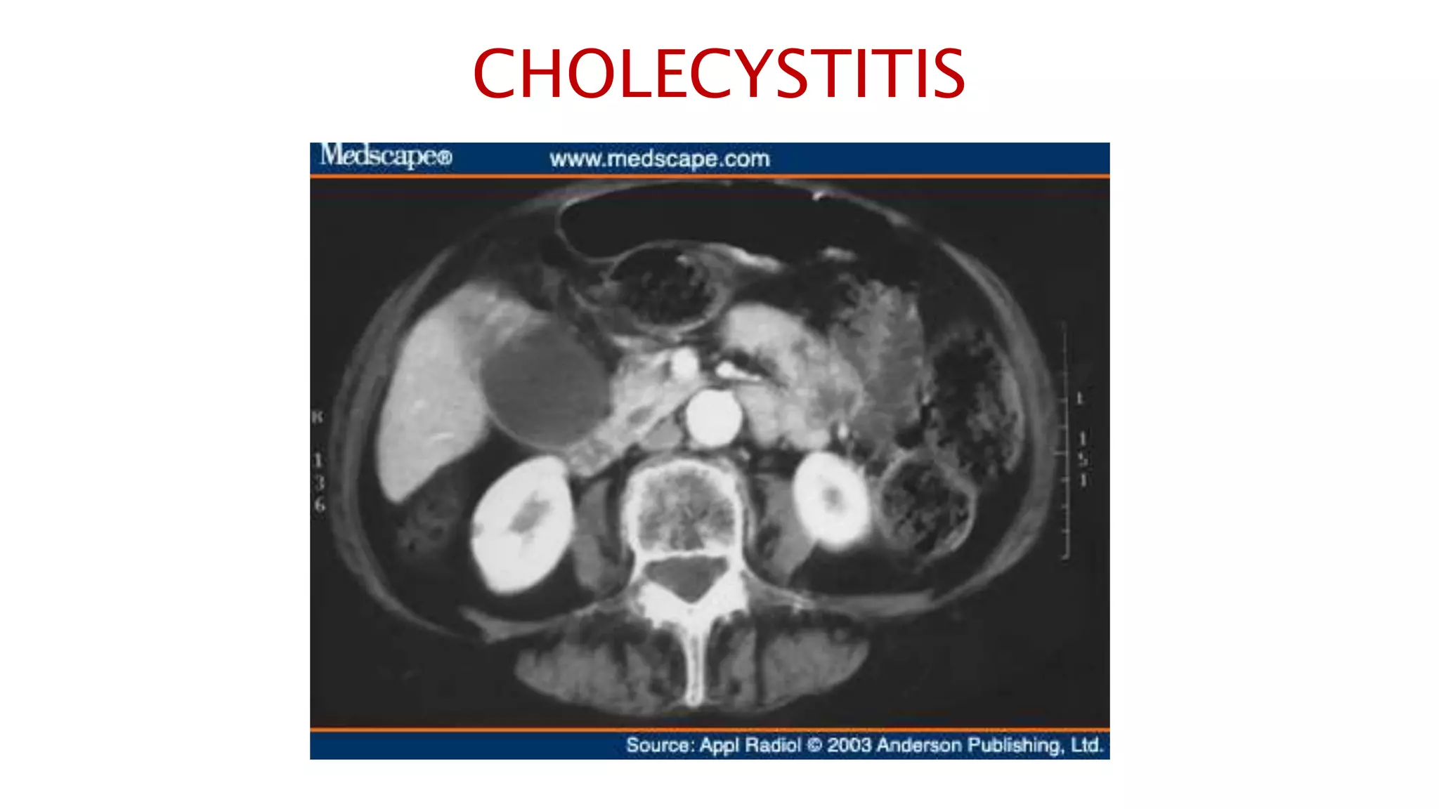
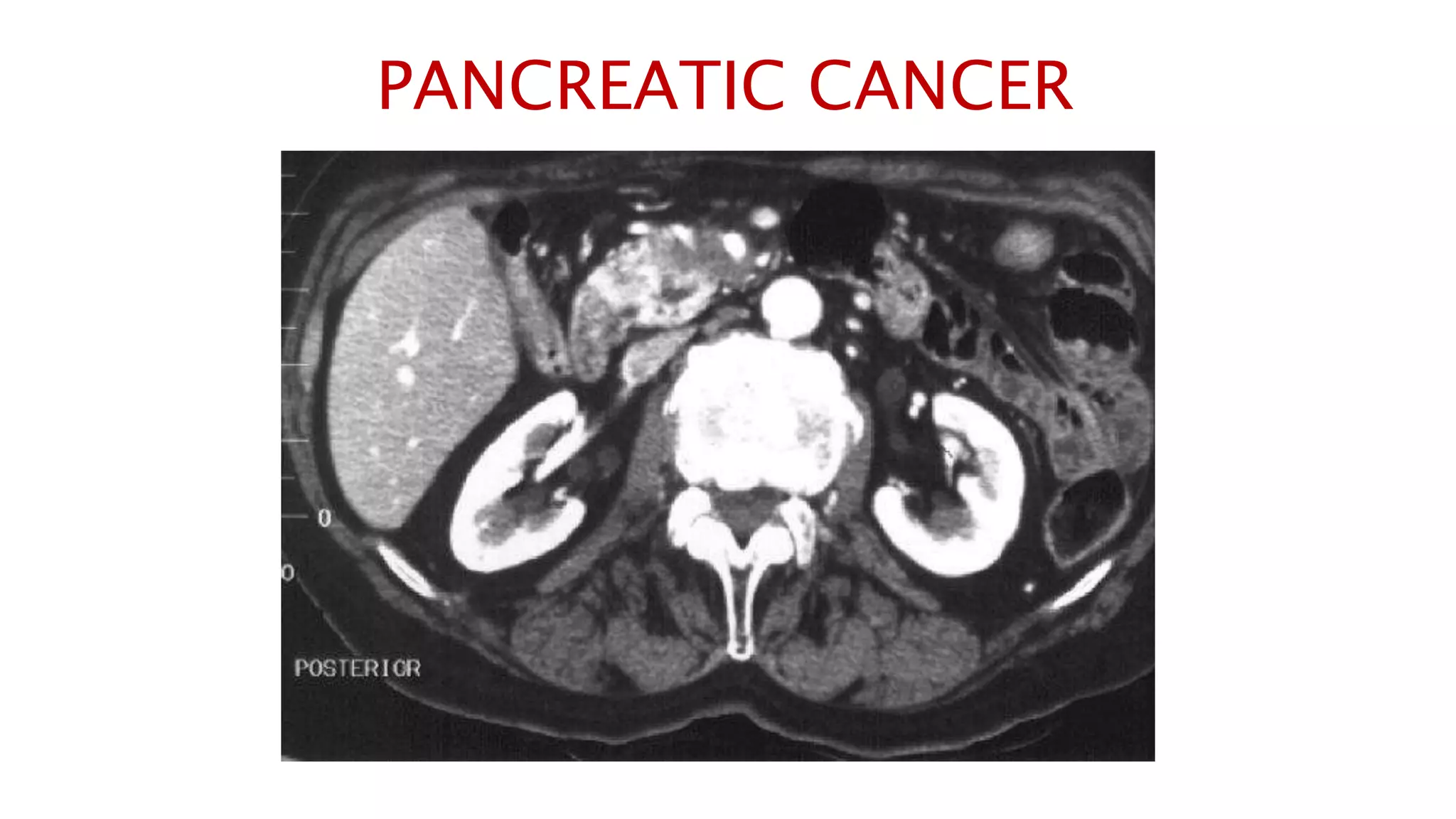
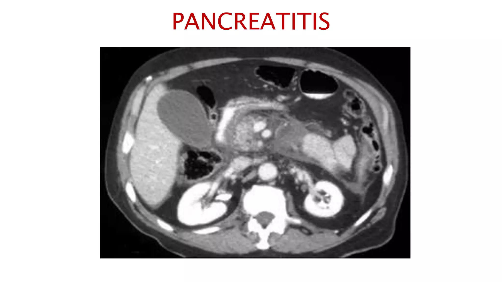
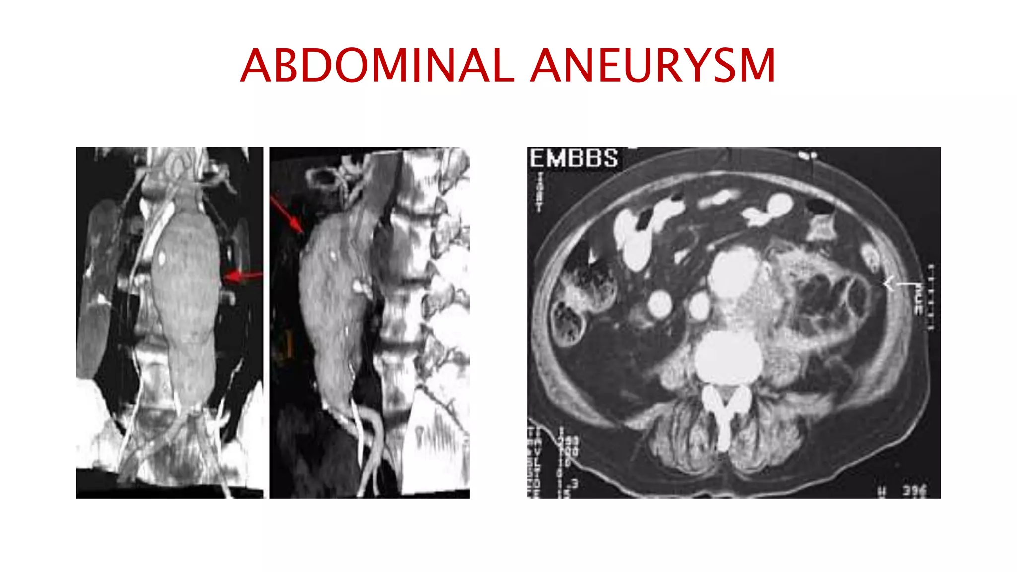
An abdominal CT scan uses x-rays to create detailed cross-sectional images of the abdomen. During the test, the patient lies still on a table that slides into a scanner, which rotates an x-ray beam around the body. Images are created as "slices" and can be combined to form 3D models. An abdominal CT scan is used to detect various abdominal abnormalities such as masses, tumors, infections, kidney stones, and issues affecting the liver, gallbladder, or pancreas. Abnormal results could indicate cancers, organ problems, appendicitis, aneurysms, or other issues requiring follow-up.
2. An abdominal CT scan is an imaging method that uses x-rays to create cross-
sectional pictures of the belly area. CT stands for computed tomography.
How the Test is Performed
You will lie on a narrow table that slides into the center of the CT scanner. Most
often, you will lie on your back with your arms raised above your head.
Once you are inside the scanner, the machine's x-ray beam rotates around you.
Modern spiral scanners can perform the exam without stopping.
A computer creates separate images of the belly area. These are called slices. These
images can be stored, viewed on a monitor, or printed on film. Three-dimensional
models of the belly area can be made by stacking the slices together.
You must be still during the exam, because movement causes blurred images. You
may be told to hold your breath for short periods of time.
3. Why the Test is Performed
An abdominal CT scan makes detailed pictures of the structures inside your belly
(abdomen) very quickly.
This test may be used to look for:
Cause of abdominal pain or swelling
Hernia
Cause of a fever
Masses and tumors, including cancer
Infections or injury
Kidney stones
Appendicitis
4. What Abnormal Results Mean
The abdominal CT scan may show some cancers, including:
Cancer of the renal pelvis or ureter
Colon cancer
Hepatocellular carcinoma
Lymphoma
Melanoma
Ovarian cancer
Pancreatic cancer
Pheochromocytoma
Renal cell carcinoma (kidney cancer)
Testicular cancer
5. The abdominal CT scan may show problems with the gallbladder, liver, or
pancreas, including:
Acute cholecystitis
Alcoholic liver disease
Cholelithiasis
Pancreatic abscess
Pancreatic pseudocyst
Pancreatitis
Blockage of bile ducts
6. The abdominal CT scan may reveal the following kidney problems:
Acute bilateral obstructive uropathy
Acute unilateral obstructive uropathy
Chronic bilateral obstructive uropathy
Chronic unilateral obstructive uropathy
Complicated UTI (pyelonephritis)
Kidney stones
Kidney or ureter damage
Polycystic kidney disease
7. Abnormal results may also be due to:
Abdominal aortic aneurysm
Abscesses
Appendicitis
Bowel wall thickening
Retroperitoneal fibrosis
Renal artery stenosis
Renal vein thrombosis
8. Studying the CT image
In this sequence of images, we will label the abdominal vasculature.
The CT images are 5mm slices with soft tissue window settings. IV
and oral contrast have been administered which causes the vessels
and GI tract to appear hyperdense (white). Some images will contain
labels to assist with tracking the vessels.
IMAGES ARE VIEWED AS LOOKING FROM THE FEET
RIGHT LEFT
9.
10. Follow the IV contrast filled Aorta as we descend caudally. Branches and points
of interest will be noted.
15. This is an excellent image of the right, middle and left hepatic veins draining into the Inferior
Vena Cava. Don’t confuse this structure with the IVC, this is the esophagus at the level of the
Lower esophageal sphincter, page up and down to confirm this.
16. The outline of the Inferior Vena Cava is more
distinct in this image.
26. You can see the Celiac artery starting to branch from the
Aorta. You can follow this down in the next four images
Proper Hepatic Artery is labeled in the upper right The splenic
vein and artery are in the lower left
27. Proper Hepatic Artery and Splenic Artery
(the splenic artery is the circle).
Splenic Vein
28. Here the Splenic Vein is emptying into the portal vein.
Follow this up and down.
Pancreas
29.
30. This is the Superior Mesenteric Artery branching
off the Aorta.
32. Renal Veins emptying into the IVC. We also see the right renal artery branching
off the Aorta, follow it down till you see it enter the right kidney. The Superior
Mesenteric Vein is outlined on the top of this image. If you follow the SMV up, you
will see it empty into the Portal Vein.
33. Here we see the right and left renal vein entering into the Inferior Vena Cava. We
Also see the left renal artery branching off the aorta and heading toward the left
kidney. Page up and down to trace these vessels.
49. Note inferior mesenteric artery emerging from aorta
Inferior mesenteric vein extends cephalad to join smv.
50.
51.
52.
53.
54.
55. Aorta bifurcates into common illiac arteries
Appendix is noted coiling in Rt. Lower quadrant
Note air in lumen on adjacent scans
Psoas
muscles
56.
57.
58.
59.
60. ABDOMINAL CYST
An abdominal CT scan revealed a large right upper quadrant cyst measuring
14x17x21 cm ( lateral, anteroposterior and craniocaudal)There was mass effect
upon the liver and duodenum. The cyst had a thin smooth wall with internal fluid
and high density material consistent with a blood clot.
68. PHEOCHROMOCYTOMA
Pheochromocytoma is a tumor of the adrenal gland that causes
excess release of epinephrine and norepinephrine, hormones that
regulate heart rate and blood pressure