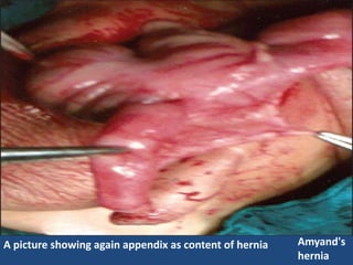
A picture showingagain appendix as content of hernia Amyand's
hernia
7.
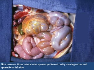
Situs Inversus: Grossnatural color opened peritoneal cavity showing cecum and
appendix on left side
8.
Acute appendicitis withyellow to tan exudate and hyperemia, including the
periappendiceal fat superiorly, rather than a smooth, glistening pale tan serosal
surface.
10.
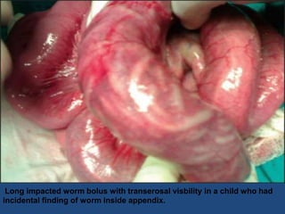
Long impacted wormbolus with transerosal visbility in a child who had
incidental finding of worm inside appendix.
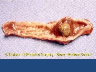
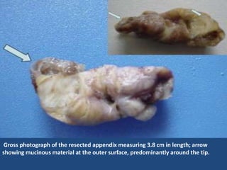
Gross photograph ofthe resected appendix measuring 3.8 cm in length; arrow
showing mucinous material at the outer surface, predominantly around the tip.
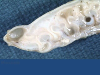
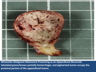
Metastatic Malignant MelanomaPresenting as an Appendiceal Mucocele,
lobulated green/brown partially hemorrhagic, and pigmented tumor occupy the
proximal portion of the appendiceal lumen.
21.
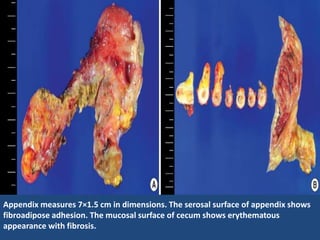
Figure 3
Gross findingof appendix. Appendix measures 7 1.5 cm in dimensions. The serosal surface of appendix shows fibroadipose
adhesion. The mucosal surface of cecum shows erythematous appearance with fibrosis.
Appendix measures 7×1.5 cm in dimensions. The serosal surface of appendix shows
fibroadipose adhesion. The mucosal surface of cecum shows erythematous
appearance with fibrosis.
22.
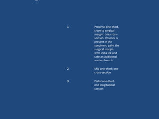
1 Proximal one-third,
close to surgical
margin: one cross-
section. If tumor is
present in the
specimen, paint the
surgical margin
with India ink and
take an additional
section from it
2 Mid one-third: one
cross-section
3 Distal one-third:
one longitudinal
section