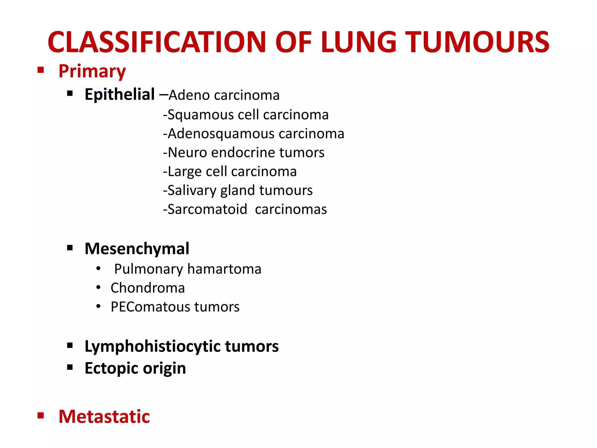
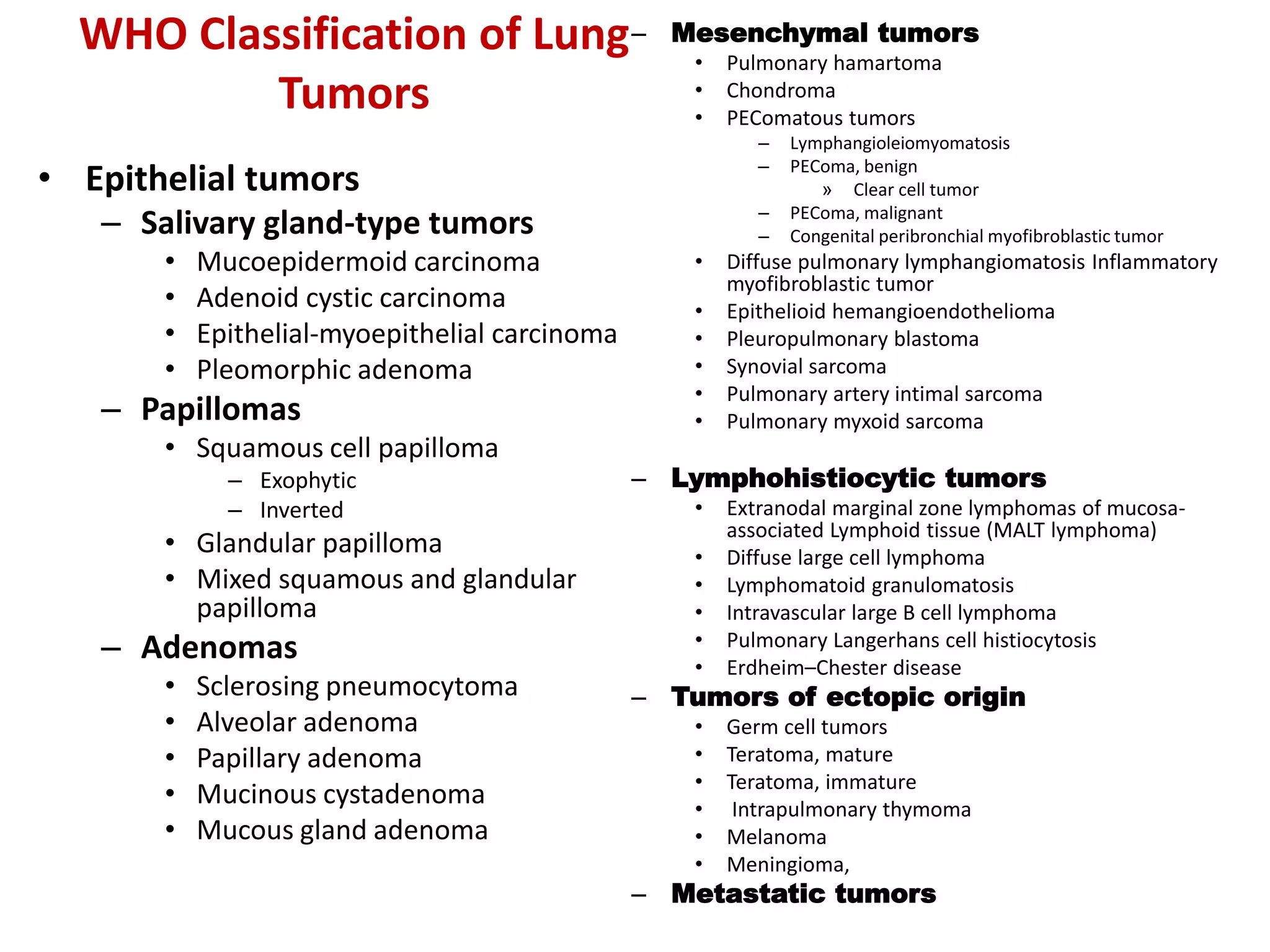
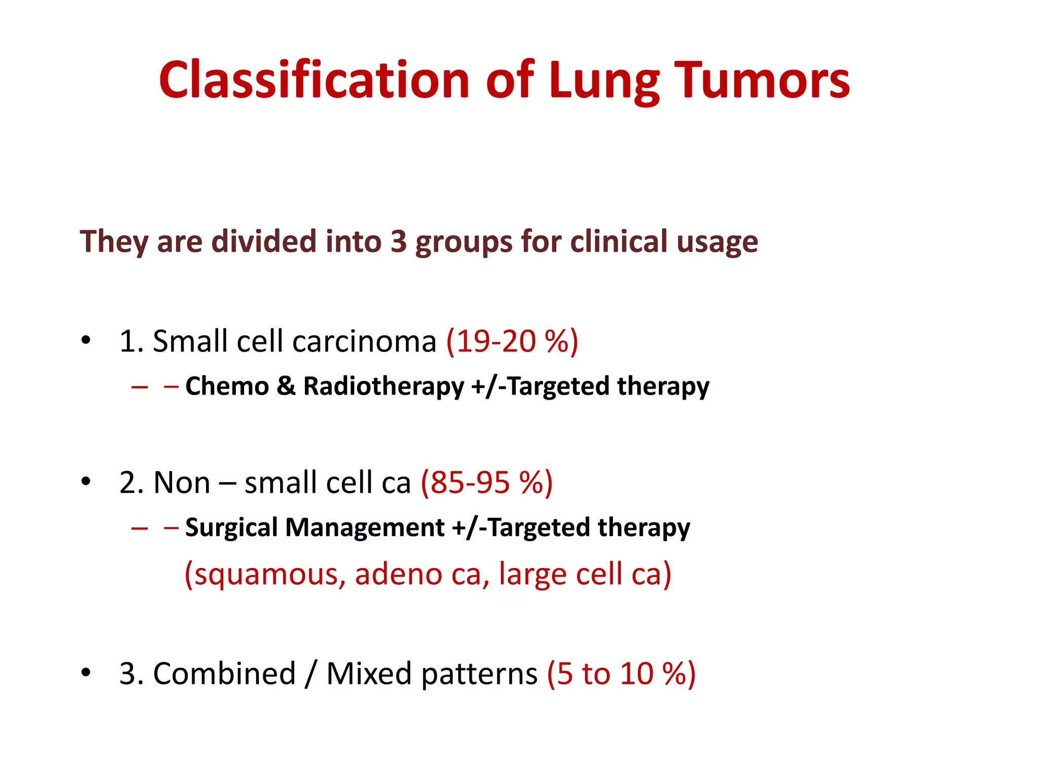
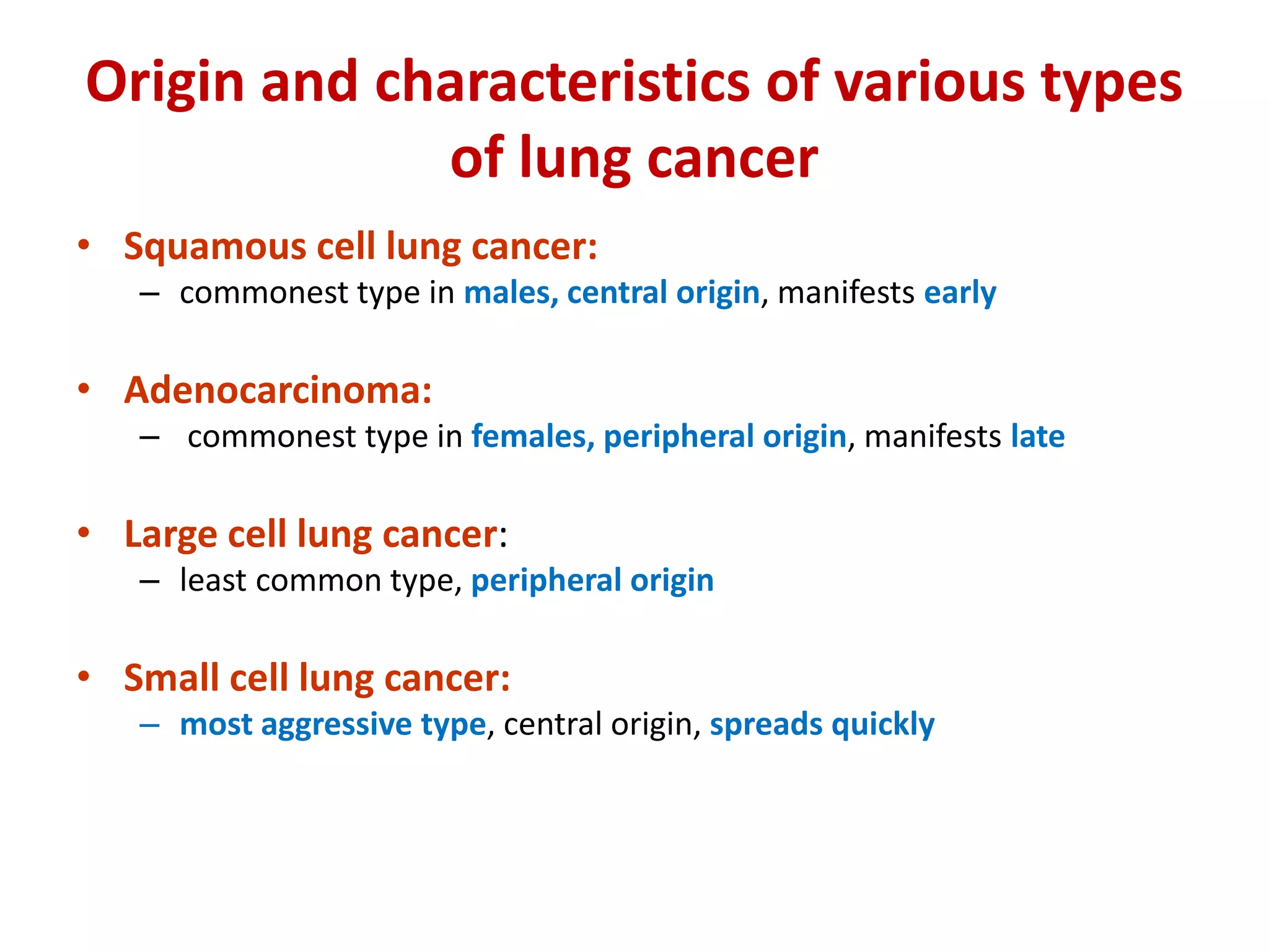
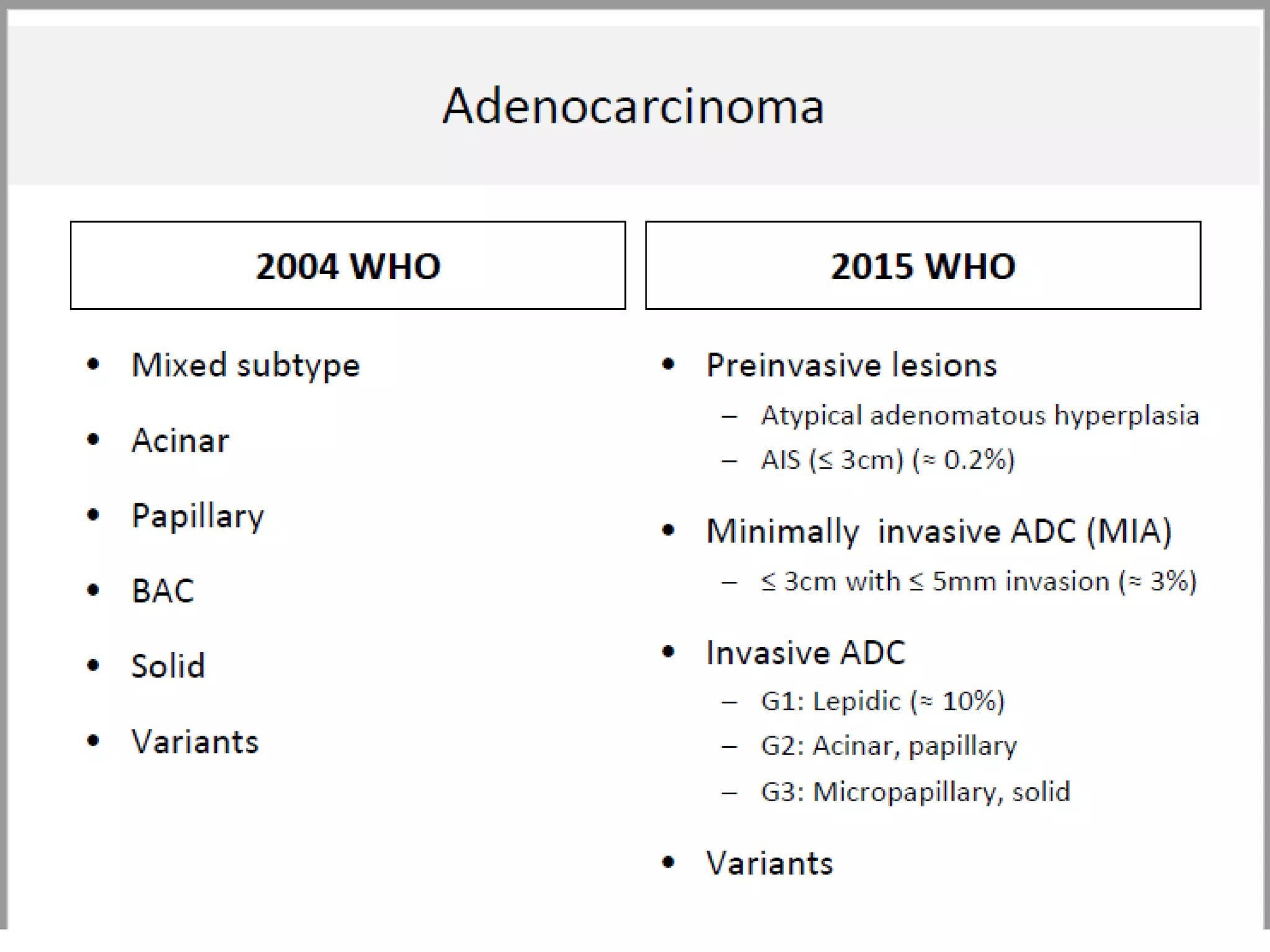
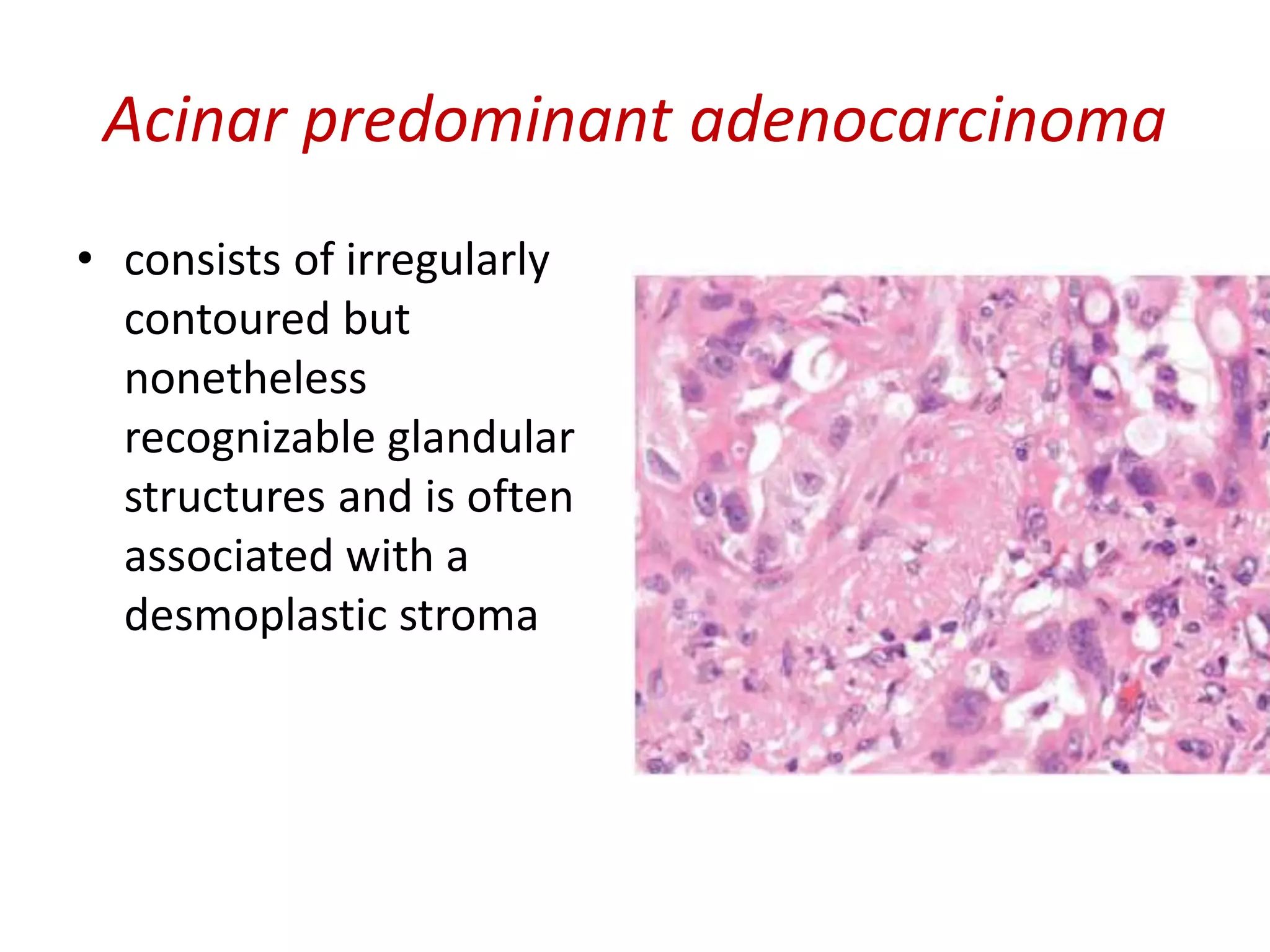
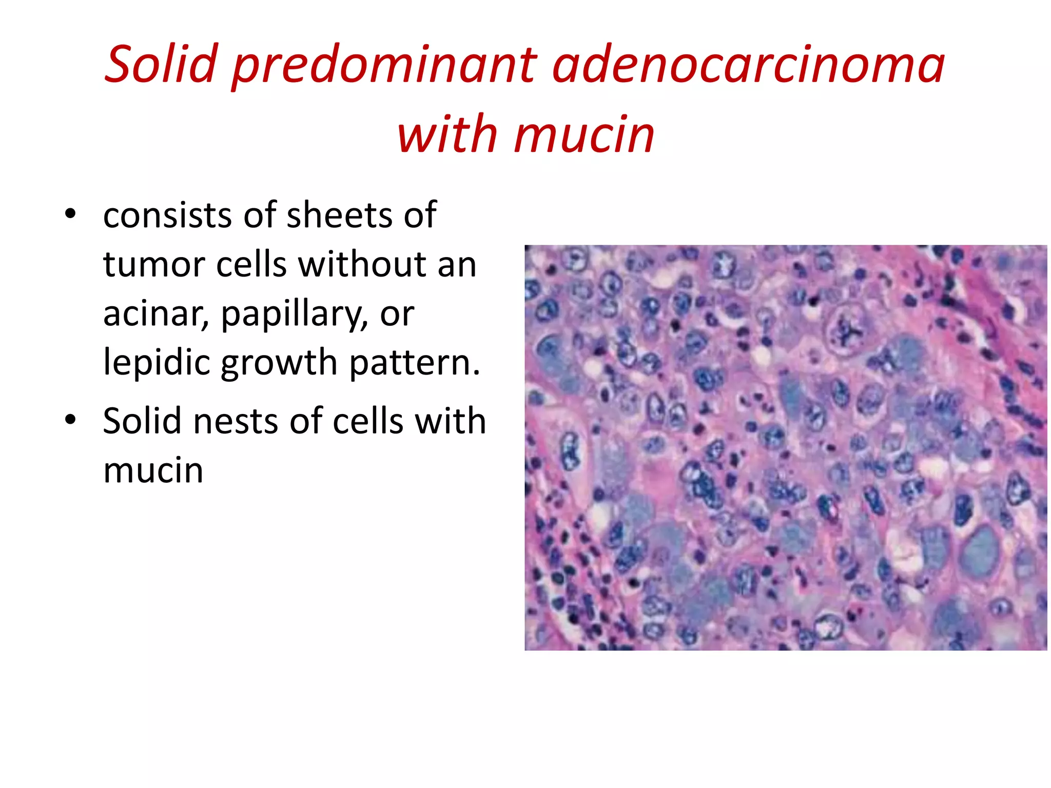
Classification of Lung tumors
Dr Kamal Bharathi. S
Department of Pulmonary Medicine
Sri Manakula Vinayagar Medical college and Hospital
CLASSIFICATION OF LUNG TUMOURS
 Primary
 Epithelial –Adeno carcinoma
-Squamous cell carcinoma
-Adenosquamous carcinoma
-Neuro endocrine tumors
-Large cell carcinoma
-Salivary gland tumours
-Sarcomatoid carcinomas
 Mesenchymal
• Pulmonary hamartoma
• Chondroma
• PEComatous tumors
 Lymphohistiocytic tumors
 Ectopic origin
 Metastatic
WHO Classification of Lung
Tumors
• Epithelial tumors
– Adenocarcinoma
– Lepidic adenocarcinomae
– Acinar adenocarcinoma
– Papillary adenocarcinoma
– Micropapillary adenocarcinoma
– Solid adenocarcinoma
– Invasive mucinous adenocarcinoma
• Mixed invasive mucinous
• nonmucinous adenocarcinoma
– Colloid adenocarcinoma
– Fetal adenocarcinoma
– Enteric adenocarcinomae
– Minimally invasive adenocarcinoma
• Nonmucinous
• Mucinous
– Preinvasive lesions
• Atypical adenomatous hyperplasia
• Adenocarcinoma in situe
– Nonmucinous
– Mucinous
– Squamous cell carcinoma
• Keratinizing squamous cell carcinoma
• Nonkeratinizing squamous cell carcinoma
• Basaloid squamous cell carcinoma
• Preinvasive lesion
– Squamous cell carcinoma in situ
– Neuroendocrine tumors
• Small cell carcinoma
– Combined small cell carcinoma
• Large cell neuroendocrine carcinoma
– Combined large cell neuroendocrine
carcinoma
• Carcinoid tumors
– Typical carcinoid tumor
– Atypical carcinoid tumor
• Preinvasive lesion
– Diffuse idiopathic pulmonary
neuroendocrine cell hyperplasia
-Large cell carcinoma
-Adenosquamous carcinoma
-Sarcomatoid carcinomas
– Pleomorphic carcinoma
– Spindle cell carcinoma
– Giant cell carcinoma
– Carcinosarcoma
– Pulmonary blastoma
WHO Classification of Lung
Tumors
• Epithelial tumors
– Salivary gland-type tumors
• Mucoepidermoid carcinoma
• Adenoid cystic carcinoma
• Epithelial-myoepithelial carcinoma
• Pleomorphic adenoma
– Papillomas
• Squamous cell papilloma
– Exophytic
– Inverted
• Glandular papilloma
• Mixed squamous and glandular
papilloma
– Adenomas
• Sclerosing pneumocytoma
• Alveolar adenoma
• Papillary adenoma
• Mucinous cystadenoma
• Mucous gland adenoma
– Mesenchymal tumors
• Pulmonary hamartoma
• Chondroma
• PEComatous tumors
– Lymphangioleiomyomatosis
– PEComa, benign
» Clear cell tumor
– PEComa, malignant
– Congenital peribronchial myofibroblastic tumor
• Diffuse pulmonary lymphangiomatosis Inflammatory
myofibroblastic tumor
• Epithelioid hemangioendothelioma
• Pleuropulmonary blastoma
• Synovial sarcoma
• Pulmonary artery intimal sarcoma
• Pulmonary myxoid sarcoma
– Lymphohistiocytic tumors
• Extranodal marginal zone lymphomas of mucosa-
associated Lymphoid tissue (MALT lymphoma)
• Diffuse large cell lymphoma
• Lymphomatoid granulomatosis
• Intravascular large B cell lymphoma
• Pulmonary Langerhans cell histiocytosis
• Erdheim–Chester disease
– Tumors of ectopic origin
• Germ cell tumors
• Teratoma, mature
• Teratoma, immature
• Intrapulmonary thymoma
• Melanoma
• Meningioma,
– Metastatic tumors
Classification of Lung Tumors
They are divided into 3 groups for clinical usage
• 1. Small cell carcinoma (19-20 %)
– – Chemo & Radiotherapy +/-Targeted therapy
• 2. Non – small cell ca (85-95 %)
– – Surgical Management +/-Targeted therapy
(squamous, adeno ca, large cell ca)
• 3. Combined / Mixed patterns (5 to 10 %)
Origin and characteristics of various types
of lung cancer
• Squamous cell lung cancer:
– commonest type in males, central origin, manifests early
• Adenocarcinoma:
– commonest type in females, peripheral origin, manifests late
• Large cell lung cancer:
– least common type, peripheral origin
• Small cell lung cancer:
– most aggressive type, central origin, spreads quickly
Adenocarcinoma
• Most common lung cancer in nonsmokers and females
• Activating mutation of K-Ras
• Associated with hypertrophic osteoarthropathy “clubbing”
• Located peripherally with central scar
• Early metastasis
• Histological examination:
• Acinar (gland forming)
• Papillary
• Mucinous
• Solid
• Atypical adenomatous hyperplasia (AAH) >
Adenocarcinoma in situ (AIS) > minimally invasive
adenocarcinoma > invasive adenocarcinoma
• AAH: well demarcated focus of epithelial proliftation 5 mm
or less composed of cuboidal to low-columnar cells with
cytological atypia.
• AIS: (bronchoalveolar carcinoma) involve peripheral parts
of the lung. Diameter of 3 cm or less, growth along
preexisting structures & preservation of alveolar
architecture.
• Can be nonmucinous, mucinous or mixed.
Adenocarcinoma precursor
(AAH)
Atypical adenomatous
hyperplasia
• Focal, 5.0 mm or less
with defined borders
• Alveoli lined by cuboidal
to low columnar cells
with variable atypia
• Alveolar walls may be
slightly thickened
• Non-mucinous
Adenocarcinoma In Situ
• The 2011 classification completely discards
the term BAC in favor of “adenocarcinoma in
situ” (AIS).
• small (≤3 cm) solitary AC with predominant
lepidic growth and small foci of invasion
measuring ≤0.5 cm, the term “minimally
invasive adenocarcinoma” is recommended.
• typically present as pure GGO without a solid
component on CT scan
AAH vs AIS
Atypical Adenomatous Hyperplasia Adenocarcinoma in situ
(Mucinous)
a less common type of
adenocarcinoma -
bronchioloalveolar carcinoma
ADENOCARCINOMA IN SITU
(bronchioloalveolar carcinoma) - well-
differentiated columnar cells proliferate
along the framework of alveolar septae
- better prognosis than other primary
lung cancers
Invasive Adenocarcinomas
• Lepidic predominant,
• Acinar predominant,
• Papillary predominant,
• Micropapillary predominant,
• Solid predominant with mucin production.
Lepidic predominant adenocarcinoma
• consists mainly of tumor cells growing along
alveolar septa (previously referred to as a
nonmucinous BAC pattern) but with greater
than 5 mm invasion.
Acinar predominant adenocarcinoma
• consists of irregularly
contoured but
nonetheless
recognizable glandular
structures and is often
associated with a
desmoplastic stroma
Papillary predominant
adenocarcinoma
• consists of malignant
cuboidal or columnar
cells that line the
surface of fibrovascular
cores
Micropapillary
predominant adenocarcinoma
• consists of small
papillary clusters of
glandular cells growing
within airspaces
Solid predominant adenocarcinoma
with mucin
• consists of sheets of
tumor cells without an
acinar, papillary, or
lepidic growth pattern.
• Solid nests of cells with
mucin
Variants of invasive adenocarcinoma
• Invasive mucinous adenocarcinoma (formerly
mucinous BAC)
• Colloid adenocarcinoma
• Fetal adenocarcinoma (low & high grade)
• Enteric adenocarcinoma
CLEAR CELL
ADENOCARCINOMA
Mucinous (“colloid”)
carcinoma
IHC
• Immunohistochemistry
Positive for
– TTF - 1
– Napsin - A
• Differential Diagnosis
• Metastatic Adenocarcinoma
• 65 years old female
presented with
breathlessness 1 week
duration
• Right mass lesion in the
periphery of lung
• Bronchial wash & Lung
Biopsy done Posteroanterior radiograph of chest showing
a non-cavitated opaque density in the left
upper lung field
Bronchial
Wash
group of large pleomorphic
cells with high nuclear
cytoplasmic ratio and vague
glandular formations
TTF -1 NUCLEAR
POSITIVITY Chromogranin was negative
Diagnosis
• Adenocarcinoma
Squamous cell carcinoma
• commonly in men, cigarette smoking.
• 2/3 occur centrally.
• Endobronchial growth pattern- associated
with bronchial obstruction and
postobstructive pneumonia.
• Cavitation is seen more frequently.
Definition
• Squamous cell carcinoma is defined as a
malignant epithelial tumor showing
keratinization and/or intercellular bridges.
squamous cell carcinoma
arising centrally, obstructing
the right main bronchus
sqamous cell carcinoma extends
from hilum to pleura
•Squamous cell carcinoma
in which a portion of the
tumor demonstrates
central cavitation
•Probably because the
tumor outgrew its blood
supply.
•Squamous cell
carcinomas are one of the
more common primary
malignancies of lung
Central type (hilar type) Diffuse type
Peripheral type
(nodular type)
Gross types
Histologically: Squamous Cell CA
Keratin pearls
Intercellular bridges
Histologic variants
• Papillary- exophytic growth pattern and
papillary cores
• Clear cell- clear cell cytoplasm
• Small cell- lack the nuclear characteristics of
small cell ca and may show focal
keratinization.
• Basaloid patterns- Very aggressive clinical
course , peripheral palisading of cells.
SCC WITH PAPILLARY GROWTH PATTERN
Small cell variant of squamous cell carcinoma
small cell variant of
squamous cell carcinoma
can only be distinguished
from true small cell
carcinomas by means
of immunohistochemical
markers.
Clear cell variant
squamous cell
carcinoma shows
cells with
cytoplasmic
clearing as well as
groups of cells
with features of
keratinization
Basaloid variant
• Preinvasive lesion
Squamous cell carcinoma
in situ
IHC
• Immunohistochemistry
– Positive for
P40
P63
– CK 5/6
• Differential Diagnosis
• Metastatic Squamous cell carcinoma
• 46 years male
• Known Smoker for
past 16 years
• Bronchial wash &
Lung Biopsy taken Posteroanterior radiograph of chest showing
large cavitated lesion in the left mid upper
lung field.
Bronchial wash
elongated cluster of large cells with
increased nuclear cytoplasmic ratio.
Necrotic debris in the background
string-like arrangement
of tumor cell showing very dense
nuclear chromatin.
Diagnosis
• Squamous cell Carcinoma
Neuroendocrine tumors
• Small cell carcinoma
• Large cell neuroendocrine carcinoma
• Carcinoid tumors
• Sarcomatoid carcinomas
• Salivary gland-type tumors
Small Cell Carcinoma
• Arises from “Kulchtisky cells”
• Centrally located masses , extension to lung
parenchyma
• Involvement of hilar & mediastinal nodes
• Amplicfication of myc oncogen is common
Usually respond to chemo
and radiotherapy
Clinical Features
Paraneoplastic syndrome due to abnormal production of:
• Hormones:
– ACTH (Cushing's syndrome)
– ADH (hyponatremia)
• Auto-antibodies:
– Encephalomyelitis
– Lambert-Eaton syndrome
– Subacute sensory neuropathy
• Main line of treatment: chemotherapy, radiation
– SCLC is rarely resected
– Small cell carcinoma is very aggressive tumor with early
distant metastasis
Small cell anaplastic (oat cell) carcinoma
small cell anaplastic (oat cell)
carcinoma –
small dark blue cells with minimal
cytoplasm Ill defined borders salt-pepper
nuclear chromatin pattern
Microscopy
Crushed Blue Cells “Crush artifact” is
common SCLC, especially in biopsies
rather than large specimens.
– “chromatin smearing or streaming”
Azzopardi phenomena:
• basophilic nuclear DNA from necrotic
tumor cells get deposited in the walls of
vessels and connective tissue
• +ve for feulgen reaction
• Also seen in lymphomas, seminomas
Differential diagnosis
• Other neuroendocrine tumors:
– Carcinoid tumors
– Large cell Neuroendocrine carcinoma
• 40 years old Female
• Complaints of breathlessness
during night
• Endoscopy, bronchial wash
and lung Biopsy taken
Hilar and perihilar masses in continuity with
mediastinal lymphadenopathy
Bronchial wash
cluster of small very dark cells showing scant
cytoplasm and hyperchromatic nuclei.
Chromogranin Positive
Diagnosis
• Small cell carcinoma
Large cell carcinoma
• “an undifferentiated malignant epithelial
tumor that lacks the cytologic features of
small-cell carcinoma and glandular or
squamous differentiation.”
• 58 years old male
presented with
Breathlessness,
hemoptysis for past 3
days duration
• Chest X-ray was taken
• Bronchial wash & Lung
Biopsy done Posteroanterior radiograph of chest showing
large nodular density with relatively well-
defined borders in the left upper lobe.
Bronchial
Wash
Dispersed tumor cell population
with predominance of larger
cells with very dense nuclear
chromatin and lack of
squamous or glandular features
Few lymphocytes can be seen in
the background
Biopsy
Diagnosis
• Large cell carcinoma
Carcinoid tumor
• Typical Carcinoid tumor- central & peripheral
variants.
• Mostly asymptomatic.
• Central carcinoids- present with recurrent
pneumonias or hemoptysis.
• association with (MEN)1 syndrome in 5% ppl.
• grossly appear as yellow
or fleshy, polypoid
masses.
• tumor can infiltrate
between cartilaginous
rings to extensively
involve the bronchial
submucosa.
• tumor cells are uniform
in appearance
• low nuclear:cytoplasmic
ratio
• characteristic
neuroendocrine tumor
chromatin that is finely
granular or classically
described as “salt and
pepper.”
• carcinoid tumors have a generally excellent
prognosis with reported 5-year survival rates
of 87% to 100%
Sarcomatoid carcinomas
- by definition poorly differentiated non–small-cell
carcinomas that have a histologic appearance that
suggests mesenchymal differentiation.
• Pleomorphic carcinoma
• Spindle cell carcinoma
• Giant cell carcinoma
• Carcinosarcoma
• Pulmonary blastoma
Spindle cell carcinoma
 Subtype of Sarcomatoid
carcinoma (WHO)
 < 1% of all primary lung
carcinomas
 Majority of cases are diagnosed
in adults over the age of 65 years
 Carcinoma composed exclusively
of spindle-shaped tumor cells
 Tumor cells often obliterate
vessels
Carcinosarcoma
 Rare, 0.1% of all lung cancers
 M > F, most with smoking history,
average age 60
 Sites - Large airways and peripheral
lung
 Biphasic tumor consisting of a non-
small cell carcinoma with
heterologous sarcomatoid
differentiation
 Rare tumor with poor prognosis
mixture of well differentiated adenocarcinoma
and heterologous elements of cartilage
Pulmonary Blastoma
 Rare tumor composed of mixture of immature
epithelial and mesenchymal tissue resembling
fetal lung tissue – Embryoma
 Usually adults (mean age 43 years)
 Microscopy
 Biphasic tumor in which epithelial and
mesenchymal components have a primitive,
"fetal-type" appearance
 Well formed tubular glands surrounded by
cellular stroma of "embryonal" appearance
 Morules with ground glass nuclei are
common
 Positive stains
 PAS (glycogen in epithelial cells)
 Molecular Genetics
 CTNNB1 mutation
Mesenchymal tumors
Pulmonary
Hamartoma
Pulmonary
Chondroma
Perivascular epithelioid cell tumor
(PEComa)
 Also called- Clear cell (sugar) tumor
 Extremely rare, benign pulmonary tumor
derived from perivascular epithelioid cells
 Generally older adults >40 years old
 No gender predilection
• Microscopy :
– Clear to eosinophilic, finely granular
cytoplasm containing abundant PAS+
glycogen
– Small, uniform, rounded nuclei with small
nucleoli, variably prominent sclerotic
vasculature
– “Spider cells” with nuclear condensation of
eosinophilic cytoplasm with extensions to
the cell membrane
• Positive stains
– HMB45, MART1/MelanA, SMA, desmin
Synovial sarcoma
• Rare tumor of lung
• Associated with chest pain,
hemoptysis, dyspnea, cough, fever
• Monophasic spindle cells or biphasic
with epithelial and spindle cell
component
• Death due to metastases to bone,
CNS, liver, or invasion of adjacent
organs
• Positive stains
– TLE1 highly specific
• Negative stains
– CD117
• Molecular cytogenetics:
– t(X;18)(p11.2;icq11.2)
– SYT-SSX1 or SYT-SSX2 fusion genes
Salivary gland-type tumorsAdenoid cystic Carcinoma Acinic Cell Carcinoma
Epithelial Myoepithelial Carcinoma Mucoepidermoid Carcinoma
LYMPHOMA
 Types:
• Extranodal marginal zone
lymphomas of mucosa-associated
Lymphoid tissue (MALT
lymphoma)
• Diffuse large B-cell lymphoma
• Intravascular large B cell
lymphoma
 Presents as large parenchymal solitary
discrete mass, occasionally multiple
nodules
 Median age 68 years (range 34 - 88)
 Monotonous population of lymphocytes
with germinal centers that infiltrate
overlying epithelium (lymphoepithelial
lesions) and around vessels, pleura and
alveolar septa
 5 year survival is 50%
Tumors of Ectopic Origin
Mediastinal GERM CELL TUMOUR
• Primary extragonadal germ cell
tumors comprise 2% to 5% of all
germ cell tumors
• Approximately two thirds of these
tumors occur in the mediastinum
• The mediastinum is the most
common site of primary
extragonadal germ cell tumors in
young adults
• Represent 10-15% of adult antero-
superior mediastinal tumors
Pulmonary meningioma
• Very rare tumour, - presents as a
solitary pulmonary nodule
• Age range is 41 to 75, with a
mean of 52 years.
• The lesions are usually
asymptomatic.
• The appearances are the same as
those of meningioma of the
central nervous system
• Vimentin & EMA positive
Mediastinal Thymoma
• Most common primary anterior
mediastinal neoplasm
• Commonly ages 49-62 years
• Associated with Autoimmune-
mediated disorders
• Gross –
– 80% encapsulated - Multinodular,
yellow-gray with Sharp lobulations due
to fibrous bands
• Microscopy:
– Spindle cell histologic patterns with
cytologically bland epithelial cells and
non-neoplastic lymphocytes and thick
capsule. No well formed Hassalls
corpuscles
• Positive stains
– CEA, CD3, EMA, keratin, S100, PAX8
• Negative stains
– Vimentin
• 58 years old female with
past history of mastectomy
for CA breast 15 years back
• Presented with weight loss
and dyspnoea for 1 month
duration
• Chest X-ray was taken
• Bronchial wash & Lung
Biopsy done
Posteroanterior radiograph of chest showing
multiple large nodular density both right and
left lobe of lung.
Bronchial wash
Biopsy
small nodular aggregates of tumor cells
suggestive of a mammary origin
IHC
• Mammaglobin +ve
Diagnosis
Carcinoma breast metastasis to lung
Metastatic carcinoma from colon
Metastatic renal cell carcinoma
Metastatic melanoma
Metastatic follicular carcinoma thyroid
Metastatic
cholangiocarcinoma
Metastatic carcinoma endometrium
Common Sites of Metastasis of Lung Cancer
1. Lymph node (most cases)
2. Liver 30-50%
3. Adrenal >50%
4. Bone 15-20%
5. Brain 20%
6. Kidney 15%
7. Spleen 5%
95
Metastatic Tumors into the Lungs
• Prostate
• Colon
• Cervix
• Breast
• Bone
• Bladder
• 68 years old male presented
with Progressive shortness
of breath for past 2 years
• History of worked in
Asbestos manufacturing
company
• Chest X-ray was taken
• Biopsy done
Posteroanterior radiograph of chest showing
multiple large nodular density both right and
left lobe of lung.
Pleural Fluid
Biopsy
Enlarged nuclei with prominent nucleoli
represent malignant cells that resemble
mesothelial cells in the background of
numerous lymphocytes
Diagnosis
• Mesothelioma
Gross
Both lungs with tumor in pleural space around right lung and
parenchyma extension at diaphragm
Microscopy
• Three broad histopathological features
– Epithelioid: includes tubulopapillary, deciduoid,
clear cell, and small cell types
– Sarcomatoid: desmoplastic and
lymphohistiocytoid types
– Biphasic / mixed
• Stromal or fat invasion is helpful in diagnosis
Differential diagnosis
• Adenocarcinoma
• Atypical mesothelial hyperplasia
synovial sarcoma
Mesothelioma vs Carcinoma
Antigen Mesothelioma % Carcinoma %
WT 1 100 5
Thrombomodulin 100 8
Calretinin 90 30
104
Thank you…!!!

CLASSIFICATION OF LUNG TUMORS

  • 1.
    Classification of Lungtumors Dr Kamal Bharathi. S Department of Pulmonary Medicine Sri Manakula Vinayagar Medical college and Hospital
  • 2.
    CLASSIFICATION OF LUNGTUMOURS  Primary  Epithelial –Adeno carcinoma -Squamous cell carcinoma -Adenosquamous carcinoma -Neuro endocrine tumors -Large cell carcinoma -Salivary gland tumours -Sarcomatoid carcinomas  Mesenchymal • Pulmonary hamartoma • Chondroma • PEComatous tumors  Lymphohistiocytic tumors  Ectopic origin  Metastatic
  • 3.
    WHO Classification ofLung Tumors • Epithelial tumors – Adenocarcinoma – Lepidic adenocarcinomae – Acinar adenocarcinoma – Papillary adenocarcinoma – Micropapillary adenocarcinoma – Solid adenocarcinoma – Invasive mucinous adenocarcinoma • Mixed invasive mucinous • nonmucinous adenocarcinoma – Colloid adenocarcinoma – Fetal adenocarcinoma – Enteric adenocarcinomae – Minimally invasive adenocarcinoma • Nonmucinous • Mucinous – Preinvasive lesions • Atypical adenomatous hyperplasia • Adenocarcinoma in situe – Nonmucinous – Mucinous – Squamous cell carcinoma • Keratinizing squamous cell carcinoma • Nonkeratinizing squamous cell carcinoma • Basaloid squamous cell carcinoma • Preinvasive lesion – Squamous cell carcinoma in situ – Neuroendocrine tumors • Small cell carcinoma – Combined small cell carcinoma • Large cell neuroendocrine carcinoma – Combined large cell neuroendocrine carcinoma • Carcinoid tumors – Typical carcinoid tumor – Atypical carcinoid tumor • Preinvasive lesion – Diffuse idiopathic pulmonary neuroendocrine cell hyperplasia -Large cell carcinoma -Adenosquamous carcinoma -Sarcomatoid carcinomas – Pleomorphic carcinoma – Spindle cell carcinoma – Giant cell carcinoma – Carcinosarcoma – Pulmonary blastoma
  • 4.
    WHO Classification ofLung Tumors • Epithelial tumors – Salivary gland-type tumors • Mucoepidermoid carcinoma • Adenoid cystic carcinoma • Epithelial-myoepithelial carcinoma • Pleomorphic adenoma – Papillomas • Squamous cell papilloma – Exophytic – Inverted • Glandular papilloma • Mixed squamous and glandular papilloma – Adenomas • Sclerosing pneumocytoma • Alveolar adenoma • Papillary adenoma • Mucinous cystadenoma • Mucous gland adenoma – Mesenchymal tumors • Pulmonary hamartoma • Chondroma • PEComatous tumors – Lymphangioleiomyomatosis – PEComa, benign » Clear cell tumor – PEComa, malignant – Congenital peribronchial myofibroblastic tumor • Diffuse pulmonary lymphangiomatosis Inflammatory myofibroblastic tumor • Epithelioid hemangioendothelioma • Pleuropulmonary blastoma • Synovial sarcoma • Pulmonary artery intimal sarcoma • Pulmonary myxoid sarcoma – Lymphohistiocytic tumors • Extranodal marginal zone lymphomas of mucosa- associated Lymphoid tissue (MALT lymphoma) • Diffuse large cell lymphoma • Lymphomatoid granulomatosis • Intravascular large B cell lymphoma • Pulmonary Langerhans cell histiocytosis • Erdheim–Chester disease – Tumors of ectopic origin • Germ cell tumors • Teratoma, mature • Teratoma, immature • Intrapulmonary thymoma • Melanoma • Meningioma, – Metastatic tumors
  • 6.
    Classification of LungTumors They are divided into 3 groups for clinical usage • 1. Small cell carcinoma (19-20 %) – – Chemo & Radiotherapy +/-Targeted therapy • 2. Non – small cell ca (85-95 %) – – Surgical Management +/-Targeted therapy (squamous, adeno ca, large cell ca) • 3. Combined / Mixed patterns (5 to 10 %)
  • 7.
    Origin and characteristicsof various types of lung cancer • Squamous cell lung cancer: – commonest type in males, central origin, manifests early • Adenocarcinoma: – commonest type in females, peripheral origin, manifests late • Large cell lung cancer: – least common type, peripheral origin • Small cell lung cancer: – most aggressive type, central origin, spreads quickly
  • 8.
    Adenocarcinoma • Most commonlung cancer in nonsmokers and females • Activating mutation of K-Ras • Associated with hypertrophic osteoarthropathy “clubbing” • Located peripherally with central scar • Early metastasis • Histological examination: • Acinar (gland forming) • Papillary • Mucinous • Solid
  • 10.
    • Atypical adenomatoushyperplasia (AAH) > Adenocarcinoma in situ (AIS) > minimally invasive adenocarcinoma > invasive adenocarcinoma • AAH: well demarcated focus of epithelial proliftation 5 mm or less composed of cuboidal to low-columnar cells with cytological atypia. • AIS: (bronchoalveolar carcinoma) involve peripheral parts of the lung. Diameter of 3 cm or less, growth along preexisting structures & preservation of alveolar architecture. • Can be nonmucinous, mucinous or mixed.
  • 12.
    Adenocarcinoma precursor (AAH) Atypical adenomatous hyperplasia •Focal, 5.0 mm or less with defined borders • Alveoli lined by cuboidal to low columnar cells with variable atypia • Alveolar walls may be slightly thickened • Non-mucinous
  • 13.
    Adenocarcinoma In Situ •The 2011 classification completely discards the term BAC in favor of “adenocarcinoma in situ” (AIS). • small (≤3 cm) solitary AC with predominant lepidic growth and small foci of invasion measuring ≤0.5 cm, the term “minimally invasive adenocarcinoma” is recommended. • typically present as pure GGO without a solid component on CT scan
  • 15.
    AAH vs AIS AtypicalAdenomatous Hyperplasia Adenocarcinoma in situ (Mucinous)
  • 16.
    a less commontype of adenocarcinoma - bronchioloalveolar carcinoma ADENOCARCINOMA IN SITU (bronchioloalveolar carcinoma) - well- differentiated columnar cells proliferate along the framework of alveolar septae - better prognosis than other primary lung cancers
  • 17.
    Invasive Adenocarcinomas • Lepidicpredominant, • Acinar predominant, • Papillary predominant, • Micropapillary predominant, • Solid predominant with mucin production.
  • 19.
    Lepidic predominant adenocarcinoma •consists mainly of tumor cells growing along alveolar septa (previously referred to as a nonmucinous BAC pattern) but with greater than 5 mm invasion.
  • 20.
    Acinar predominant adenocarcinoma •consists of irregularly contoured but nonetheless recognizable glandular structures and is often associated with a desmoplastic stroma
  • 21.
    Papillary predominant adenocarcinoma • consistsof malignant cuboidal or columnar cells that line the surface of fibrovascular cores
  • 22.
    Micropapillary predominant adenocarcinoma • consistsof small papillary clusters of glandular cells growing within airspaces
  • 23.
    Solid predominant adenocarcinoma withmucin • consists of sheets of tumor cells without an acinar, papillary, or lepidic growth pattern. • Solid nests of cells with mucin
  • 24.
    Variants of invasiveadenocarcinoma • Invasive mucinous adenocarcinoma (formerly mucinous BAC) • Colloid adenocarcinoma • Fetal adenocarcinoma (low & high grade) • Enteric adenocarcinoma
  • 25.
  • 27.
    IHC • Immunohistochemistry Positive for –TTF - 1 – Napsin - A • Differential Diagnosis • Metastatic Adenocarcinoma
  • 29.
    • 65 yearsold female presented with breathlessness 1 week duration • Right mass lesion in the periphery of lung • Bronchial wash & Lung Biopsy done Posteroanterior radiograph of chest showing a non-cavitated opaque density in the left upper lung field
  • 30.
    Bronchial Wash group of largepleomorphic cells with high nuclear cytoplasmic ratio and vague glandular formations
  • 32.
    TTF -1 NUCLEAR POSITIVITYChromogranin was negative
  • 33.
  • 34.
    Squamous cell carcinoma •commonly in men, cigarette smoking. • 2/3 occur centrally. • Endobronchial growth pattern- associated with bronchial obstruction and postobstructive pneumonia. • Cavitation is seen more frequently.
  • 35.
    Definition • Squamous cellcarcinoma is defined as a malignant epithelial tumor showing keratinization and/or intercellular bridges.
  • 36.
    squamous cell carcinoma arisingcentrally, obstructing the right main bronchus sqamous cell carcinoma extends from hilum to pleura
  • 37.
    •Squamous cell carcinoma inwhich a portion of the tumor demonstrates central cavitation •Probably because the tumor outgrew its blood supply. •Squamous cell carcinomas are one of the more common primary malignancies of lung
  • 38.
    Central type (hilartype) Diffuse type Peripheral type (nodular type) Gross types
  • 39.
    Histologically: Squamous CellCA Keratin pearls Intercellular bridges
  • 40.
    Histologic variants • Papillary-exophytic growth pattern and papillary cores • Clear cell- clear cell cytoplasm • Small cell- lack the nuclear characteristics of small cell ca and may show focal keratinization. • Basaloid patterns- Very aggressive clinical course , peripheral palisading of cells.
  • 41.
    SCC WITH PAPILLARYGROWTH PATTERN
  • 42.
    Small cell variantof squamous cell carcinoma small cell variant of squamous cell carcinoma can only be distinguished from true small cell carcinomas by means of immunohistochemical markers.
  • 43.
    Clear cell variant squamouscell carcinoma shows cells with cytoplasmic clearing as well as groups of cells with features of keratinization
  • 44.
  • 45.
    • Preinvasive lesion Squamouscell carcinoma in situ
  • 46.
    IHC • Immunohistochemistry – Positivefor P40 P63 – CK 5/6 • Differential Diagnosis • Metastatic Squamous cell carcinoma
  • 47.
    • 46 yearsmale • Known Smoker for past 16 years • Bronchial wash & Lung Biopsy taken Posteroanterior radiograph of chest showing large cavitated lesion in the left mid upper lung field.
  • 48.
    Bronchial wash elongated clusterof large cells with increased nuclear cytoplasmic ratio. Necrotic debris in the background string-like arrangement of tumor cell showing very dense nuclear chromatin.
  • 51.
  • 52.
    Neuroendocrine tumors • Smallcell carcinoma • Large cell neuroendocrine carcinoma • Carcinoid tumors • Sarcomatoid carcinomas • Salivary gland-type tumors
  • 53.
    Small Cell Carcinoma •Arises from “Kulchtisky cells” • Centrally located masses , extension to lung parenchyma • Involvement of hilar & mediastinal nodes • Amplicfication of myc oncogen is common Usually respond to chemo and radiotherapy
  • 54.
    Clinical Features Paraneoplastic syndromedue to abnormal production of: • Hormones: – ACTH (Cushing's syndrome) – ADH (hyponatremia) • Auto-antibodies: – Encephalomyelitis – Lambert-Eaton syndrome – Subacute sensory neuropathy • Main line of treatment: chemotherapy, radiation – SCLC is rarely resected – Small cell carcinoma is very aggressive tumor with early distant metastasis
  • 55.
    Small cell anaplastic(oat cell) carcinoma small cell anaplastic (oat cell) carcinoma – small dark blue cells with minimal cytoplasm Ill defined borders salt-pepper nuclear chromatin pattern
  • 56.
    Microscopy Crushed Blue Cells“Crush artifact” is common SCLC, especially in biopsies rather than large specimens. – “chromatin smearing or streaming” Azzopardi phenomena: • basophilic nuclear DNA from necrotic tumor cells get deposited in the walls of vessels and connective tissue • +ve for feulgen reaction • Also seen in lymphomas, seminomas
  • 57.
    Differential diagnosis • Otherneuroendocrine tumors: – Carcinoid tumors – Large cell Neuroendocrine carcinoma
  • 58.
    • 40 yearsold Female • Complaints of breathlessness during night • Endoscopy, bronchial wash and lung Biopsy taken Hilar and perihilar masses in continuity with mediastinal lymphadenopathy
  • 59.
    Bronchial wash cluster ofsmall very dark cells showing scant cytoplasm and hyperchromatic nuclei.
  • 62.
  • 63.
  • 64.
    Large cell carcinoma •“an undifferentiated malignant epithelial tumor that lacks the cytologic features of small-cell carcinoma and glandular or squamous differentiation.”
  • 66.
    • 58 yearsold male presented with Breathlessness, hemoptysis for past 3 days duration • Chest X-ray was taken • Bronchial wash & Lung Biopsy done Posteroanterior radiograph of chest showing large nodular density with relatively well- defined borders in the left upper lobe.
  • 67.
    Bronchial Wash Dispersed tumor cellpopulation with predominance of larger cells with very dense nuclear chromatin and lack of squamous or glandular features Few lymphocytes can be seen in the background
  • 68.
  • 69.
  • 70.
    Carcinoid tumor • TypicalCarcinoid tumor- central & peripheral variants. • Mostly asymptomatic. • Central carcinoids- present with recurrent pneumonias or hemoptysis. • association with (MEN)1 syndrome in 5% ppl.
  • 71.
    • grossly appearas yellow or fleshy, polypoid masses. • tumor can infiltrate between cartilaginous rings to extensively involve the bronchial submucosa.
  • 72.
    • tumor cellsare uniform in appearance • low nuclear:cytoplasmic ratio • characteristic neuroendocrine tumor chromatin that is finely granular or classically described as “salt and pepper.”
  • 73.
    • carcinoid tumorshave a generally excellent prognosis with reported 5-year survival rates of 87% to 100%
  • 74.
    Sarcomatoid carcinomas - bydefinition poorly differentiated non–small-cell carcinomas that have a histologic appearance that suggests mesenchymal differentiation. • Pleomorphic carcinoma • Spindle cell carcinoma • Giant cell carcinoma • Carcinosarcoma • Pulmonary blastoma
  • 75.
    Spindle cell carcinoma Subtype of Sarcomatoid carcinoma (WHO)  < 1% of all primary lung carcinomas  Majority of cases are diagnosed in adults over the age of 65 years  Carcinoma composed exclusively of spindle-shaped tumor cells  Tumor cells often obliterate vessels
  • 76.
    Carcinosarcoma  Rare, 0.1%of all lung cancers  M > F, most with smoking history, average age 60  Sites - Large airways and peripheral lung  Biphasic tumor consisting of a non- small cell carcinoma with heterologous sarcomatoid differentiation  Rare tumor with poor prognosis mixture of well differentiated adenocarcinoma and heterologous elements of cartilage
  • 77.
    Pulmonary Blastoma  Raretumor composed of mixture of immature epithelial and mesenchymal tissue resembling fetal lung tissue – Embryoma  Usually adults (mean age 43 years)  Microscopy  Biphasic tumor in which epithelial and mesenchymal components have a primitive, "fetal-type" appearance  Well formed tubular glands surrounded by cellular stroma of "embryonal" appearance  Morules with ground glass nuclei are common  Positive stains  PAS (glycogen in epithelial cells)  Molecular Genetics  CTNNB1 mutation
  • 78.
  • 79.
  • 80.
    Perivascular epithelioid celltumor (PEComa)  Also called- Clear cell (sugar) tumor  Extremely rare, benign pulmonary tumor derived from perivascular epithelioid cells  Generally older adults >40 years old  No gender predilection • Microscopy : – Clear to eosinophilic, finely granular cytoplasm containing abundant PAS+ glycogen – Small, uniform, rounded nuclei with small nucleoli, variably prominent sclerotic vasculature – “Spider cells” with nuclear condensation of eosinophilic cytoplasm with extensions to the cell membrane • Positive stains – HMB45, MART1/MelanA, SMA, desmin
  • 81.
    Synovial sarcoma • Raretumor of lung • Associated with chest pain, hemoptysis, dyspnea, cough, fever • Monophasic spindle cells or biphasic with epithelial and spindle cell component • Death due to metastases to bone, CNS, liver, or invasion of adjacent organs • Positive stains – TLE1 highly specific • Negative stains – CD117 • Molecular cytogenetics: – t(X;18)(p11.2;icq11.2) – SYT-SSX1 or SYT-SSX2 fusion genes
  • 82.
    Salivary gland-type tumorsAdenoidcystic Carcinoma Acinic Cell Carcinoma Epithelial Myoepithelial Carcinoma Mucoepidermoid Carcinoma
  • 83.
    LYMPHOMA  Types: • Extranodalmarginal zone lymphomas of mucosa-associated Lymphoid tissue (MALT lymphoma) • Diffuse large B-cell lymphoma • Intravascular large B cell lymphoma  Presents as large parenchymal solitary discrete mass, occasionally multiple nodules  Median age 68 years (range 34 - 88)  Monotonous population of lymphocytes with germinal centers that infiltrate overlying epithelium (lymphoepithelial lesions) and around vessels, pleura and alveolar septa  5 year survival is 50%
  • 84.
  • 85.
    Mediastinal GERM CELLTUMOUR • Primary extragonadal germ cell tumors comprise 2% to 5% of all germ cell tumors • Approximately two thirds of these tumors occur in the mediastinum • The mediastinum is the most common site of primary extragonadal germ cell tumors in young adults • Represent 10-15% of adult antero- superior mediastinal tumors
  • 86.
    Pulmonary meningioma • Veryrare tumour, - presents as a solitary pulmonary nodule • Age range is 41 to 75, with a mean of 52 years. • The lesions are usually asymptomatic. • The appearances are the same as those of meningioma of the central nervous system • Vimentin & EMA positive
  • 87.
    Mediastinal Thymoma • Mostcommon primary anterior mediastinal neoplasm • Commonly ages 49-62 years • Associated with Autoimmune- mediated disorders • Gross – – 80% encapsulated - Multinodular, yellow-gray with Sharp lobulations due to fibrous bands • Microscopy: – Spindle cell histologic patterns with cytologically bland epithelial cells and non-neoplastic lymphocytes and thick capsule. No well formed Hassalls corpuscles • Positive stains – CEA, CD3, EMA, keratin, S100, PAX8 • Negative stains – Vimentin
  • 88.
    • 58 yearsold female with past history of mastectomy for CA breast 15 years back • Presented with weight loss and dyspnoea for 1 month duration • Chest X-ray was taken • Bronchial wash & Lung Biopsy done Posteroanterior radiograph of chest showing multiple large nodular density both right and left lobe of lung.
  • 89.
  • 90.
    Biopsy small nodular aggregatesof tumor cells suggestive of a mammary origin
  • 91.
  • 92.
  • 93.
    Metastatic carcinoma fromcolon Metastatic renal cell carcinoma
  • 94.
    Metastatic melanoma Metastatic follicularcarcinoma thyroid Metastatic cholangiocarcinoma Metastatic carcinoma endometrium
  • 95.
    Common Sites ofMetastasis of Lung Cancer 1. Lymph node (most cases) 2. Liver 30-50% 3. Adrenal >50% 4. Bone 15-20% 5. Brain 20% 6. Kidney 15% 7. Spleen 5% 95
  • 96.
    Metastatic Tumors intothe Lungs • Prostate • Colon • Cervix • Breast • Bone • Bladder
  • 97.
    • 68 yearsold male presented with Progressive shortness of breath for past 2 years • History of worked in Asbestos manufacturing company • Chest X-ray was taken • Biopsy done Posteroanterior radiograph of chest showing multiple large nodular density both right and left lobe of lung.
  • 98.
  • 99.
    Biopsy Enlarged nuclei withprominent nucleoli represent malignant cells that resemble mesothelial cells in the background of numerous lymphocytes
  • 100.
  • 101.
    Gross Both lungs withtumor in pleural space around right lung and parenchyma extension at diaphragm
  • 102.
    Microscopy • Three broadhistopathological features – Epithelioid: includes tubulopapillary, deciduoid, clear cell, and small cell types – Sarcomatoid: desmoplastic and lymphohistiocytoid types – Biphasic / mixed • Stromal or fat invasion is helpful in diagnosis
  • 103.
    Differential diagnosis • Adenocarcinoma •Atypical mesothelial hyperplasia synovial sarcoma
  • 104.
    Mesothelioma vs Carcinoma AntigenMesothelioma % Carcinoma % WT 1 100 5 Thrombomodulin 100 8 Calretinin 90 30 104
  • 105.