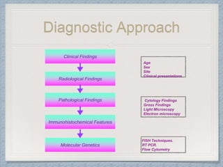
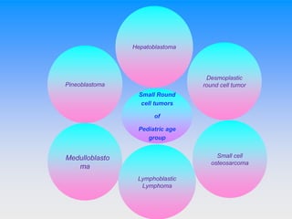
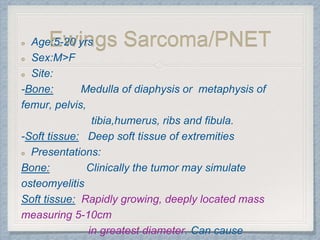
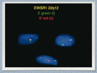
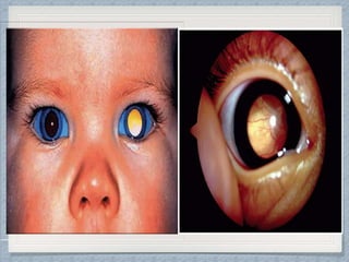
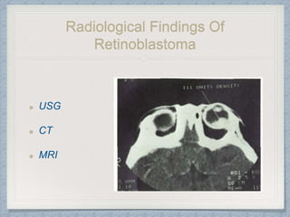
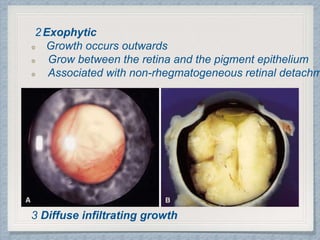
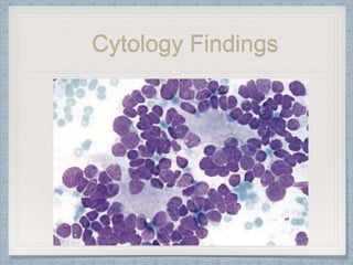
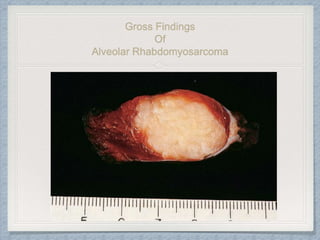
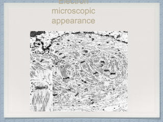
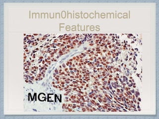
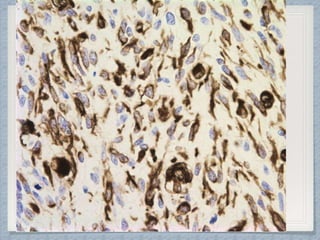
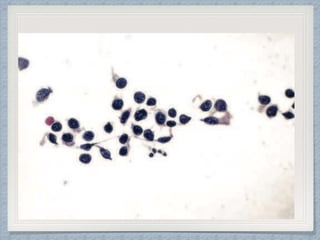
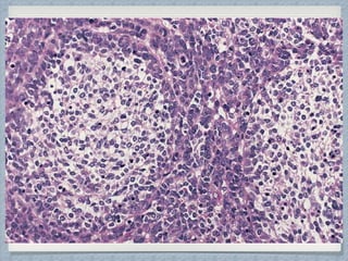
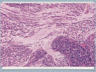
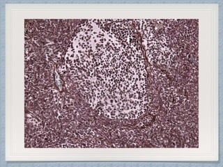
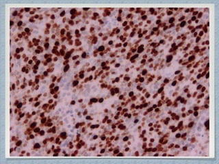
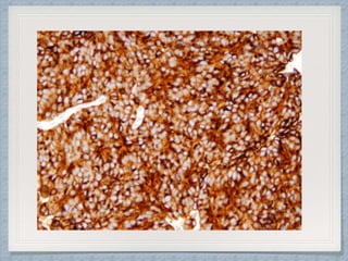
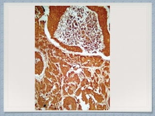
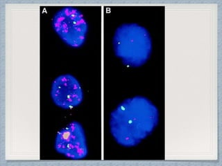
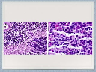
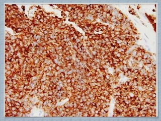
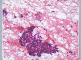
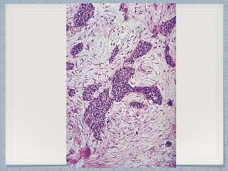
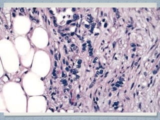
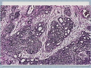
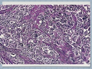
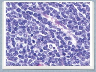
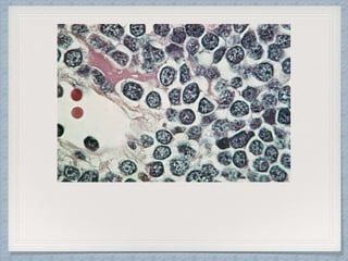
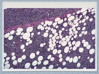
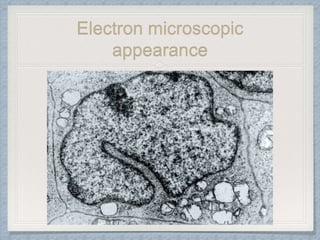
SMALL ROUND CELL
TUMOR
DIAGNOSTIC APPROACH
Dr Niharika Singh
MD Pathology II yearResident
Gandhi Medical College. Bhopal
Definition
Group of highly aggressive malignant neoplasm
which are composed predominantly of small,
round and monotonous undifferentiated cells
with high
nuclear-cytoplasmic ratio.
Diagnostic Approach
Clinical Findings
Radiological Findings
Pathological Findings
Molecular Genetics
Immunohistochemical Features
Age
Sex
Site
Clinical presentations
Cytology Findings
Gross Findings
Light Microscopy
Electron microscopy
FISH Techniques.
RT PCR.
Flow Cytometry
Ewing’s
Sarcoma/PNET
Nephroblastoma
Rhabdomyosarcoma
Neuroblastoma
Retinoblastoma Small Round
cell tumors
of
Pediatric age
group
Medulloblasto
ma
Desmoplastic
round cell tumor
Small cell
osteosarcoma
Hepatoblastoma
Pineoblastoma
Small Round
cell tumors
of
Pediatric age
group
Lymphoblastic
Lymphoma
Primitive Neuroectodermal Tumors
(Neural crest tumor)
Batsakis et al(1996)
CNS PNETs
Tumors derived
from the
central nervous system
Tumors derived from the
autonomic nervous system
Neuroblastoma
Tumors derived from
tissues outside the
central and
autonomic nervous system
Peripheral PNETs/
Ewing Family of tumors
Based on the tissue of origin
Ewing sarcoma Malignant peripheral PNETs Askin tumor *Other
*Neuroectodermal tumor, ectomesenchymoma, peripheral medulloepithelioma. These tumors were thought to arise directly from nerves.
Ewings Sarcoma/PNETAge:5-20 yrs
Sex:M>F
Site:
-Bone: Medulla of diaphysis or metaphysis of
femur, pelvis,
tibia,humerus, ribs and fibula.
-Soft tissue: Deep soft tissue of extremities
Presentations:
Bone: Clinically the tumor may simulate
osteomyelitis
Soft tissue: Rapidly growing, deeply located mass
measuring 5-10cm
in greatest diameter. Can cause
Radiological Findings
Ewing’s Sarcoma
1
2
3
Cytology Findings
Gross Findings
Ewing’s Sarcoma
Electron
microscopic
appearance
Positive for
Vimentin.
Neuron specific enolase
Neurofilament
Leu 7
CD -99.
Immun0histochemical
Features
Immun0histochemical Features
Molecular Genetics
Over 95% show reciprocal translocation of chromosome 11 : 22
(q24 : q 12).
This leads to fusion of EWS gene with FLI-1.
Detected by the
• RT-PCR
• Fluorescent in situ hybridization (FISH) technique
*in cytologic preparations, frozen sections, or paraffin sections.
Retinoblastoma
Age:16 months- 2yrs.
Sex:M=F.
Site:Intraocular
Presentations:Leukocoria (characterstic)
Strabisumus(Less often esp.in macula).
Rarely, extraocular extension with the format
an orbital mass is the presenting manifestati
Bilateral in 30% of all cases and in over
90% familial cases.
Trilateral retinoblastoma
Radiological Findings Of
Retinoblastoma
USG
CT
MRI
Cytology Findings
Gross Findings
Of Retinoblastoma
Gross:May be flat and diffuse or elevated, and
may show multicentric foci of origin, especially
in the hereditary type.
Three patterns of growth are recognised :
1Endophytic:growth occurs
inwards into the vitreous.
Cell clusters may detach and
float in the vitreous
(vitreous seeding)
2Exophytic
Growth occurs outwards
Grow between the retina and the pigment epithelium
Associated with non-rhegmatogeneous retinal detachm
3 Diffuse infiltrating growth
Electron
microscopic
appearance
Immun0histochemical
Features
Reactivity for
• Neuron-specic enolase
• Synaptophysin
• S-100 protein
• Glial fibrillary acidic protein
• Myelin basic protein
• Leu7 (CD57)
Immun0histochemical
Features
Specific markers of retinal differentiation
• Retinal-binding protein
• Retinal S-antigen
• Interphotoreceptor retinal-binding protein
• Cone opsin
• Rod opsin
• MLGAPC
Loss-of-function mutations in the RB
gene
Molecular Genetics
Neuroblastoma
Age: Under 4 yrs (21 month)
Sex:M=F
Site: Retroperitoneum (70%), and the majority of these involve the
adrenal gland.
Other sites-along sympathetic chain, posterior mediastinum, neck,
brain
Presentations
• An abdominal mass first noted by the parents.
• Rarely watery diarrhea,Cushing syndrome,heterochromia iridis and
horner syndrome (in cervical or mediastinal tumors) and
opsoclonus/myoclonus
Radiological Findings
Neuroblastoma
Cytology Findings
Pathological Findings
Of Neuroblastoma
Electron microscopic
appearance
Immun0histochemical
Features
Reactivity for
• Neuron-specic enolase
• NB-84(anti neuroblastoma antibody)
• S-100
• Insulin-like growth factor 2
• Neurofilament
• Chromogranin
• Synaptophysin
• Secretogranin II
• Vasoactive intestinal peptide
• Microtubule-associated proteins
• Growth factor receptors, and other neural-related
antigens, including the cell surface ganglioside GD2.
Molecular Genetics
Amplification of the N-myc oncogene detected by FISH techniqu
Catecholamines demonstration in sections or touch preparations
formaldehyde-induced fluorescence technique.
Expression of genes involved in catecholamine biosynthesis can
with
the polymerase chain reaction (PCR) technique.
Trk receptor protein by Northern analysis and immunohistochem
Nephroblastoma
Age:Infants (primarily), less than 3 years ( 50%) & less than 6
years (90%)
Sex:M=F
Site: Kidney;
Presentations:Large abdominal mass
Hematuria
Pain in abdomen
Hypertension
Conditions associated with wilms’ tumor are
• WAGR syndrome
• Beckwith wiedemann Syndrome
• Denys Drash Syndrome
Radiological Findings
Nephroblastoma
Intravenous pyelography
Ultrasonography
CT scan
MRI
Cytology Findings
Gross Findings
Of Nephroblastoma
Electron
microscopic
appearance
Ultrastructurally, the cells of Wilms tumor
closely resemble those of the developing metanep
Immun0histochemical
Features
Reactivity for
• Vimentin
• Keratin
• Epithelial membrane antigen
• Myogenin
• Desmin
• Neuron specific enolase
• S-100
• Glial fibrillary acid protein
• WT-1(90%)
• CD56(96%)
• Type I insulin-like growth factor
Blastematous elements
Epithelial elements
Mesenchymal /Stromal
elements
Neural elements
Molecular GeneticsGenetic loci predisposing to wilms’
tumor are
• WT1 ( located on chromosome 11p 13
)
• WT2 ( located on chromosome
11p15.5)
• Mutations of B catenin gene-14-20%
Rhabdomyosarcoma
Age:
• Embryonal:3-12 yrs;
• Alveolar:10-25 yr
Sex:M>F
Site: Embryonal: Unsegmented and undifferentiated mesoderm
Common in the
- Head and neck region (particularly the orbit, nasopharynx, middle ear, and oral cavity),
- Retroperitoneum
-Urogenital tract
-Extremities
-Skin.
Alveolar:
• Mostly extremities (forearm, arm)
-Perirectal & perineal region
Presentations:
• Based on location.
• Usually manifests as an expanding mass.
• Hypercalcemia or with elevated parathormone levels in some cases.
Radiological Findings
Embryonal Rhabdomyosarcoma
Computed tomography (CT) scanning
Magnetic resonance imaging (MRI)
Bone scanning
Ultrasonography
Cytology Findings
Embryonal Rhabdomyosarcoma
Pathological Findings
Of
Embryonal Rhabdomyosarcoma
Cytology Findings
Alveolar Rhabdomyosarcoma
Gross Findings
Of
Alveolar Rhabdomyosarcoma
Electron
microscopic
appearance
Histochemical studies
PTAH
Masson’s trichome
Silver impregnation technique
Immun0histochemical
Features
Immun0histochemical
Features
Reactivity for
• Vimentin
• Enzymes( creatine kinase)
• ILGF-2
• Neurofilament
• S-100 protein
• CARP- cardiac ankyrin related protein
Molecular Genetics
Translocation t(2;13) or t(1;13), which results in
the
gene fusions PAX3–FOXO1A and PAX7–
FOXO1A,
respectively.
Translocations can be detected in paraffin
embedded tissue with the FISH technique.
MYCN amplification in close alveolar
rhabdomyosarcomas.
Medulloblastoma
Age: 5-10 yr
Sex:M>F
Site: Commonly arise from Cerebellar vermis.
Presentations:
• Truncal ataxia
• Disturbed gait
• Lethargy
• Headache
• Morning emesis
Mostly sporadic.
Heritable cases are associated with:
• Type 2 Turcot syndrome
• Nevoid basal cell carcinoma – or ‘Gorlin’ – syndrome
• Li–Fraumeni syndrome
• Rubinstein–Taybi syndrome
• Coffin–Siris syndrome
Radiological Finding
Medulloblastoma
CT
MRI
Cytology Findings
Gross Findings
Of Medulloblastoma
Microscopic variants
of
Medulloblastomas:
Classical.
Desmoplastic/Nodular.
Medulloblastoma with extensive nodularity.
Large cell variant/ Anaplastic.
Melanotic medulloblastoma.
Medullomyoblastoma.
Microscopic Findings
Of Medulloblastoma
microscopic
appearance
Immun0histochemical
Features
Reactivity for
• Class III beta tubulin
• microtubule-associated protein 2
• Hu and NeuN
• TrkA and TrkC neurotrophin
receptors.
• Photoreceptor-associated proteins
-(rod-opsin and retinal S-antigen)
• Neurofilament
• Vimentin
• Desmin
• Myogenin
• NSE
Molecular Genetics
Chromosome 17q
Wnt Cascade
Patched gene mutation
SUFU gene mutation
INI1 gene.
MYC gene Amplification
PineoblastomaAge:Children and adolescents
Sex:M>F
Site:Parenchyma of pineal gland
Presentations:
• Obstructive hydrocephalus
• Parinaud syndrome
• CSF seeding in 15% of patients at the time of
diagnosis.
• Trilateral retinoblastoma
• Association with familial adenomatous colonic
polyposis as a possible variant of type 2 Turcot
syndrome.
Radiological Finding
Pineoblastoma
Cytology Findings
Pathological Findings
Of Pineoblastoma
Electron microscopic
appearance
Immun0histochemical
Features
Reactivity for
• Synaptophysin
• Rhodopsin
• Retinal S antigen
• Arrestin
• Ki-67
• NSE
Molecular Genetics
Most frequent rearrangement is that of
chromosome 1
RB1 gene on Chromosome 13 germline
deletion
DICER1 gene mutation
Hepatoblastoma
Age:. Primarily in infants, but isolated instances in older childr
Sex:M>F.
Site: Liver cells( Rt lobe)
Presentations:
• A large mass in the abdomen and swollen abdomen
• Decreased appetite and weight loss
• Vomiting
• Jaundice (yellowing of eyes and skin) and Itchy skin
• Anemia
• Back pain
• Virilization
Hepatoblastoma
Associated with a variety of congenital abnormalties
• Particularly hemihypertrophy
• Wilms tumor of kidney
• Glycogen storage disease
• Familial colonic polyposis.
Radiological Finding
Hepatoblastoma
Hepatic angiography
CT scans
Cytology Findings
Pathological Findings
Of Hepatoblastoma
Microscopic Findings Of
Hepatoblastoma
Electron microscopic
appearance
Immun0histochemical
Features
Reactivity for
• β-catenin
• Glypican-3
• Polyclonal CEA
• Keratin
• EMA
• HepPar-1
• a1-antitrypsin
• Vimentin
• AFP
• HCG
Immun0histochemical
Features
Reactivity for
• Transferrin receptor
• Delta-like protein
• Neuron specific enolase
• Chromogranin
• Somatostatin
• Serotonin
• HMB-45
• Laminin, type IV collagen, and fibronectin.
Molecular Genetics
Trisomies 2, 8, and 20, and rearrangements of
1q.
CTNNB1 (β-catenin) gene.
CGH analysis :High frequency of X-chromosome
gains.
Evaluation of DNA content by flow cytometry.
Desmoplastic small round cell
tumor
Age:Adolescents and young adults
Sex:M>F
Site:Intra Abdominal: Pelvic region and sometimes the entire
tumor is limited to the paratesticular region
• Other sites:bone, kidney, lung, ovary, pancreas, pleura,
spermatic cord, uterus,
central nervous system, sinonasal region and salivary glands.
Presentation:Intra Abdominal:Characteristically presents as a
single mass or multiple nodules within the abdominal cavity.
• Accompanying ascites is the rule;
Association with Peutz–Jeghers syndrome
Radiological Finding
Desmoplastic small round cell
tumor
Cytology Findings
Pathological Findings
Desmoplastic small round cell
tumor
Electron microscopic appearance
Immun0histochemical
Features
Keratin
Desmin
Immun0histochemical
Features
Reactivity for
• Neural (neuron-specific enolase) markers
• Leu-7
• WT-1
• Vimentin
Molecular Genetics
Lymphoblastic Lymphoma
Age:Primarily in children and adolescents, but it also occurs in
adults
Sex:M>F
Site: Mediastinum( Thymus).
Presentations:
• Typically presents with acute respiratory distress in teenager.
• Extremely aggressive, with rapid multisystem dissemination.
• Leukemic blood picture (acute lymphoblastic leukemia).
• Death after a few months.
Radiological Finding
Lymphoblastic Lymphoma
Cytological Findings
Pathological Findings
Lymphoblastic Lymphoma
Electron microscopic
appearance
Histochemical studies
Acid phosphatase
β- glucuronidase
α- naphthyl acetate esterase
TdT
Immun0histochemical
Features
Reactivity for
• TdT
• CD1
• CD2
• CD3
• CD43
• CD99
• bcl2
• CD71
• LCA+
Molecular Genetics
Translocation
alpha & delta T-cell
receptor loci
beta T-cell receptor
loci
gamma T-cell receptor
loci
14q11.2 7q35 7p14–15
Dysregulation of transcription
Partner genes (MYC, TAL1, RBTN1, RBTN2, and HOX11)
Small round cell tumors

Small round cell tumors