Downloaded 176 times


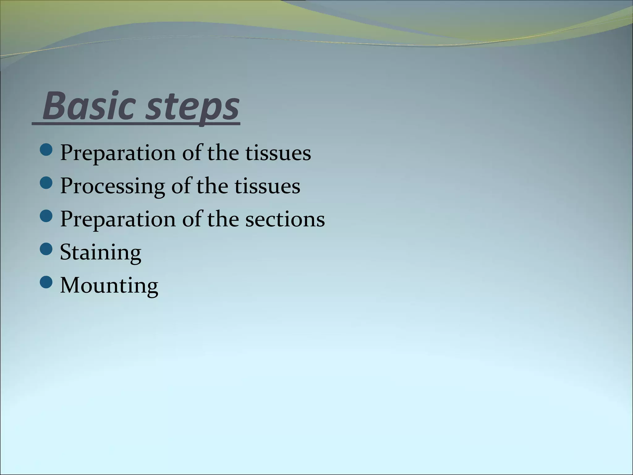

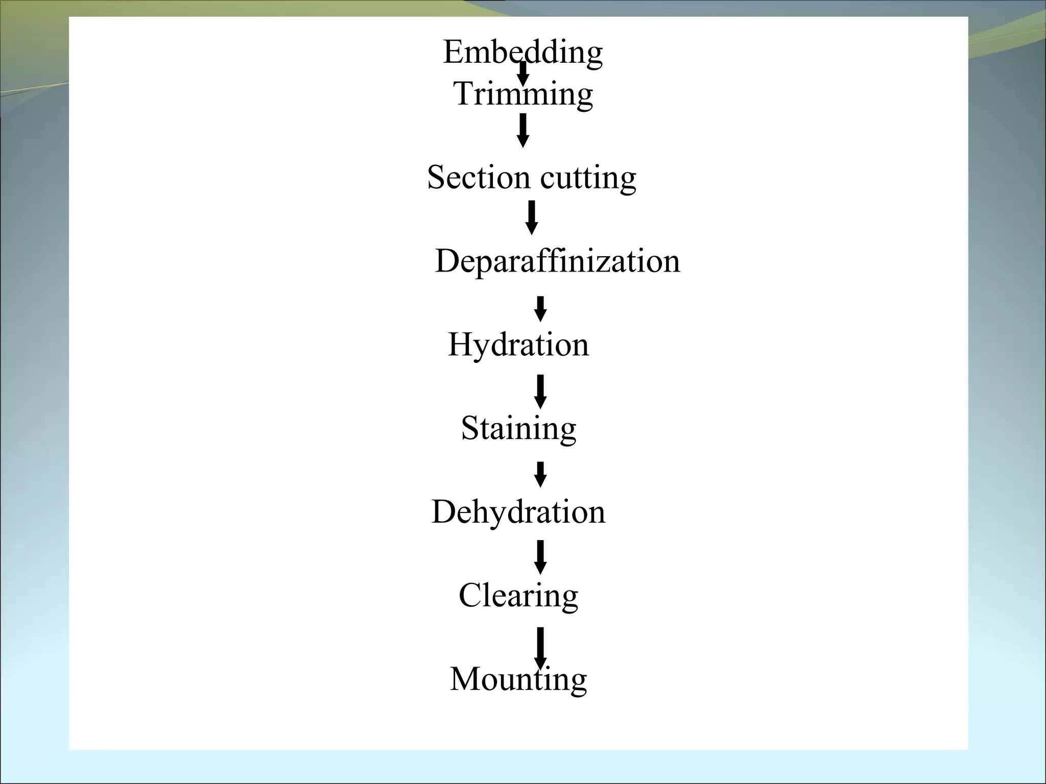
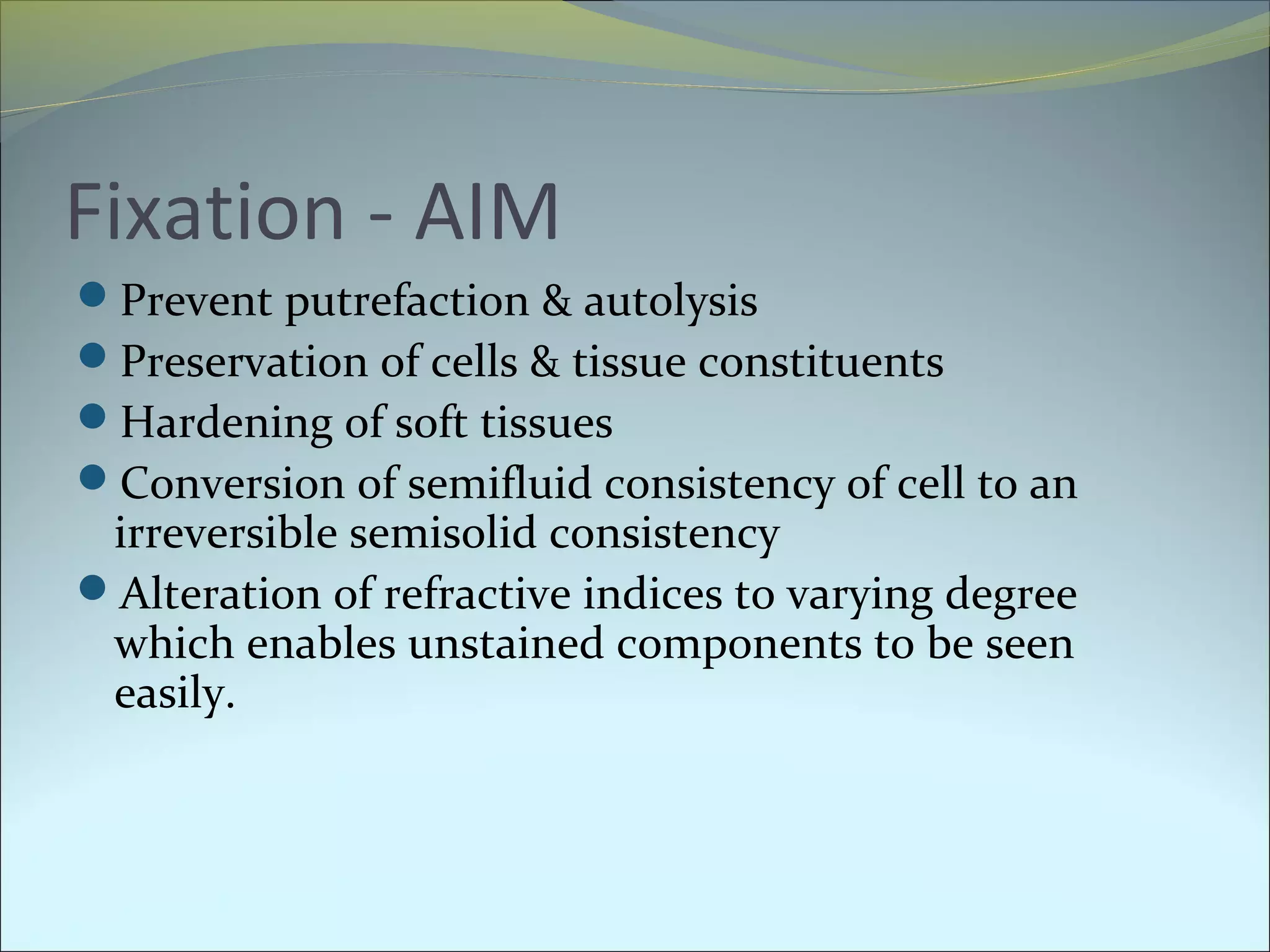


This document discusses the process of histology, which involves preparing tissue samples for microscopic examination. Specimens should be transported in fixatives like formalin or refrigerated. There are several steps involved including fixation, processing, sectioning, staining, and mounting. Tissue processors are used to dehydrate samples through a series of alcohols and xylene before infiltrating and embedding them in paraffin wax for sectioning with a microtome. Sections are then stained using techniques like H&E staining before examination under a microscope. Frozen sectioning and newer techniques using resins and microwaves can also be used to prepare samples for histological analysis.