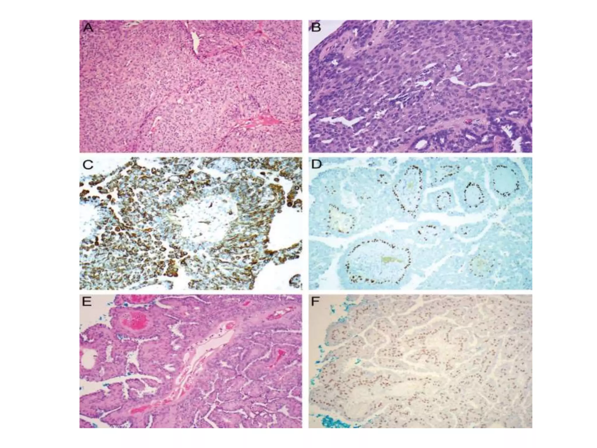
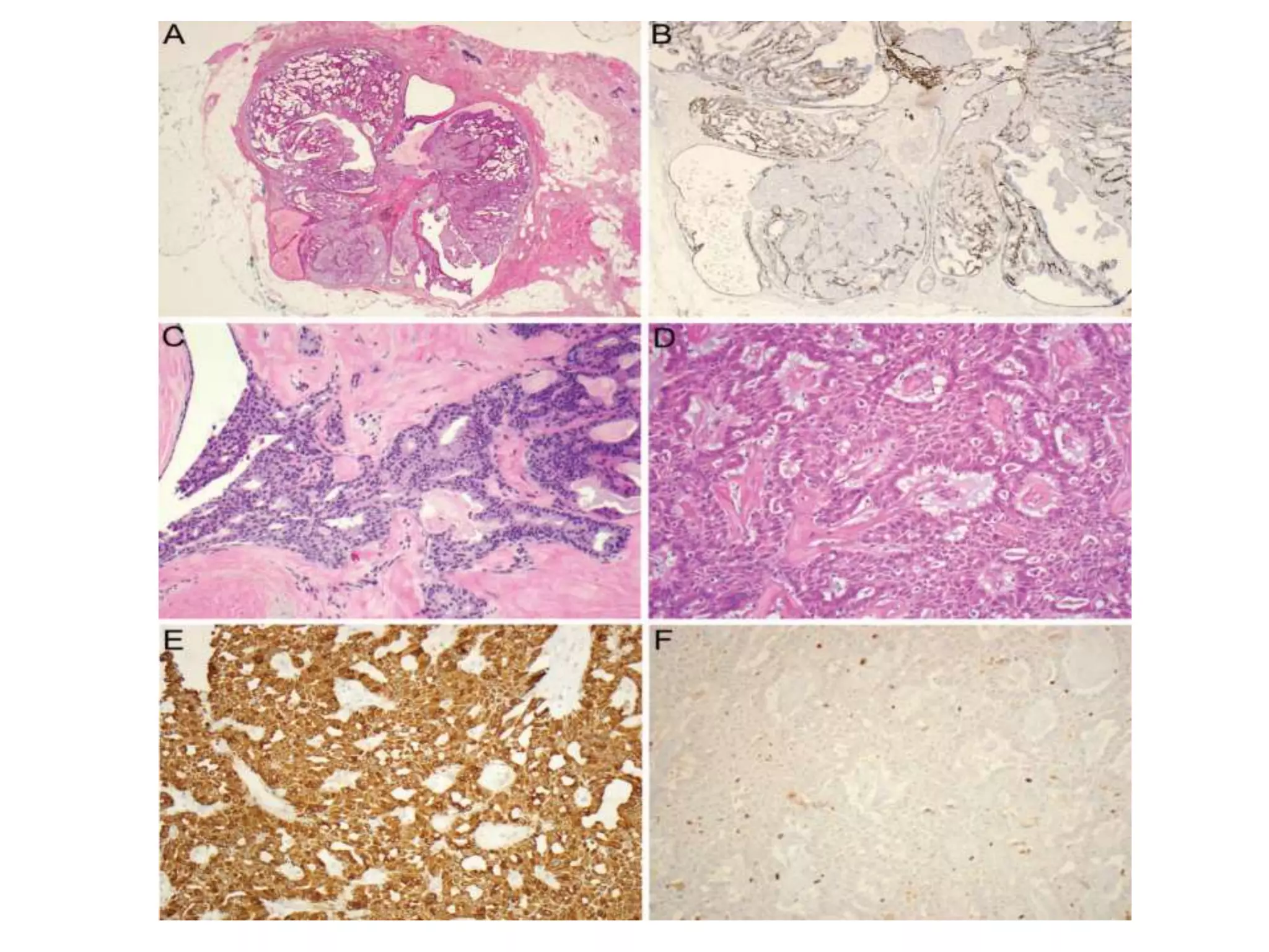
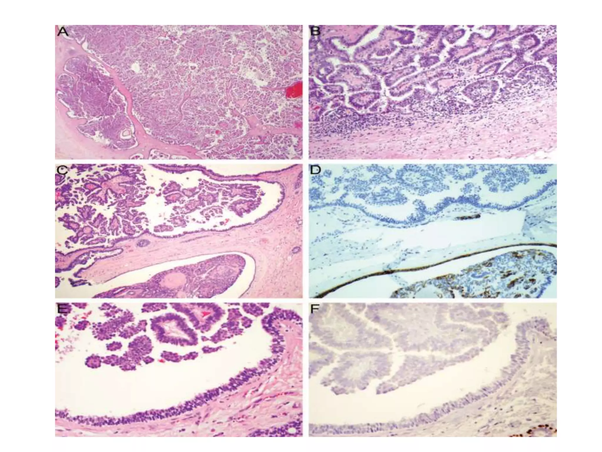
Papillary Lesions of the Breast
An Update
BY MELESE A.(R3)
Papillary Lesions of the Breast
• Are characterized by the presence of arborescent
fibrovascular cores that support epithelial proliferation
• are exclusively intraductal neoplasms
• The spectrum of intraductal papillary lesions comprises
 intraductal papilloma
 papilloma with atypical ductal hyperplasia (ADH)
 papilloma with ductal carcinoma in situ (DCIS), and
 intraductal papillary carcinoma
INTRADUCTAL PAPILLOMA
• Are characterized by the presence of proliferating arborescent
fibrovascular cores lined by an outer epithelial layer and an
inner myoepithelial layer
• They can be divided into
 central papilloma (large-duct papilloma) and
 peripheral papilloma (small-duct papilloma)
Central papillomas
• originate in the segmental or subsegmental duct
• are typically solitary
• They present with serous or serosanguineous nipple discharge
On gross examination
=> often visible and appear as circumscribed nodules in a
cystically dilated duct space
Peripheral papillomas
• arise in the terminal duct lobular units (clinically occult)
• primarily a disease of middle-aged women
• are typically grossly unidentifiable
Histopathology
• The fibrovascular stalks are typically broad
• An intervening myoepithelial cell layer invariably resides in
the papillary fronds
• The luminal epithelial component may comprise a single layer
of columnar or cuboidal cells
• A variable degree of epithelial proliferation
• The proliferating epithelial cells
=> heterogeneous and overlapping or irregularly spaced, and
have indistinct cell borders and variably sized nuclei with
frequent nuclear grooves and pseudonuclear inclusions
• Focal apocrine metaplasia is frequently seen within intraductal
papillomas
• Other reported metaplastic features include
 squamous, mucinous, clear cell, and sebaceous metaplasia
• Collagenous spherulosis can involve papillomas
• Areas of hemorrhage or infarction are not uncommon
• Rarely, the entire lesion may be infarcted
 In cases of no residual viable tissue, the tumor should be
designated as an infarcted papillary lesion
• A variable amount of stromal fibrosis is another common
finding
PAPILLOMA WITH ADH & PAPILLOMA WITH DCIS
• Some intraductal papillomas may have areas of atypical
ductal epithelial proliferation
• These lesions are designated as
 papilloma with ADH
 papilloma with DCIS
• The atypical features include
 the presence of evenly spaced, small, regular cells with round,
monotonous nuclei
 complex architecture patterns (ie, solid, cribriform, or
micropapillary)
• These foci
 Have scant or no myoepithelial cells
 lack expression of high molecular-weight keratins
 show strong staining for estrogen and progesterone receptors
A pitfall:- malignant cells with squamous differentiation
 tend to have strong immunoreactivity with CK5/6
 p63 is a superior marker in this setting
• There is no universally accepted criteria as to the distinction
between papilloma with ADH and papilloma with DCIS.
• Extent/size-based criteria
 Atypical epithelial proliferation <3 mm =>papilloma with ADH
 Atypical epithelial proliferation >3 mm =>papilloma with DCIS
• when the atypical epithelial proliferation is of intermediate or high
nuclear grade => papilloma with DCIS
• The presence of DCIS in neighboring breast tissue is particularly
supportive of the diagnosis of DCIS within a papilloma
INTRADUCTAL PAPILLARY CARCINOMA
• AKA papillary DCIS and noninvasive papillary carcinoma
• Is distinguished by the complete or near-complete absence of
myoepithelial cells
• Nipple discharge or a palpable mass may be the presenting
symptom, depending on the location and size of the lesion
• These lesions may appear as
 an irregular intraductal/ intracystic mass with increased vascularity on
imaging studies, or
 present with mammographic calcifications
• There are no distinguishable macroscopic features.
Histology
 variably dilated ductal-lobular luminal spaces filled with
slender or branching fibrovascular stalks lined by stratified
columnar neoplastic epithelial cells
 The cytologic features of the lesional cells are typically those
of low- or intermediate-grade DCIS (rarely high-grade)
 intact myoepithelial cell layer is absent
 myoepithelial cell layer is retained at the periphery of the
involved duct wall
- may be attenuated or discontinuous in larger lesions with markedly
distended duct spaces
• A dimorphic population of malignant cells
- second population consists of cells with clear cytoplasm
• Proliferation of neoplastic epithelial cells may produce
additional architectural patterns such as
=> solid, cribriform, and micropapillations
Pitfall :- the endothelial cells juxtaposed to the neoplastic
epithelial cells in the papillary fronds
- p63, CK5/6, CK14, 34bE12/CK903
ENCAPSULATED (INTRACYSTIC) PAPILLARY
CARCINOMA
• Frequently occurs in elderly patients (average 65yrs)
• The tumor often presents as a
- retroareolar well-defined mass on mammography
- cystic lesion with solid components on ultrasound
• On gross examination=> circumscribed friable mass within a
cystic cavity
• Morphologically similar to intraductal papillary carcinoma
• The myoepithelial cells are absent at the surrounding thick
fibrous capsule
• Has a favorable prognosis with adequate local therapy alone.
Histologically
• One or more well-circumscribed nodular masses
within a cystically dilated duct surrounded by a thick
fibrous capsule
• The duct spaces are filled by slender fibrovascular
stalks devoid of myoepithelial cells
• The papillary fronds are lined by neoplastic epithelial
proliferation arranged in various patterns, including
solid and cribriform
• The neoplastic cells typically have low- or
intermediate-grade nuclei
• The neoplastic cells typically show strong ER and PR
expression
• A layer of myoepithelial cells is absent at the periphery of
the lesion(both on H&E and IHC)
• Rarely, the lining cells may take on a columnar cell
appearance that contains subnuclear vacuoles and shows
reverse polarity, thus simulating piano keys
• A small proportion may be associated with frankly
invasive carcinoma.
• The invasive component is typically of no special type,
with no papillary features
SOLID PAPILLARY CARCINOMA
• Is an intraductal papillary carcinoma that
 grows in a solid papillary pattern
 displays low-grade cytologic features
 often has neuroendocrine differentiation
 Intracellular or extracellular mucin production
Grossly
=> fleshy, firm or soft, well circumscribed nodular mass with a
tan-white or yellowish brown appearance
• These lesions occur mostly in elderly women(7th decade of
life)
Microscopically
=> multiple solidified circumscribed nodules
=> delicate underlying fibrovascular stromal network
devoid of a myoepithelial cell layer
=> densely cellular neoplastic cells which appear to be
streaming
• Intracellular and/or extracellular mucin production
• The proliferating tumor cells are
=> bland and cohesive, have oval or spindled nuclei,
and exhibit low to intermediate nuclear grade
• The fibrovascular cores can be variably collagenized,
with peripheral palisading of tumor cells forming
perivascular pseudorosettes
• The lesional cells often demonstrate neuroendocrine
features, including finely granular eosinophilic
cytoplasm and salt-and-pepper nuclei
• Mitotic figures are visible, but not brisk and with no
atypical forms
• Metastases from such tumors are rare but do occur
• The nodules of solid papillary carcinoma
frequently have a geographic jigsaw pattern
and irregular peripheries, thus suggesting
invasion.
• The cells of solid papillary carcinoma typically
show strong ER and PR expression, and lack
HER2 overexpression/gene amplification.
• Rarely, solid papillary carcinoma and
encapsulated papillary carcinoma may present
as a composite tumor
INVASIVE PAPILLARY CARCINOMA
• invasive adenocarcinomas with an exclusively (>90%)
papillary histomorphology
• extremely rare, with a reported incidence of
approximately 0.5% of all IBC
• an invasive nonpapillary carcinoma arising from
encapsulated or solid papillary carcinoma should not
be categorized as invasive papillary carcinoma
EPITHELIAL DISPLACEMENT: A PAPILLARY
PHENOMENON
• Mechanical displacement of epithelium may occur
following a variety of needling procedures, including
=> core needle biopsy, fine-needle aspiration, anesthetic
injection, suture placement, and wire localization
• Artifactual epithelial displacement occurs in
association with underlying papillary neoplasms as
these lesions are inherently friable
• The displaced epithelium may produce a pattern
closely simulating an invasive carcinoma
• present in the form of isolated, small fragments of
epithelium in artificial spaces within the mammary
stroma
• These epithelial cell clusters are typically associated
with a spectrum of biopsy site changes such as
 organizing hemorrhage
 acute and chronic inflammation
 foreign body reaction (foamy histiocytes, giant cells,
cholesterol clefts)
 fat necrosis
 hemosiderin deposition
 granulation tissue, or scarring
papillary lesions of the breast.pptx

papillary lesions of the breast.pptx

  • 1.
    Papillary Lesions ofthe Breast An Update BY MELESE A.(R3)
  • 2.
    Papillary Lesions ofthe Breast • Are characterized by the presence of arborescent fibrovascular cores that support epithelial proliferation • are exclusively intraductal neoplasms • The spectrum of intraductal papillary lesions comprises  intraductal papilloma  papilloma with atypical ductal hyperplasia (ADH)  papilloma with ductal carcinoma in situ (DCIS), and  intraductal papillary carcinoma
  • 3.
    INTRADUCTAL PAPILLOMA • Arecharacterized by the presence of proliferating arborescent fibrovascular cores lined by an outer epithelial layer and an inner myoepithelial layer • They can be divided into  central papilloma (large-duct papilloma) and  peripheral papilloma (small-duct papilloma)
  • 4.
    Central papillomas • originatein the segmental or subsegmental duct • are typically solitary • They present with serous or serosanguineous nipple discharge On gross examination => often visible and appear as circumscribed nodules in a cystically dilated duct space Peripheral papillomas • arise in the terminal duct lobular units (clinically occult) • primarily a disease of middle-aged women • are typically grossly unidentifiable
  • 5.
    Histopathology • The fibrovascularstalks are typically broad • An intervening myoepithelial cell layer invariably resides in the papillary fronds • The luminal epithelial component may comprise a single layer of columnar or cuboidal cells • A variable degree of epithelial proliferation • The proliferating epithelial cells => heterogeneous and overlapping or irregularly spaced, and have indistinct cell borders and variably sized nuclei with frequent nuclear grooves and pseudonuclear inclusions
  • 7.
    • Focal apocrinemetaplasia is frequently seen within intraductal papillomas • Other reported metaplastic features include  squamous, mucinous, clear cell, and sebaceous metaplasia • Collagenous spherulosis can involve papillomas • Areas of hemorrhage or infarction are not uncommon • Rarely, the entire lesion may be infarcted  In cases of no residual viable tissue, the tumor should be designated as an infarcted papillary lesion • A variable amount of stromal fibrosis is another common finding
  • 11.
    PAPILLOMA WITH ADH& PAPILLOMA WITH DCIS • Some intraductal papillomas may have areas of atypical ductal epithelial proliferation • These lesions are designated as  papilloma with ADH  papilloma with DCIS • The atypical features include  the presence of evenly spaced, small, regular cells with round, monotonous nuclei  complex architecture patterns (ie, solid, cribriform, or micropapillary)
  • 12.
    • These foci Have scant or no myoepithelial cells  lack expression of high molecular-weight keratins  show strong staining for estrogen and progesterone receptors A pitfall:- malignant cells with squamous differentiation  tend to have strong immunoreactivity with CK5/6  p63 is a superior marker in this setting
  • 14.
    • There isno universally accepted criteria as to the distinction between papilloma with ADH and papilloma with DCIS. • Extent/size-based criteria  Atypical epithelial proliferation <3 mm =>papilloma with ADH  Atypical epithelial proliferation >3 mm =>papilloma with DCIS • when the atypical epithelial proliferation is of intermediate or high nuclear grade => papilloma with DCIS • The presence of DCIS in neighboring breast tissue is particularly supportive of the diagnosis of DCIS within a papilloma
  • 16.
    INTRADUCTAL PAPILLARY CARCINOMA •AKA papillary DCIS and noninvasive papillary carcinoma • Is distinguished by the complete or near-complete absence of myoepithelial cells • Nipple discharge or a palpable mass may be the presenting symptom, depending on the location and size of the lesion • These lesions may appear as  an irregular intraductal/ intracystic mass with increased vascularity on imaging studies, or  present with mammographic calcifications • There are no distinguishable macroscopic features.
  • 17.
    Histology  variably dilatedductal-lobular luminal spaces filled with slender or branching fibrovascular stalks lined by stratified columnar neoplastic epithelial cells  The cytologic features of the lesional cells are typically those of low- or intermediate-grade DCIS (rarely high-grade)  intact myoepithelial cell layer is absent  myoepithelial cell layer is retained at the periphery of the involved duct wall - may be attenuated or discontinuous in larger lesions with markedly distended duct spaces
  • 18.
    • A dimorphicpopulation of malignant cells - second population consists of cells with clear cytoplasm • Proliferation of neoplastic epithelial cells may produce additional architectural patterns such as => solid, cribriform, and micropapillations Pitfall :- the endothelial cells juxtaposed to the neoplastic epithelial cells in the papillary fronds - p63, CK5/6, CK14, 34bE12/CK903
  • 20.
    ENCAPSULATED (INTRACYSTIC) PAPILLARY CARCINOMA •Frequently occurs in elderly patients (average 65yrs) • The tumor often presents as a - retroareolar well-defined mass on mammography - cystic lesion with solid components on ultrasound • On gross examination=> circumscribed friable mass within a cystic cavity • Morphologically similar to intraductal papillary carcinoma • The myoepithelial cells are absent at the surrounding thick fibrous capsule • Has a favorable prognosis with adequate local therapy alone.
  • 21.
    Histologically • One ormore well-circumscribed nodular masses within a cystically dilated duct surrounded by a thick fibrous capsule • The duct spaces are filled by slender fibrovascular stalks devoid of myoepithelial cells • The papillary fronds are lined by neoplastic epithelial proliferation arranged in various patterns, including solid and cribriform • The neoplastic cells typically have low- or intermediate-grade nuclei • The neoplastic cells typically show strong ER and PR expression
  • 22.
    • A layerof myoepithelial cells is absent at the periphery of the lesion(both on H&E and IHC) • Rarely, the lining cells may take on a columnar cell appearance that contains subnuclear vacuoles and shows reverse polarity, thus simulating piano keys • A small proportion may be associated with frankly invasive carcinoma. • The invasive component is typically of no special type, with no papillary features
  • 24.
    SOLID PAPILLARY CARCINOMA •Is an intraductal papillary carcinoma that  grows in a solid papillary pattern  displays low-grade cytologic features  often has neuroendocrine differentiation  Intracellular or extracellular mucin production Grossly => fleshy, firm or soft, well circumscribed nodular mass with a tan-white or yellowish brown appearance • These lesions occur mostly in elderly women(7th decade of life)
  • 25.
    Microscopically => multiple solidifiedcircumscribed nodules => delicate underlying fibrovascular stromal network devoid of a myoepithelial cell layer => densely cellular neoplastic cells which appear to be streaming • Intracellular and/or extracellular mucin production • The proliferating tumor cells are => bland and cohesive, have oval or spindled nuclei, and exhibit low to intermediate nuclear grade
  • 26.
    • The fibrovascularcores can be variably collagenized, with peripheral palisading of tumor cells forming perivascular pseudorosettes • The lesional cells often demonstrate neuroendocrine features, including finely granular eosinophilic cytoplasm and salt-and-pepper nuclei • Mitotic figures are visible, but not brisk and with no atypical forms • Metastases from such tumors are rare but do occur
  • 28.
    • The nodulesof solid papillary carcinoma frequently have a geographic jigsaw pattern and irregular peripheries, thus suggesting invasion. • The cells of solid papillary carcinoma typically show strong ER and PR expression, and lack HER2 overexpression/gene amplification. • Rarely, solid papillary carcinoma and encapsulated papillary carcinoma may present as a composite tumor
  • 32.
    INVASIVE PAPILLARY CARCINOMA •invasive adenocarcinomas with an exclusively (>90%) papillary histomorphology • extremely rare, with a reported incidence of approximately 0.5% of all IBC • an invasive nonpapillary carcinoma arising from encapsulated or solid papillary carcinoma should not be categorized as invasive papillary carcinoma
  • 34.
    EPITHELIAL DISPLACEMENT: APAPILLARY PHENOMENON • Mechanical displacement of epithelium may occur following a variety of needling procedures, including => core needle biopsy, fine-needle aspiration, anesthetic injection, suture placement, and wire localization • Artifactual epithelial displacement occurs in association with underlying papillary neoplasms as these lesions are inherently friable • The displaced epithelium may produce a pattern closely simulating an invasive carcinoma • present in the form of isolated, small fragments of epithelium in artificial spaces within the mammary stroma
  • 35.
    • These epithelialcell clusters are typically associated with a spectrum of biopsy site changes such as  organizing hemorrhage  acute and chronic inflammation  foreign body reaction (foamy histiocytes, giant cells, cholesterol clefts)  fat necrosis  hemosiderin deposition  granulation tissue, or scarring

Editor's Notes

  • #7 Intraductal papilloma. A, The fibrovascular stalks are typically broad with variable degree of stromal fibrosis. B, An outer luminal epithelial cell layer and an inner myoepithelial cell layer overlie the papillary fronds. C, Thin papillae can be seen in areas and may cause confusion with intraductal papillary carcinoma. D, The presence of the intervening myoepithelial cell layer in the papillae shown in C is highlighted by p63 immunostaining (hematoxylin-eosin, original magnifications340 [A],3200 [B], and3100 [C]; original magnification3100 [D]).
  • #9 Intraductal papilloma with epithelial hyperplasia. A, Intraductal papilloma with florid ductal epithelial hyperplasia. B, High-power view of epithelial hyperplasia demonstrates overlapping, heterogeneous cells forming secondary lumina, with frequent nuclear grooves and pseudoinclusions typical of usual ductal hyperplasia. C, An immunostain for cytokeratin 5/6 shows a patchy staining pattern in the proliferating epithelial cells in addition to its reactivity with basal myoepithelial cells. D, The p63 stain highlights only the myoepithelial cells and is negative in ductal epithelium. E, Intraductal papilloma with focal myoepithelial cell hyperplasia. F, A p63 stain highlights the proliferating myoepithelial cells in the lesion shown in E (hematoxylin-eosin, original magnifications3100 [A] and3200 [B and E]; original magnifications3100 [C and D] and3200 [F]).
  • #10 Figure 3. Various appearances of intraductal papilloma. A, Focal apocrine metaplasia is frequently present in benign papillomas but is almost always absent in malignant lesions. B, The proliferating epithelial cells with apocrine metaplasia are negative for cytokeratin 5/6, thus are not to be confused with intraductal papillary carcinoma. C, Collagenous spherulosis involving a papilloma may give rise to an appearance of a cribriform ductal carcinoma in situ on low-power view. D, Higher magnification shows tubular structures containing intraluminal basement membrane material surrounded by flattened myoepithelial cells in collagenous spherulosis. E, Papilloma with infarction. F, Papilloma with squamous metaplasia (hematoxylin-eosin, original magnifications340 [A, C, and E] and3200 [D and F]; original magnification3100 [B]).
  • #11 Figure 4. Sclerosing intraductal papilloma. A, Markedly sclerosed papilloma with minimal epithelial elements. B, Areas of papilloma with extensive stromal fibrosis that obscures the underlying papillary configuration. C, The pronounced sclerosis results in extensively compressed and distorted glandular structures, thus creating a pseudoinfiltrative pattern mimicking invasive carcinoma (hematoxylineosin, original magnifications340 [A] and3100 [B and C]).
  • #14 Papilloma with atypical ductal hyperplasia (ADH)/ductal carcinoma in situ (DCIS). A, A papilloma with areas of monotonous cellular proliferation. B, A cytokeratin 5/6 [CK5/6] stain in the lesion shown in A demonstrates lack of a mosaic staining pattern in the proliferating cells, thus rendering it a papilloma with DCIS according to the extent-/size-based criteria, given the area of interest is greater than 3 mm. C, A sclerosing papilloma with ADH showing focal cellular proliferation with small, round, uniform, and evenly spaced nuclei characteristic of those in ADH and low-grade DCIS. D, Papilloma with DCIS showing prominent squamous differentiation. E, The neoplastic cells shown in D are strongly immunoreactive with CK5/6. F, A p63 stain highlights rare myoepithelial cells in the lesion shown in D (hematoxylin-eosin, original magnifications 320 [A],3100 [C], and3200 [D]; original magnifications340 [B] and3200 [E and F]
  • #20 Figure 6. Intraductal papillary carcinoma. A, The papillae are typically thin and delicate, and are lined by stratified columnar neoplastic epithelial cells devoid of a myoepithelial cell layer; the latter is retained at the periphery of the involved duct wall. B, A dimorphic population of malignant cells in the papillae, in which the cells in the basal location show clear cell features, thus resembling myoepithelial cells. C, The neoplastic epithelial proliferation results in a solid growth pattern that obliterates the spaces between the papillae. D, Scant myoepithelial cells may be present within the papillae or in the epithelial proliferation, as shown in this cytokeratin 5/6 immunostain (hematoxylin-eosin, original magnification3200 [A through C]; original magnification3200 [D]).
  • #24 Encapsulated papillary carcinoma. A, The tumor often presents with one or more well-circumscribed nodular masses within a cystically dilated duct. B, The tumor nodule is surrounded by a thick fibrous capsule with variable degree of chronic inflammation. C, The periphery may be lined by stratified epithelial cells similar to those in the papillary fronds, in contrast to myoepithelial cells lining the periphery of benign papillomas that are typically flattened, cuboidal, or rounded. D, A cytokeratin 5/6 stain confirms the absence of myoepithelial lining at the periphery of the encapsulated papillary carcinoma shown in C, whereas it highlights the myoepithelial cells in the adjacent papilloma. E, The peripheral lining cells of encapsulated papillary carcinoma may rarely take on the appearance of piano key–like columnar cells with subnuclear vacuoles and reverse polarity. F, The nonmyoepithelial nature of the lining cells shown in E is demonstrated by p63 immunostaining (hematoxylin-eosin, original magnifications320 [A],3100 [B and C], and3400 [E]; original magnifications3200 [D] and3400 [F]).
  • #28 Solid papillary carcinoma. A, Multiple solid, circumscribed nodules without apparent papillary architecture are characteristic on lowpower view. B, The absence of peripheral myoepithelium is demonstrated by a cytokeratin 5/6 [CK5/6] stain, which highlights the myoepithelial cells in the adjacent normal duct. C, An intact myoepithelial cell layer may be seen in some of the tumoral nests as confirmed by this CK5/6 stain. D, Mucin production is not uncommon in solid papillary carcinoma. E, The neoplastic cells typically have low- to intermediate-grade oval or spindled nuclei. F, The lesional cells often exhibit neuroendocrine features, with granular eosinophilic cytoplasm and salt-and-pepper nuclei, and form perivascular pseudorosettes in the variably collagenized fibrovascular cores (hematoxylin-eosin, original magnifications340 [A],3100 [D],3200 [E], and3400 [F]; original magnification340 [B and C]).
  • #30 Solid papillary carcinoma merged with invasive carcinoma. A, The nodules of solid papillary carcinoma merge with invasive carcinoma (lower left). B, The neuroendocrine differentiation is demonstrated by an immunostain for synaptophysin in this invasive carcinoma arising from a solid papillary carcinoma (hematoxylin-eosin, original magnification340 [A]; original magnification3100 [B]).
  • #31 Figure 10. Composite encapsulated papillary carcinoma and solid papillary carcinoma. A, A low-power view shows a tumor with 2 distinct morphologies characteristic of encapsulated papillary carcinoma and solid papillary carcinoma. B, The absence of myoepithelium within the papillae and at the periphery of both components is demonstrated by this cytokeratin 5/6 immunostaining. C, The 2 components intermingle in one tumor nodule, imparting the appearance of one tumor growing into the other (hematoxylin-eosin, original magnifications340 [A] and3100 [C]; original magnification3200 [B]).
  • #37 Displacement of epithelium. A, The displaced epithelial clusters from an intraductal papilloma reside in the stroma with myofibroblastic proliferation and chronic inflammation typical of a needle tract. B, Displaced epithelium from a sclerosing papilloma in a circumscribed area with hemorrhage, hemosiderin deposition, chronic inflammation, and cholesterol clefts characteristic of a biopsy site. C, Displacement of epithelium following core biopsy of a micropapillary ductal carcinoma in situ (DCIS) is confined to the traumatized area of the needle tract. D, Displaced malignant epithelial cells surrounded by abundant foamy histiocytes in the subcapsular sinus of a sentinel lymph node in a patient with micropapillary DCIS (hematoxylin-eosin, original magnifications3100 [A],340 [B and C], and3200 [D]).