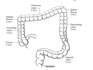
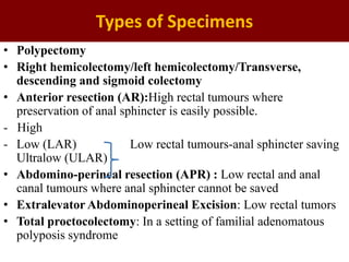
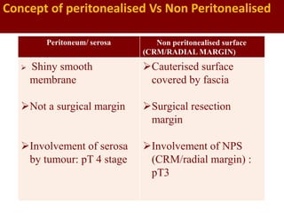
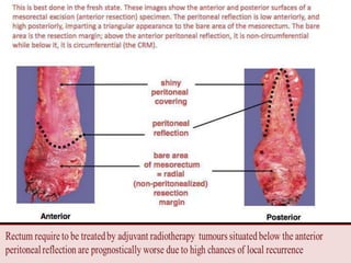
CAP Protocol-Colorectum
Types of Specimens
• Polypectomy
• Right hemicolectomy/left hemicolectomy/Transverse,
descending and sigmoid colectomy
• Anterior resection (AR):High rectal tumours where
preservation of anal sphincter is easily possible.
- High
- Low (LAR) Low rectal tumours-anal sphincter saving
Ultralow (ULAR)
• Abdomino-perineal resection (APR) : Low rectal and anal
canal tumours where anal sphincter cannot be saved
• Extralevator Abdominoperineal Excision: Low rectal tumors
• Total proctocolectomy: In a setting of familial adenomatous
polyposis syndrome
-
Peritoneum/ serosa Non peritonealised surface
(CRM/RADIAL MARGIN)
 Shiny smooth
membrane
Not a surgical margin
Involvement of serosa
by tumour: pT 4 stage
Cauterised surface
covered by fascia
Surgical resection
margin
Involvement of NPS
(CRM/radial margin) :
pT3
Concept of peritonealised Vs Non Peritonealised
Total Mesorectal Excision
• Thick blanket of fatty tissue encasing rectum-Mesorectum
• Contains the blood vessels and lymph nodes draining the rectum
• Invested by endopelvic fascia
Removal of the mesorectum en bloc with the rectal tumour
:Total mesorectal excision (TME)
 Goals of TME for rectal cancer
- Adequate lymphadenectomy
- Achieving maximum lateral resection margin
- Reduces the possibility of loco regional relapse
Pathological Assessment of Quality of TME
• Asses local recurrence rates
• Increase corresponding survival ~20%
• Feedback for the radiologist : To improve accuracy
of pre-operative prediction
• Feedback to the surgeon : Indicator of quality of
surgery
Complete TME (Grade 3) Nearly complete TME
(Grade 2)
Incomplete TME (Grade 1)
•Mesorectal fascial plane
•Mesorectum is intact &
bulky with a smooth
surface
•Minor irregularities in the
mesorectal surface(surface
defects <5 mm in depth
•No coning towards the
distal margin of the
specimen
•Smooth CRM on
transverse section
•Mesorectum
•Bulk of mesorectum is
moderate
•Surface defects >5 mm in
depth but none extending to
the muscularis propria
• Moderate coning
seen towards the distal
margin of the specimen
•Moderate irregularity of
CRM seen in transverse
• Muscularis propria
• Little bulk of mesorectum
is present
•Defects in the mesorectum
expose the muscularis
propria
•Very irregular CRM in
transverse section
Quirke’s Method
Other Margins
• In segments completely encased by peritoneum (eg,
transverse colon):Mesenteric resection margin
 Involvement of this margin should be reported even if tumor
does not penetrate the serosal surface
• Proximal and distal resection margin:
-Distal resection margin of 2 cm is adequate
-Anastomotic recurrences are rare: distance to the closest
transverse margin is >5 cm
• In a background of inflammatory bowel disease, proximal
and distal resection margins to be evaluated for dysplasia
and active inflammation
A. Mesenteric margin in portion of colon completely encased by peritoneum (dotted line)
B. Circumferential margin/ radial margin (dotted line) in portion of colon incompletely
encased by peritoneum
C. Circumferential margin (dotted line) in rectum, completely unencased by peritoneum
Tumor site perforation
• Poor prognostic factor
• Peritonitis and sepsis
• Access to tumour cells to the peritoneal cavity- pT 4 in TNM
classification
• Presence of exudates over the serosal surface indicates sealed-off
perforation to be confirmed on subsequent microscopy
Lymph nodes
Nodal yield < 12 is considered inadequate
•Lymph nodes ~ 2 mm, may also contain metastasis
•Peritumoral lymphnodes/proximal and distal parts of the colon
•Vascular pedicle is a good guide to search for the nodes: Apical
lymph node is the first node one encounters from the highest
vascular tie
•Examine the rest of the bowel segment:
Synchronous carcinoma/Inflammatory bowel disease/ polyps etc
•Sample mesorectum/peri-colonic fat to confirm presence or
absence of extramural venous invasion
•Specimen obtained after treatment and tumor is not grossly
evident- entire area of fibrosis/ induration/ ulceration is submitted-
A complete pathologic response (no residual tumor on
pathologic examination) is seen in 10% to 30% of patients, so
the entire area needs to be submitted to find microscopic
residual disease
• Gross description of the polyp:
1.Polyp stalk present or absent - sessile or pedunculated
2.Diameter of head and length of the stalk
3. Surface - smooth or papillary
4. Surface ulceration- present or absent
5. Cross-section of polyp - any cyst, mucoid areas, haemorrhage
6. Appearance of stalk - normal or abnormal
Grossing of a colorectal polyp
- Pedunculated: Base of the stalk is inked-2mm thick end is
sampled as excision margin
-Broad based,sessile polyp-Entire base is inked. Serial parallel
sections each containing the inked base(Excision margin in a
sessile polyp is sampled in a perpendicular manner)
Important to reinforce the ink at the margin in both types of
polyps :Invasive carcinoma is found within the polyp, then the
distance of carcinoma from the excision margin is to be
measured
•Reporting :
 Specimen Integrity :
-Intact
-Fragmented
 Polyp Size :
 Polyp Configuration :Pedunculated with stalk /Sessile
 Tumor Extension : -Tumor invades lamina propria
-Tumor invades muscularis mucosae
-Tumor invades submucosa
-Tumor invades muscularis propria
 Margins : Deep Margin (Stalk Margin)
Mucosal Margin
 Type of Polyp :in Which Invasive Carcinoma Arose
Synoptic Reporting Format:CAP Based
RESECTION SPECIMEN
• Procedure:
• Tumor Site:
• Tumor Location (applicable only to rectal primaries)
- Entirely above the anterior peritoneal reflection
- Entirely below the anterior peritoneal reflection
- Straddles the anterior peritoneal reflection
• Tumor Size:
• Macroscopic Tumor Perforation:
• Macroscopic Evaluation of Mesorectum (required for
rectal cancers)
• Histologic type:
• Histologic Grade:
• Tumor Extension:
• Margins :Invasive carcinoma, high grade dysplasia / intramucosal
carcinoma/low grade dysplasia
 Proximal Margin/Distal Margin/Radial (circumferential)/ Mesenteric
Margin
-Tumor is located <1 mm from the nonperitonealized surface, :Radial
margin is positive
 Assessed only in the primary tumor
 Lymph node metastases should not be included in the assessment
 Acellular pools of mucin in specimens following neoadjuvant therapy are
considered to represent completely eradicated tumor and are not used to assign
pT stage or counted as positive lymph nodes
• Treatment Effect :
-No known presurgical therapy
-Present +
Vascular Invasion
Small vessel invasion
-Involvement of thin-walled structures lined
by endothelium (No identifiable smooth
muscle layer or elastic lamina)
-lymphatics, capillaries and postcapillary
venules
-Associated with lymph node metastasis
-Independent indicator of adverse outcome
in several studies
-Importance of anatomic location in small
vessel invasion (extramural or intramural) is
not well defined
Venous (large vessel) invasion
-Involvement of endothelium-lined spaces
with an identifiable smooth muscle layer or
elastic lamina
-Extramural (beyond muscularis propria) or
intramural (submucosa or muscularis
propria)
- Tumor deposits adjacent to arteries(“orphan artery” sign)
-Elongated tumor nodules extending into pericolic fat from the muscularis propria
(“protruding tongue” sign)
-Elastic stain can lead to 2- to 3-fold increase in the detection of venous invasion
Schneider N, Langner C. Prognostic stratification of colorectal cancer patients: current
perspectives. Cancer Manag Res. 2014;6:291-300
Hwang C, Lee S, Kim A, Kim YG, Ahn SJ, Park DY. Venous Invasion in Colorectal Cancer: Impact of Morphologic Findings on Detection
Rate. Cancer Res Treat. 2016;48(4):1222-1228.
Elongated tumor nodules extending into pericolic fat from the muscularis propria (“protruding
tongue” sign (A)
Tumor deposits adjacent to arteries(“orphan artery” sign) (B)
Tumor Budding
• Single cells or small clusters <5 cells at the
advancing front of the tumor: Peritumoral budding
 High tumor budding in adenocarcinoma arising in
polyp-Nodal involvement
Chemotherapy decisions for stage II patients
An international tumor budding consensus conference (ITBCC) in 2016
recommended the following criteria:
(1) Done on H&E sections
(2) Select a “hotspot” chosen after review of all available slides with invasive
tumor.Reported in an area measuring 0.785 mmsq
= 20x field in some microscopes.
(3) Both total number of buds and a three-tier score:
-Low (0-4 buds)
-Intermediate (5-9 buds)
-High (10 or more buds)
Mitrovic, B., Schaeffer, D., Riddell, R. et al. Tumor budding in colorectal carcinoma: time to take
notice. Mod Pathol 25, 1315–1325 (2012).
Tumor Deposits
• Tumor focus in the pericolic/perirectal fat/in adjacent mesentery (mesocolic or
rectal fat) within the lymph drainage area of the primary tumor, but without
identifiable lymph node tissue or vascular structure
• The presence of tumor deposits in the absence of any regional node involvement is
categorized as N1c ( irrespective of T category)-Adjuvant therapy
• If tumor deposits are accompanied by identifiable lymph node metastasis (including
micrometastasis), it does not affect the N category
• Preoperative or neoadjuvant therapy:The tumor foci may represent residual primary
tumor with incomplete response
Direct invasion in T4 :
-Invasion of other organs or other segments of the colorectum due to direct extension
through the serosa, microscopically (eg- invasion of the sigmoid colon by a carcinoma
cecum)
or
-Direct invasion of other organs or structures by virtue of extension beyond the
muscularis propria (a tumor on the posterior wall of the descending colon invading the
left kidney or lateral abdominal wall; or a mid or distal rectal cancer with invasion of
prostate, seminal vesicles, cervix, or vagina)
-Tumor that is adherent to other organs or structures-grossly, is classified T4b. (For
rectal tumors, invasion of the external sphincter and/or levator ani muscle )
-If no tumor is present in the adhesion, microscopically, the classification should be
pT1-4a depending on the anatomical depth of wall invasion
a b
b
b
a
N Category:
•Cecum: Pericolic, ileocolic, right colic
•Ascending colon: Pericolic, ileocolic, right colic, right branch of middle
colic
•Hepatic flexure: Pericolic, ileocolic, middle colic, right colic
Transverse colon: Pericolic, middle colic
•Splenic flexure: Pericolic, middle colic, left colic
•Descending colon: Pericolic, left colic, inferior mesenteric, sigmoid
•Sigmoid colon: Pericolic, sigmoid, inferior mesenteric, superior rectal
(hemorrhoidal)
•Rectosigmoid: Pericolic, sigmoid, superior rectal
• Rectum: Mesorectal, superior rectal, inferior mesenteric, internal iliac,
inferior rectal
For rectal cancers, metastasis in the external iliac or common iliac
nodes is classified as distant metastasis
• pNX: Regional lymph nodes cannot be assessed
• pN0: No regional lymph node metastasis
• pN1:
- pN1a: 1 regional lymph node is positive
- pN1b: 2-3 regional lymph nodes are positive
- pN1c: No regional lymph nodes are positive, but there are
tumor deposits in the subserosa, mesentery/nonperitonealized
pericolic, or perirectal/mesorectal tissues
• pN2:
- pN2a: 4 to 6 regional lymph nodes are positive
- pN2b: > 7 regional lymph nodes are positive
Metastatic deposits 0.2 mm-2.0 mm:Micrometastasis
M Category:
Distant Metastasis (pM) :
pM1:
-pM1a:Metastasis to 1 site or organ identified without
peritoneal metastasis
-pM1b: Metastasis to >2 sites or organs identified
without peritoneal metastasis
-pM1c: Metastasis to the peritoneal surface identified
alone or with other site or organ metastases
Thankyou

Grossing colon.pptx

  • 1.
  • 3.
    Types of Specimens •Polypectomy • Right hemicolectomy/left hemicolectomy/Transverse, descending and sigmoid colectomy • Anterior resection (AR):High rectal tumours where preservation of anal sphincter is easily possible. - High - Low (LAR) Low rectal tumours-anal sphincter saving Ultralow (ULAR) • Abdomino-perineal resection (APR) : Low rectal and anal canal tumours where anal sphincter cannot be saved • Extralevator Abdominoperineal Excision: Low rectal tumors • Total proctocolectomy: In a setting of familial adenomatous polyposis syndrome
  • 4.
    - Peritoneum/ serosa Nonperitonealised surface (CRM/RADIAL MARGIN)  Shiny smooth membrane Not a surgical margin Involvement of serosa by tumour: pT 4 stage Cauterised surface covered by fascia Surgical resection margin Involvement of NPS (CRM/radial margin) : pT3 Concept of peritonealised Vs Non Peritonealised
  • 7.
    Total Mesorectal Excision •Thick blanket of fatty tissue encasing rectum-Mesorectum • Contains the blood vessels and lymph nodes draining the rectum • Invested by endopelvic fascia Removal of the mesorectum en bloc with the rectal tumour :Total mesorectal excision (TME)  Goals of TME for rectal cancer - Adequate lymphadenectomy - Achieving maximum lateral resection margin - Reduces the possibility of loco regional relapse
  • 8.
    Pathological Assessment ofQuality of TME • Asses local recurrence rates • Increase corresponding survival ~20% • Feedback for the radiologist : To improve accuracy of pre-operative prediction • Feedback to the surgeon : Indicator of quality of surgery
  • 9.
    Complete TME (Grade3) Nearly complete TME (Grade 2) Incomplete TME (Grade 1) •Mesorectal fascial plane •Mesorectum is intact & bulky with a smooth surface •Minor irregularities in the mesorectal surface(surface defects <5 mm in depth •No coning towards the distal margin of the specimen •Smooth CRM on transverse section •Mesorectum •Bulk of mesorectum is moderate •Surface defects >5 mm in depth but none extending to the muscularis propria • Moderate coning seen towards the distal margin of the specimen •Moderate irregularity of CRM seen in transverse • Muscularis propria • Little bulk of mesorectum is present •Defects in the mesorectum expose the muscularis propria •Very irregular CRM in transverse section Quirke’s Method
  • 11.
    Other Margins • Insegments completely encased by peritoneum (eg, transverse colon):Mesenteric resection margin  Involvement of this margin should be reported even if tumor does not penetrate the serosal surface • Proximal and distal resection margin: -Distal resection margin of 2 cm is adequate -Anastomotic recurrences are rare: distance to the closest transverse margin is >5 cm • In a background of inflammatory bowel disease, proximal and distal resection margins to be evaluated for dysplasia and active inflammation
  • 12.
    A. Mesenteric marginin portion of colon completely encased by peritoneum (dotted line) B. Circumferential margin/ radial margin (dotted line) in portion of colon incompletely encased by peritoneum C. Circumferential margin (dotted line) in rectum, completely unencased by peritoneum
  • 13.
    Tumor site perforation •Poor prognostic factor • Peritonitis and sepsis • Access to tumour cells to the peritoneal cavity- pT 4 in TNM classification • Presence of exudates over the serosal surface indicates sealed-off perforation to be confirmed on subsequent microscopy
  • 14.
    Lymph nodes Nodal yield< 12 is considered inadequate •Lymph nodes ~ 2 mm, may also contain metastasis •Peritumoral lymphnodes/proximal and distal parts of the colon •Vascular pedicle is a good guide to search for the nodes: Apical lymph node is the first node one encounters from the highest vascular tie
  • 15.
    •Examine the restof the bowel segment: Synchronous carcinoma/Inflammatory bowel disease/ polyps etc •Sample mesorectum/peri-colonic fat to confirm presence or absence of extramural venous invasion •Specimen obtained after treatment and tumor is not grossly evident- entire area of fibrosis/ induration/ ulceration is submitted- A complete pathologic response (no residual tumor on pathologic examination) is seen in 10% to 30% of patients, so the entire area needs to be submitted to find microscopic residual disease
  • 16.
    • Gross descriptionof the polyp: 1.Polyp stalk present or absent - sessile or pedunculated 2.Diameter of head and length of the stalk 3. Surface - smooth or papillary 4. Surface ulceration- present or absent 5. Cross-section of polyp - any cyst, mucoid areas, haemorrhage 6. Appearance of stalk - normal or abnormal Grossing of a colorectal polyp
  • 17.
    - Pedunculated: Baseof the stalk is inked-2mm thick end is sampled as excision margin -Broad based,sessile polyp-Entire base is inked. Serial parallel sections each containing the inked base(Excision margin in a sessile polyp is sampled in a perpendicular manner) Important to reinforce the ink at the margin in both types of polyps :Invasive carcinoma is found within the polyp, then the distance of carcinoma from the excision margin is to be measured
  • 19.
    •Reporting :  SpecimenIntegrity : -Intact -Fragmented  Polyp Size :  Polyp Configuration :Pedunculated with stalk /Sessile  Tumor Extension : -Tumor invades lamina propria -Tumor invades muscularis mucosae -Tumor invades submucosa -Tumor invades muscularis propria  Margins : Deep Margin (Stalk Margin) Mucosal Margin  Type of Polyp :in Which Invasive Carcinoma Arose
  • 20.
    Synoptic Reporting Format:CAPBased RESECTION SPECIMEN • Procedure: • Tumor Site: • Tumor Location (applicable only to rectal primaries) - Entirely above the anterior peritoneal reflection - Entirely below the anterior peritoneal reflection - Straddles the anterior peritoneal reflection • Tumor Size: • Macroscopic Tumor Perforation: • Macroscopic Evaluation of Mesorectum (required for rectal cancers)
  • 21.
    • Histologic type: •Histologic Grade: • Tumor Extension: • Margins :Invasive carcinoma, high grade dysplasia / intramucosal carcinoma/low grade dysplasia  Proximal Margin/Distal Margin/Radial (circumferential)/ Mesenteric Margin -Tumor is located <1 mm from the nonperitonealized surface, :Radial margin is positive
  • 22.
     Assessed onlyin the primary tumor  Lymph node metastases should not be included in the assessment  Acellular pools of mucin in specimens following neoadjuvant therapy are considered to represent completely eradicated tumor and are not used to assign pT stage or counted as positive lymph nodes • Treatment Effect : -No known presurgical therapy -Present +
  • 23.
    Vascular Invasion Small vesselinvasion -Involvement of thin-walled structures lined by endothelium (No identifiable smooth muscle layer or elastic lamina) -lymphatics, capillaries and postcapillary venules -Associated with lymph node metastasis -Independent indicator of adverse outcome in several studies -Importance of anatomic location in small vessel invasion (extramural or intramural) is not well defined Venous (large vessel) invasion -Involvement of endothelium-lined spaces with an identifiable smooth muscle layer or elastic lamina -Extramural (beyond muscularis propria) or intramural (submucosa or muscularis propria) - Tumor deposits adjacent to arteries(“orphan artery” sign) -Elongated tumor nodules extending into pericolic fat from the muscularis propria (“protruding tongue” sign) -Elastic stain can lead to 2- to 3-fold increase in the detection of venous invasion
  • 24.
    Schneider N, LangnerC. Prognostic stratification of colorectal cancer patients: current perspectives. Cancer Manag Res. 2014;6:291-300
  • 25.
    Hwang C, LeeS, Kim A, Kim YG, Ahn SJ, Park DY. Venous Invasion in Colorectal Cancer: Impact of Morphologic Findings on Detection Rate. Cancer Res Treat. 2016;48(4):1222-1228. Elongated tumor nodules extending into pericolic fat from the muscularis propria (“protruding tongue” sign (A) Tumor deposits adjacent to arteries(“orphan artery” sign) (B)
  • 26.
    Tumor Budding • Singlecells or small clusters <5 cells at the advancing front of the tumor: Peritumoral budding  High tumor budding in adenocarcinoma arising in polyp-Nodal involvement Chemotherapy decisions for stage II patients An international tumor budding consensus conference (ITBCC) in 2016 recommended the following criteria: (1) Done on H&E sections (2) Select a “hotspot” chosen after review of all available slides with invasive tumor.Reported in an area measuring 0.785 mmsq = 20x field in some microscopes. (3) Both total number of buds and a three-tier score: -Low (0-4 buds) -Intermediate (5-9 buds) -High (10 or more buds)
  • 28.
    Mitrovic, B., Schaeffer,D., Riddell, R. et al. Tumor budding in colorectal carcinoma: time to take notice. Mod Pathol 25, 1315–1325 (2012).
  • 29.
    Tumor Deposits • Tumorfocus in the pericolic/perirectal fat/in adjacent mesentery (mesocolic or rectal fat) within the lymph drainage area of the primary tumor, but without identifiable lymph node tissue or vascular structure • The presence of tumor deposits in the absence of any regional node involvement is categorized as N1c ( irrespective of T category)-Adjuvant therapy • If tumor deposits are accompanied by identifiable lymph node metastasis (including micrometastasis), it does not affect the N category • Preoperative or neoadjuvant therapy:The tumor foci may represent residual primary tumor with incomplete response
  • 30.
    Direct invasion inT4 : -Invasion of other organs or other segments of the colorectum due to direct extension through the serosa, microscopically (eg- invasion of the sigmoid colon by a carcinoma cecum) or -Direct invasion of other organs or structures by virtue of extension beyond the muscularis propria (a tumor on the posterior wall of the descending colon invading the left kidney or lateral abdominal wall; or a mid or distal rectal cancer with invasion of prostate, seminal vesicles, cervix, or vagina) -Tumor that is adherent to other organs or structures-grossly, is classified T4b. (For rectal tumors, invasion of the external sphincter and/or levator ani muscle ) -If no tumor is present in the adhesion, microscopically, the classification should be pT1-4a depending on the anatomical depth of wall invasion
  • 31.
  • 32.
    N Category: •Cecum: Pericolic,ileocolic, right colic •Ascending colon: Pericolic, ileocolic, right colic, right branch of middle colic •Hepatic flexure: Pericolic, ileocolic, middle colic, right colic Transverse colon: Pericolic, middle colic •Splenic flexure: Pericolic, middle colic, left colic •Descending colon: Pericolic, left colic, inferior mesenteric, sigmoid •Sigmoid colon: Pericolic, sigmoid, inferior mesenteric, superior rectal (hemorrhoidal) •Rectosigmoid: Pericolic, sigmoid, superior rectal • Rectum: Mesorectal, superior rectal, inferior mesenteric, internal iliac, inferior rectal For rectal cancers, metastasis in the external iliac or common iliac nodes is classified as distant metastasis
  • 33.
    • pNX: Regionallymph nodes cannot be assessed • pN0: No regional lymph node metastasis • pN1: - pN1a: 1 regional lymph node is positive - pN1b: 2-3 regional lymph nodes are positive - pN1c: No regional lymph nodes are positive, but there are tumor deposits in the subserosa, mesentery/nonperitonealized pericolic, or perirectal/mesorectal tissues • pN2: - pN2a: 4 to 6 regional lymph nodes are positive - pN2b: > 7 regional lymph nodes are positive Metastatic deposits 0.2 mm-2.0 mm:Micrometastasis
  • 34.
    M Category: Distant Metastasis(pM) : pM1: -pM1a:Metastasis to 1 site or organ identified without peritoneal metastasis -pM1b: Metastasis to >2 sites or organs identified without peritoneal metastasis -pM1c: Metastasis to the peritoneal surface identified alone or with other site or organ metastases
  • 35.