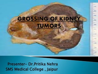
Histopathological Grossing of Kidney Tumors with the common gross differentials encountered,
reference - TATA memorial grossing techniques , Rosai and ackerman surgical pathology , Fletcher , Springer histopathology Specimen
This is a presentation on the topic of cytology of the breast, prepared by Dr Ashish Jawarkar, he is MD in pathology and a teacher at Parul institute of Medical sciences and research Vadodara.
Fluid cytology in serous cavity effusionstashagarwal
The intrathoracic and intraperitoneal organs are covered by a single layer of mesothelial cells, which is continuous with the lining of the thoracic and peritoneal cavities. The potential space between the two layers of epithelium contains a small amount of lubricating fluid.
Serous fluid lies between the membranes lining the body cavities(parietal) and those covering the organs within the cavities(visceral).
Production and reabsorption are normally at a constant rate. They are influenced by
Changes in osmotic and hydrostatic pressure in the blood.
Concentration of chemical constituents in the plasma
Permeability of blood vessels and membranes.
An accumulation of fluid, called an effusion, results from an imbalance of fluid production and reabsorption. This fluid accumulation in the pleural, pericardial, and peritoneal cavities is known as serous effusion.
A malignant neoplasm that contains elements of carcinoma (cancer of epithelial tissue, which is skin and tissue that lines or covers the internal organs) and sarcoma (cancer of connective tissue, such as bone, cartilage, and fat) so extensively intermixed as to indicate neoplasia of epithelial and mesenchymal tissue.
A Rare Case Report of Angiomyolipoma Kidney Associated with Tuberous Sclerosisiosrphr_editor
The IOSR Journal of Pharmacy (IOSRPHR) is an open access online & offline peer reviewed international journal, which publishes innovative research papers, reviews, mini-reviews, short communications and notes dealing with Pharmaceutical Sciences( Pharmaceutical Technology, Pharmaceutics, Biopharmaceutics, Pharmacokinetics, Pharmaceutical/Medicinal Chemistry, Computational Chemistry and Molecular Drug Design, Pharmacognosy & Phytochemistry, Pharmacology, Pharmaceutical Analysis, Pharmacy Practice, Clinical and Hospital Pharmacy, Cell Biology, Genomics and Proteomics, Pharmacogenomics, Bioinformatics and Biotechnology of Pharmaceutical Interest........more details on Aim & Scope).
Histopathological Correlation of Lymph Nodes Imprintsiosrjce
IOSR Journal of Dental and Medical Sciences is one of the speciality Journal in Dental Science and Medical Science published by International Organization of Scientific Research (IOSR). The Journal publishes papers of the highest scientific merit and widest possible scope work in all areas related to medical and dental science. The Journal welcome review articles, leading medical and clinical research articles, technical notes, case reports and others.
Endometrial Ca classification and histopathological features , CAP protocol for reporting , grading and staging tumors
Reference - Robbins , Rosai & Ackerman , Sternberg ,Fletcher ,WHO classification of tumors of female reproductive system, CAP
Gastric Cancer - Deifinition , epidemiology , histological types and molecular genetics and WHO update
Reference - WHO Classificiation of tumors of Digestive system
Rosai and Ackermann
Deep Behavioral Phenotyping in Systems Neuroscience for Functional Atlasing a...Ana Luísa Pinho
Functional Magnetic Resonance Imaging (fMRI) provides means to characterize brain activations in response to behavior. However, cognitive neuroscience has been limited to group-level effects referring to the performance of specific tasks. To obtain the functional profile of elementary cognitive mechanisms, the combination of brain responses to many tasks is required. Yet, to date, both structural atlases and parcellation-based activations do not fully account for cognitive function and still present several limitations. Further, they do not adapt overall to individual characteristics. In this talk, I will give an account of deep-behavioral phenotyping strategies, namely data-driven methods in large task-fMRI datasets, to optimize functional brain-data collection and improve inference of effects-of-interest related to mental processes. Key to this approach is the employment of fast multi-functional paradigms rich on features that can be well parametrized and, consequently, facilitate the creation of psycho-physiological constructs to be modelled with imaging data. Particular emphasis will be given to music stimuli when studying high-order cognitive mechanisms, due to their ecological nature and quality to enable complex behavior compounded by discrete entities. I will also discuss how deep-behavioral phenotyping and individualized models applied to neuroimaging data can better account for the subject-specific organization of domain-general cognitive systems in the human brain. Finally, the accumulation of functional brain signatures brings the possibility to clarify relationships among tasks and create a univocal link between brain systems and mental functions through: (1) the development of ontologies proposing an organization of cognitive processes; and (2) brain-network taxonomies describing functional specialization. To this end, tools to improve commensurability in cognitive science are necessary, such as public repositories, ontology-based platforms and automated meta-analysis tools. I will thus discuss some brain-atlasing resources currently under development, and their applicability in cognitive as well as clinical neuroscience.
What is greenhouse gasses and how many gasses are there to affect the Earth.moosaasad1975
What are greenhouse gasses how they affect the earth and its environment what is the future of the environment and earth how the weather and the climate effects.
Comparing Evolved Extractive Text Summary Scores of Bidirectional Encoder Rep...University of Maribor
Slides from:
11th International Conference on Electrical, Electronics and Computer Engineering (IcETRAN), Niš, 3-6 June 2024
Track: Artificial Intelligence
https://www.etran.rs/2024/en/home-english/
Nutraceutical market, scope and growth: Herbal drug technologyLokesh Patil
As consumer awareness of health and wellness rises, the nutraceutical market—which includes goods like functional meals, drinks, and dietary supplements that provide health advantages beyond basic nutrition—is growing significantly. As healthcare expenses rise, the population ages, and people want natural and preventative health solutions more and more, this industry is increasing quickly. Further driving market expansion are product formulation innovations and the use of cutting-edge technology for customized nutrition. With its worldwide reach, the nutraceutical industry is expected to keep growing and provide significant chances for research and investment in a number of categories, including vitamins, minerals, probiotics, and herbal supplements.
Earliest Galaxies in the JADES Origins Field: Luminosity Function and Cosmic ...Sérgio Sacani
We characterize the earliest galaxy population in the JADES Origins Field (JOF), the deepest
imaging field observed with JWST. We make use of the ancillary Hubble optical images (5 filters
spanning 0.4−0.9µm) and novel JWST images with 14 filters spanning 0.8−5µm, including 7 mediumband filters, and reaching total exposure times of up to 46 hours per filter. We combine all our data
at > 2.3µm to construct an ultradeep image, reaching as deep as ≈ 31.4 AB mag in the stack and
30.3-31.0 AB mag (5σ, r = 0.1” circular aperture) in individual filters. We measure photometric
redshifts and use robust selection criteria to identify a sample of eight galaxy candidates at redshifts
z = 11.5 − 15. These objects show compact half-light radii of R1/2 ∼ 50 − 200pc, stellar masses of
M⋆ ∼ 107−108M⊙, and star-formation rates of SFR ∼ 0.1−1 M⊙ yr−1
. Our search finds no candidates
at 15 < z < 20, placing upper limits at these redshifts. We develop a forward modeling approach to
infer the properties of the evolving luminosity function without binning in redshift or luminosity that
marginalizes over the photometric redshift uncertainty of our candidate galaxies and incorporates the
impact of non-detections. We find a z = 12 luminosity function in good agreement with prior results,
and that the luminosity function normalization and UV luminosity density decline by a factor of ∼ 2.5
from z = 12 to z = 14. We discuss the possible implications of our results in the context of theoretical
models for evolution of the dark matter halo mass function.
Observation of Io’s Resurfacing via Plume Deposition Using Ground-based Adapt...Sérgio Sacani
Since volcanic activity was first discovered on Io from Voyager images in 1979, changes
on Io’s surface have been monitored from both spacecraft and ground-based telescopes.
Here, we present the highest spatial resolution images of Io ever obtained from a groundbased telescope. These images, acquired by the SHARK-VIS instrument on the Large
Binocular Telescope, show evidence of a major resurfacing event on Io’s trailing hemisphere. When compared to the most recent spacecraft images, the SHARK-VIS images
show that a plume deposit from a powerful eruption at Pillan Patera has covered part
of the long-lived Pele plume deposit. Although this type of resurfacing event may be common on Io, few have been detected due to the rarity of spacecraft visits and the previously low spatial resolution available from Earth-based telescopes. The SHARK-VIS instrument ushers in a new era of high resolution imaging of Io’s surface using adaptive
optics at visible wavelengths.
Introduction:
RNA interference (RNAi) or Post-Transcriptional Gene Silencing (PTGS) is an important biological process for modulating eukaryotic gene expression.
It is highly conserved process of posttranscriptional gene silencing by which double stranded RNA (dsRNA) causes sequence-specific degradation of mRNA sequences.
dsRNA-induced gene silencing (RNAi) is reported in a wide range of eukaryotes ranging from worms, insects, mammals and plants.
This process mediates resistance to both endogenous parasitic and exogenous pathogenic nucleic acids, and regulates the expression of protein-coding genes.
What are small ncRNAs?
micro RNA (miRNA)
short interfering RNA (siRNA)
Properties of small non-coding RNA:
Involved in silencing mRNA transcripts.
Called “small” because they are usually only about 21-24 nucleotides long.
Synthesized by first cutting up longer precursor sequences (like the 61nt one that Lee discovered).
Silence an mRNA by base pairing with some sequence on the mRNA.
Discovery of siRNA?
The first small RNA:
In 1993 Rosalind Lee (Victor Ambros lab) was studying a non- coding gene in C. elegans, lin-4, that was involved in silencing of another gene, lin-14, at the appropriate time in the
development of the worm C. elegans.
Two small transcripts of lin-4 (22nt and 61nt) were found to be complementary to a sequence in the 3' UTR of lin-14.
Because lin-4 encoded no protein, she deduced that it must be these transcripts that are causing the silencing by RNA-RNA interactions.
Types of RNAi ( non coding RNA)
MiRNA
Length (23-25 nt)
Trans acting
Binds with target MRNA in mismatch
Translation inhibition
Si RNA
Length 21 nt.
Cis acting
Bind with target Mrna in perfect complementary sequence
Piwi-RNA
Length ; 25 to 36 nt.
Expressed in Germ Cells
Regulates trnasposomes activity
MECHANISM OF RNAI:
First the double-stranded RNA teams up with a protein complex named Dicer, which cuts the long RNA into short pieces.
Then another protein complex called RISC (RNA-induced silencing complex) discards one of the two RNA strands.
The RISC-docked, single-stranded RNA then pairs with the homologous mRNA and destroys it.
THE RISC COMPLEX:
RISC is large(>500kD) RNA multi- protein Binding complex which triggers MRNA degradation in response to MRNA
Unwinding of double stranded Si RNA by ATP independent Helicase
Active component of RISC is Ago proteins( ENDONUCLEASE) which cleave target MRNA.
DICER: endonuclease (RNase Family III)
Argonaute: Central Component of the RNA-Induced Silencing Complex (RISC)
One strand of the dsRNA produced by Dicer is retained in the RISC complex in association with Argonaute
ARGONAUTE PROTEIN :
1.PAZ(PIWI/Argonaute/ Zwille)- Recognition of target MRNA
2.PIWI (p-element induced wimpy Testis)- breaks Phosphodiester bond of mRNA.)RNAse H activity.
MiRNA:
The Double-stranded RNAs are naturally produced in eukaryotic cells during development, and they have a key role in regulating gene expression .
2. pair of organs located in the
abdominal cavity on either side
of the spine in a retroperitoneal
position.
Adrenal glands rest on top of
each kidney
Approx. at vertebral level T12 to
L3, right kidney being slightly
lower than the left.
11–12 cm in length, 5–7.5 cm in
breadth, and 2.5–3.5 cm in
thickness
weighs between 115 and 175 g.
2
3.
4.
5. dual lymphatic
drainage system.
follows the blood
vessels, from the
parenchyma to the
renal sinus,
then to the hilum, and
terminating in the
para-aortic lymph
nodes.
When tumour spreads
from the kidney, it is
initially to the hilar and
then to the para-aortic
lymph nodes.
8. Confirm that patient identification information on the requisition
form and specimen container match.
Pathology number generated should be affixed on the requisition
form and the container.
Note the condition in which specimen is received: fixed and
unfixed, fixed in inadequate or adequate formalin, or autolysed.
If there is incorrect or no identification number, mismatch in
number of specimens mentioned and received then specimens are
returned with details including reason for rejection.
10. 1. Ink the perinephric soft tissue.
2. Weigh and measure the
entire specimen.
3. Do not strip off the renal
capsule and perinephric
soft tissue until their relation
to the tumour is determined.
4. Ureter is used to orient the
specimen. it arises from
medial aspect of kidney
and points inferiorly.
Anterior to posterior (VAP)
5. Shave the ureter, renal vein and renal artery cut margins and submit
these sections.
6. Record whether tumour emboli are seen at the renal vein cut margin.
7. Open the ureter and vessels, and bivalve the kidney.
11. 8. Describe the tumour as following:-
a. Measure the size
b. Give the location
c. Attempt to determine the origin grossly
d. Describe the tumour colour
e. Note any areas of hemorrhage and necrosis
f. Mention gross capsular invasion and submit
section from this area of suspected
perinephric fat invasion.
g. Note and sample any areas with a
homogeneous, tan, bulging surface
( so called fish flesh quality),
which may represent sarcomatoid
dedifferentiation.
12. 9. Describe the hilum – vessel patency,
number and size, color and the lymph
nodes.
10. Describe perinephric fat and look for
involvement of the adrenal gland.
11. The identification of separate tumour
nodules in the perinephric fat in the
gross is diagnostic of pT3 diseases.
12. Serially section the sinus fat at 5 mm
intervals, and 2 sections must be
submitted from the interface between
the tumour and the sinus tissues.
13. Dissect out perihilar and hilar
lymph nodes.
Perinephric fat Invasion:-lost smooth interface
or irregular nodules protruding into fat
13. 14. Sections to be submitted
1) Tumour with perinephric fat and renal capsule
2) Tumour with pelvicalyceal system and hilar
structures
3) Tumour with native kidney
4)Tumor with adrenal gland
5)Renal sinus
6)Renal artery cut margin
7) Renal vein cut margin
8) Ureteric cut margin
9) Radial margin of external inked surface
10) Unremarkable renal parenchyma
11)Tumor showing fish flesh area
12) Hilar lymph nodes
14. Minimum of 5 largest tumors
(if smaller look similar)
If uncertain about histologic type or adverse
findings in remaining tumors, do additional
sampling
Largest T used – label with (m) mpT
Different subtype – separate stage
22. CLEAR CELL CARCINOMA
Are usually golden yellow to red
Spongy to firm
Occur in discrete nodules with
pushing borders.
Necrosis may be present.
Papillary renal cell carcinoma.
The tumor is well circumscribed,
is pale tan, and has a soft, friable
surface.
23. XANTHOGRANUOMATOUS
PYELONEPHRITIS
Appears as single or multiple
golden-yellow nodules in and
around the pelvis and calyces.
The nodules may rarely be
found in the renal capsule or in
adjacent fat.
The gross appearance can
mimic a renal cell carcinoma
MIMICKER OF
RCC
24. Renal Chromophobe Cell Carcinoma.
The tumor is well circumscribed and has
a light brown color.
Collecting duct carcinoma
Occur in the renal medulla and
have a hard gray/white
appearance.
25. Papillary urothelial (transitional
cell) carcinoma of renal pelvis.
Note the exophytic, multifronded
nature of the tumor.
The resection lines have been marked
with ink. Urothelial carcinomas
comprise about 5% to 10% of primary
renal tumors.
26. Pediatric kidney tumors can be broadly divided in to-
1. Wilm’s tumour
2. Non wilm’s tumours
a. Benign tumours ( mesoblastic nephroma)
b. Highly aggressive tumours ( rhabdoid tumours and
clear cell sarcoma)
Types Of Specimens -
1. Radical nephrectomy
2. Nephron sparing partial nephrectomy
27. 1. Surgical specimen should receive in fresh state along with proper
requisition stating relevant details especially the laterality of kidney
and history of neo-adjuvant therapy.
2. Orient the specimen properly by locating the hilum and identify its
contents
3. Ink the outer surface to recognize Gerota’s facia.
4. Document the weight of specimen.
5. Sample the resection margins of ureter , renal vein and renal artery.
6. Sample the hilar fat for lymph nodes and submit separately sent
nodes.
7. Open the specimen preferably through the hilar plane starting from
medial aspect continuing laterally.
8. Multiple parallel cuts may be required in case of large tumour.
9. Cut through the renal hilum will allow examination of tumour with
respect to sinus .
10. Describe the tumour
11. Document the dimensions of the whole specimen and of the
tumour.
28. 12. Insert cotton soaked with formalin in to tumour slices and immerse
the entire specimen in 10% buffered solution for overnight fixation.
13. Take out the specimen and separate the largest slice of tumour and
keep it on grossing board.
14. Orient this slice with respect to hilum and trim it further to a thickness
of 0.5 mm
15. Cut the entire slice into a grid fashion and make the
diagrammatic representation of tumour grid.
16. Give each of sections a unique no. and indicate this on diagram.
17. Carefully examine the remaining parts of capsule related to tumour
and take additional sections wherever invasion is suspected.
18. Dissect out the hilar nodes from the specimen and sample.
19. For multicentric wilm’s tumour sample each nodule.
20. Take at least one random sample of adjacent kidneys.
21. Nephrogenic rests appear paler than the adjacent kidney so
these areas must be sampled.
29. Sections to be submitted-
a. Tumour grid
b. Tumour with renal capsule
c. Tumour with Gerota’s fascia
d. Tumour with hilar structures
e. Renal sinus
f. Renal pelvis and
vessels
g. Ureteric and
vascular cut margins
h. Hilar lymph nodes
i. Adjacent kidney
30.
31. Marked nuclear pleomorphism, hyperchromasia and
large, atypical mitotic figures, present in any of the three
components of Wilm's tumour.
Marker of resistance to chemotherapy.
Presence - unfavourable histology.
classified as focal and diffuse.
EXTENSIVE SAMPLING???
32. Focal Anaplasia Diffuse Anaplasia
No anaplasia within renal vessels or
outside the kidney
Presence of anaplasia in any extrarenal
Site
Random biopsies free of anaplasia Presence of anaplasia in a random
biopsy specimen
confined to =/>1 sharply localized
regions withinmprimary intrarenal
tumour site
Unequivocal anaplasia in one region
of the tumour, coupled with extreme
nuclear unrest elsewhere in the lesion.
surrounded on all sides by
nonanaplastic tissue.
Presence of anaplasia in more than
one tumour slide from different areas
of the tumour.
remaining non-anaplastic tumour
must not show severe nuclear unrest
II to IV anaplastic Wilms tumour has
markedly diminished prognosis and
the patient will require a longer course
of chemotherapy (52 weeks as against
39 weeks).
36. WILM’S TUMOR
Solid, bulging, fleshy tan-white, and
has extended beyond the confines of
the kidney. Foci of necrosis and
hemorrhage appear as darker zones
within the tumor.
37. Clear Cell Sarcoma of Kidney
The tumor is well circumscribed
and whitish, and it bulges on the
cut surface.
Rhabdoid Tumor
Most are well defined and fleshy in
appearance.Frequent necrosis and
hemorrhage. The renal pelvis is usually
involved.
38. Proper staging depends on adequate
sampling of renal specimens
Stage is key to prognostication of renal
cancer patients