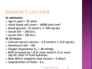
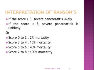
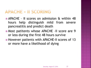
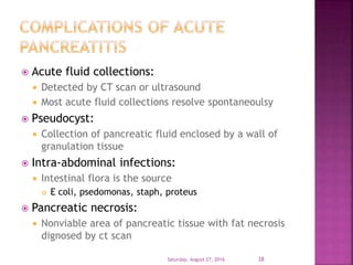
Dr. Shailendra.V.L.
Specialist in Anesthesiology
Al Bukariya general hospital
Saturday, August 27, 2016 1
Saturday, August 27, 2016 2
Saturday, August 27, 2016 3
 500 – 800 ml per day
 Enzymes: amylase, lipase & protease
 Hormones: Insulin, glucagon, somatostatin &
pancreatic polypeptide
Saturday, August 27, 2016 4
Saturday, August 27, 2016 5
 Auto-digestion of pancreas is prevented by
packaging of proteases in precursor form:
 Protease inhibitors:
 Pancreatic secretory trypsin inhibitor
 Serine pprotease inhibitor
 Low calcium levels in pancreas decrease trypsin
activity
 Loss of any of these protective mechanisms
leads to zymogen activation, auto digestion
and acute pancreatitis
Saturday, August 27, 2016 6
 Wide clinical spectrum:
Mild (acute edematous pancreatitis)
Severe (necrotizing pancreatitis)
 Course of the disease:
70% of patients have benign course
30% of patients develop complications
 50% mortality in these patients
Saturday, August 27, 2016 7
I GET SMASHED
 I – Idiopathic
 G – Gall stones
 E – Ethanol (alcohol)
 T – Trauma
 S – Steriods
 M – Mumps, Epstein barr & Cytomegalo virus
 A – Auto immune disease – SLE
 S – Scorpion bites
 H – Hypercalcemia, Hyperlipedemia,
Hypertriglyceremia
 E – ERCP following procedure 2 % develop
 D – SAND- steroids, Azothiprine,NSAIDs,Diuretics –
furesomide, thiazides
Saturday, August 27, 2016 8
 Western countries: 85 % cases due to
alcoholism & gall stones
 Eastern countries: Gall stones
 Children: Trauma
 Adoloscents & young adults: mumps
Saturday, August 27, 2016 9
 The pancreas produces a variety of
enzymes,, exocrine such as proteases, and
saccharidases. These enzymes contribute to
food digestion by breaking down food
tissues. In acute pancreatitis, the worst
offender among these enzymes is protease
trypsinogen which converts to the active
trypsin. Trypsin is responsible for auto-
digestion of the pancreas which in turn
causes the pain and complications of
pancreatitis
Saturday, August 27, 2016 10
 Cellular injury & death result in liberation of
bradykinin peptides, vasoactive substances &
histamines
 Vasodilatation, increased vascular
permeability & edema with profound effect
on lungs
 Systemic inflammatory response syndrome
(SIRS) & Acute respiratory distress syndrome
(ARDS) as well as multi organ failure occur as
result of these cascading effects
Saturday, August 27, 2016 11
Saturday, August 27, 2016 12
Saturday, August 27, 2016 13
Obstruction of pancreatic duct by stone
Ductal hypertension
Breakdown of intracellular compartments
Zymogen activation
gives rise to
Auto-digestion initiated
Saturday, August 27, 2016 14
 Signs which are less common, and indicate severe disease,
include:
 Grey-Turner’s sign(hemorrhagic discoloration of the flanks)
 Cullen’s sign (hemorrhagic discoloration of the umbilicus)
 Grünwald sign (appearance of ecchymosis around the
umbilicus due to local toxic lesion of the vessels)
 Körte's sign (pain or resistance in the zone where the head
of pancreas is located (in epigastrium, 6–7 cm above the
umbilicus)
 Kamenchik's sign (pain with pressure under the xiphoid
process)
 Mayo-Robson's sign (pain while pressing at the top of the
angle lateral to the erector spinae muscles and below the
left 12th rib (left costo-vertebral angle (CVA))[2]
Saturday, August 27, 2016 15
1. Severe abdominal pain radiating to back
2. Nausea & vomitting
3. Elevated serum levels of amylase, lipase
1. Elevated amylase is not very specific test as it
is raised in bowel perforation, ruptured
ectopic, bowel obstruction
2. No definite correlation between severity of
disease & serum amylase levels
3. Serum lipase levels elevated on first day and
persists longer than amylase level
4. CT scan (most preferred)
5. Ultrasound abdomen
Saturday, August 27, 2016 16
 Leukocytosis: occurs frequently
 Hyperglycemia: due to decreased insulin
release, increased glucogon release,
increased output of adrenal glucocorticoids &
catecholamines
 Hypocalcemia: due to
 Hypoalbuminemia
 Hypomagnesemia
 Binding of calcium by free acid albumin
complexes
 Intracellular translocation of calcium
Saturday, August 27, 2016 17
 Jaundice may or may not be present
 Present in only 10% of cases
 SGOP & SGPT levels transiently raised
 Markedly increased LDH levels indicate poor
prognosis
 Hypoxemia paO2 < 60 mm herald the onset
of ARDS
Saturday, August 27, 2016 18
 Diagnosis usually entertained when a patient
with possible predisposition to pancreatitis
presents with severe & constant abdominal
pain, nausea, emesis, fever, tachycardia
 Labs reveal leukocytosis, hypocalcemia,
hyperglycemia
 Diagnosis usually confirmed by finding of
threefold or greater elevated level of serum
amylase or and lipase
 Strong indicators include hemoconcentration
Hct > 44% and signs of organ failure
Saturday, August 27, 2016 19
1. Perforated viscus – peptic ulcer
2. Acute cholecystitis & biliary colic
3. Acute intestinal obstruction
4. Mesentric vascular obstruction
5. Renal colic
6. Myocardial infarction
7. Diabetic ketoacidosis
Saturday, August 27, 2016 20
Saturday, August 27, 2016 21
 Many scoring systems introduced prior CT
scan days
 Most are difficult to remember
 Most accepted score is
 Ranson’s early prognostic signs
 APACHE II scoring
 Imirie’s prognostic criteria
 Simplified prognostic criteria
 Glasgow criteria
Saturday, August 27, 2016 22
Saturday, August 27, 2016 23
 It is a clinical prediction rule for predicting
the severity of acute pancreatitis introduced
in 1974
 Ranson’s score less than 2 indicated mild
disease with good prognosis
 Ranson’s score more than 6 correlates with a
mortality rate of 20% and complication rate
of 80%
Saturday, August 27, 2016 24
At admission:
 age in years > 55 years
 white blood cell count > 16000 cells/mm3
 blood glucose> 10 mmol/L (> 200 mg/dL)
 Serum AST > 250 IU/L
 serum LDH > 350 IU/L
At 24 hours:
 Calcium (serum calcium < 2.0 mmol/L (< 8.0 mg/dL)
 Hemotocrit fall > 10%
 Oxygen (hypoxemia PO2 < 60 mmHg)
 BUN increased by 1.8 or more mmol/L (5 or more
mg/dL) after IV fluid hydration
 Base deficit (negative base excess) > 4 mEq/L
 Sequestration of fluids > 6 L
Saturday, August 27, 2016 25
 If the score ≥ 3, severe pancreatitis likely.
 If the score < 3, severe pancreatitis is
unlikely
Or
 Score 0 to 2 : 2% mortality
 Score 3 to 4 : 15% mortality
 Score 5 to 6 : 40% mortality
 Score 7 to 8 : 100% mortality
Saturday, August 27, 2016 26
 APACHE – II scores on admission & within 48
hours help distinguish mild from severe
pancreatitis and predict death
 Most patitents whose APACHE –II score are 9
or less during the first 48 hours survive
 However patients with APACHE-II scores of 13
or more have a likelihood of dying
Saturday, August 27, 2016 27
 Acute fluid collections:
 Detected by CT scan or ultrasound
 Most acute fluid collections resolve spontaneoulsy
 Pseudocyst:
 Collection of pancreatic fluid enclosed by a wall of
granulation tissue
 Intra-abdominal infections:
 Intestinal flora is the source
 E coli, psedomonas, staph, proteus
 Pancreatic necrosis:
 Nonviable area of pancreatic tissue with fat necrosis
dignosed by ct scan
Saturday, August 27, 2016 28
Saturday, August 27, 2016 29
 Pulmonary:
 Pleural effusion
 Atelectasis
 Mediastinal abscess
 Pneumonitis
 ARDS
 Cardiovascular:
 Hypotension
 Hypovolemia
 Sudden death
 Non specific ST-T changes
 Pericardial effusion
Saturday, August 27, 2016 30
 Hematologic:
 DIC
 Gastro intestinal hemorrhage
 Peptic ulcer disease
 Erosive gastritis
 Pancreatic necrosis with erosion of vessels
 Portal vein thrombosis
 Renal:
 Oliguria
 Azotemia
 Renal artery or vein thrombosis
 Acute tubular necrosis
Saturday, August 27, 2016 31
 Metabolic:
 Hyperglycemia
 Hypertriglyceridemia
 Hypocalcemia
 Encephalopathy
Saturday, August 27, 2016 32
 Early & aggressive resuscitation reduces
mortality rate by reducing incidence of multi
system organ failure.
1. Fluid resusitation
2. Physiologic monitoring
3. Inotropic support
4. Respiratory support
5. Renal support
6. Nutrition
Saturday, August 27, 2016 33
 Tissue fluid shifts due to release of
vasoactive toxins & retroperitoneal losses
 Replacement with crystalloid has no great
advantage over colloids
 Albumin given when levels < than 3g/L
 Blood transfused when Hb < than 10g/dl
 Measurement of CVP is a guide for
replacement
Saturday, August 27, 2016 34
 Incrementally increased infusion of
Dopamine or Dobutamine is preferred
 RESPIRATORY SUPPORT
 Hypoxemia PaO2 < 60mm Hg need early
intubation & ventilation
 Sedation, analgesia & ventilation improve cardiac
performance
Saturday, August 27, 2016 35
 Acute Tubular Necrosis (ATN) seen in acute
pancreatitis, needing temporary hemodialysis
 Prognosis for renal function is good
 Nutrition support:
 Enteral nutrition impossible as gut is rested
 Parentral nutrition is needed
 Early TPN not recommended in hemodynamically
unstable patient
 No specific outcome studies about nutrition
regimes
Saturday, August 27, 2016 36
Saturday, August 27, 2016 37

Acute pancreatitis

  • 1.
    Dr. Shailendra.V.L. Specialist inAnesthesiology Al Bukariya general hospital Saturday, August 27, 2016 1
  • 2.
  • 3.
  • 4.
     500 –800 ml per day  Enzymes: amylase, lipase & protease  Hormones: Insulin, glucagon, somatostatin & pancreatic polypeptide Saturday, August 27, 2016 4
  • 5.
  • 6.
     Auto-digestion ofpancreas is prevented by packaging of proteases in precursor form:  Protease inhibitors:  Pancreatic secretory trypsin inhibitor  Serine pprotease inhibitor  Low calcium levels in pancreas decrease trypsin activity  Loss of any of these protective mechanisms leads to zymogen activation, auto digestion and acute pancreatitis Saturday, August 27, 2016 6
  • 7.
     Wide clinicalspectrum: Mild (acute edematous pancreatitis) Severe (necrotizing pancreatitis)  Course of the disease: 70% of patients have benign course 30% of patients develop complications  50% mortality in these patients Saturday, August 27, 2016 7
  • 8.
    I GET SMASHED I – Idiopathic  G – Gall stones  E – Ethanol (alcohol)  T – Trauma  S – Steriods  M – Mumps, Epstein barr & Cytomegalo virus  A – Auto immune disease – SLE  S – Scorpion bites  H – Hypercalcemia, Hyperlipedemia, Hypertriglyceremia  E – ERCP following procedure 2 % develop  D – SAND- steroids, Azothiprine,NSAIDs,Diuretics – furesomide, thiazides Saturday, August 27, 2016 8
  • 9.
     Western countries:85 % cases due to alcoholism & gall stones  Eastern countries: Gall stones  Children: Trauma  Adoloscents & young adults: mumps Saturday, August 27, 2016 9
  • 10.
     The pancreasproduces a variety of enzymes,, exocrine such as proteases, and saccharidases. These enzymes contribute to food digestion by breaking down food tissues. In acute pancreatitis, the worst offender among these enzymes is protease trypsinogen which converts to the active trypsin. Trypsin is responsible for auto- digestion of the pancreas which in turn causes the pain and complications of pancreatitis Saturday, August 27, 2016 10
  • 11.
     Cellular injury& death result in liberation of bradykinin peptides, vasoactive substances & histamines  Vasodilatation, increased vascular permeability & edema with profound effect on lungs  Systemic inflammatory response syndrome (SIRS) & Acute respiratory distress syndrome (ARDS) as well as multi organ failure occur as result of these cascading effects Saturday, August 27, 2016 11
  • 12.
  • 13.
  • 14.
    Obstruction of pancreaticduct by stone Ductal hypertension Breakdown of intracellular compartments Zymogen activation gives rise to Auto-digestion initiated Saturday, August 27, 2016 14
  • 15.
     Signs whichare less common, and indicate severe disease, include:  Grey-Turner’s sign(hemorrhagic discoloration of the flanks)  Cullen’s sign (hemorrhagic discoloration of the umbilicus)  Grünwald sign (appearance of ecchymosis around the umbilicus due to local toxic lesion of the vessels)  Körte's sign (pain or resistance in the zone where the head of pancreas is located (in epigastrium, 6–7 cm above the umbilicus)  Kamenchik's sign (pain with pressure under the xiphoid process)  Mayo-Robson's sign (pain while pressing at the top of the angle lateral to the erector spinae muscles and below the left 12th rib (left costo-vertebral angle (CVA))[2] Saturday, August 27, 2016 15
  • 16.
    1. Severe abdominalpain radiating to back 2. Nausea & vomitting 3. Elevated serum levels of amylase, lipase 1. Elevated amylase is not very specific test as it is raised in bowel perforation, ruptured ectopic, bowel obstruction 2. No definite correlation between severity of disease & serum amylase levels 3. Serum lipase levels elevated on first day and persists longer than amylase level 4. CT scan (most preferred) 5. Ultrasound abdomen Saturday, August 27, 2016 16
  • 17.
     Leukocytosis: occursfrequently  Hyperglycemia: due to decreased insulin release, increased glucogon release, increased output of adrenal glucocorticoids & catecholamines  Hypocalcemia: due to  Hypoalbuminemia  Hypomagnesemia  Binding of calcium by free acid albumin complexes  Intracellular translocation of calcium Saturday, August 27, 2016 17
  • 18.
     Jaundice mayor may not be present  Present in only 10% of cases  SGOP & SGPT levels transiently raised  Markedly increased LDH levels indicate poor prognosis  Hypoxemia paO2 < 60 mm herald the onset of ARDS Saturday, August 27, 2016 18
  • 19.
     Diagnosis usuallyentertained when a patient with possible predisposition to pancreatitis presents with severe & constant abdominal pain, nausea, emesis, fever, tachycardia  Labs reveal leukocytosis, hypocalcemia, hyperglycemia  Diagnosis usually confirmed by finding of threefold or greater elevated level of serum amylase or and lipase  Strong indicators include hemoconcentration Hct > 44% and signs of organ failure Saturday, August 27, 2016 19
  • 20.
    1. Perforated viscus– peptic ulcer 2. Acute cholecystitis & biliary colic 3. Acute intestinal obstruction 4. Mesentric vascular obstruction 5. Renal colic 6. Myocardial infarction 7. Diabetic ketoacidosis Saturday, August 27, 2016 20
  • 21.
  • 22.
     Many scoringsystems introduced prior CT scan days  Most are difficult to remember  Most accepted score is  Ranson’s early prognostic signs  APACHE II scoring  Imirie’s prognostic criteria  Simplified prognostic criteria  Glasgow criteria Saturday, August 27, 2016 22
  • 23.
  • 24.
     It isa clinical prediction rule for predicting the severity of acute pancreatitis introduced in 1974  Ranson’s score less than 2 indicated mild disease with good prognosis  Ranson’s score more than 6 correlates with a mortality rate of 20% and complication rate of 80% Saturday, August 27, 2016 24
  • 25.
    At admission:  agein years > 55 years  white blood cell count > 16000 cells/mm3  blood glucose> 10 mmol/L (> 200 mg/dL)  Serum AST > 250 IU/L  serum LDH > 350 IU/L At 24 hours:  Calcium (serum calcium < 2.0 mmol/L (< 8.0 mg/dL)  Hemotocrit fall > 10%  Oxygen (hypoxemia PO2 < 60 mmHg)  BUN increased by 1.8 or more mmol/L (5 or more mg/dL) after IV fluid hydration  Base deficit (negative base excess) > 4 mEq/L  Sequestration of fluids > 6 L Saturday, August 27, 2016 25
  • 26.
     If thescore ≥ 3, severe pancreatitis likely.  If the score < 3, severe pancreatitis is unlikely Or  Score 0 to 2 : 2% mortality  Score 3 to 4 : 15% mortality  Score 5 to 6 : 40% mortality  Score 7 to 8 : 100% mortality Saturday, August 27, 2016 26
  • 27.
     APACHE –II scores on admission & within 48 hours help distinguish mild from severe pancreatitis and predict death  Most patitents whose APACHE –II score are 9 or less during the first 48 hours survive  However patients with APACHE-II scores of 13 or more have a likelihood of dying Saturday, August 27, 2016 27
  • 28.
     Acute fluidcollections:  Detected by CT scan or ultrasound  Most acute fluid collections resolve spontaneoulsy  Pseudocyst:  Collection of pancreatic fluid enclosed by a wall of granulation tissue  Intra-abdominal infections:  Intestinal flora is the source  E coli, psedomonas, staph, proteus  Pancreatic necrosis:  Nonviable area of pancreatic tissue with fat necrosis dignosed by ct scan Saturday, August 27, 2016 28
  • 29.
  • 30.
     Pulmonary:  Pleuraleffusion  Atelectasis  Mediastinal abscess  Pneumonitis  ARDS  Cardiovascular:  Hypotension  Hypovolemia  Sudden death  Non specific ST-T changes  Pericardial effusion Saturday, August 27, 2016 30
  • 31.
     Hematologic:  DIC Gastro intestinal hemorrhage  Peptic ulcer disease  Erosive gastritis  Pancreatic necrosis with erosion of vessels  Portal vein thrombosis  Renal:  Oliguria  Azotemia  Renal artery or vein thrombosis  Acute tubular necrosis Saturday, August 27, 2016 31
  • 32.
     Metabolic:  Hyperglycemia Hypertriglyceridemia  Hypocalcemia  Encephalopathy Saturday, August 27, 2016 32
  • 33.
     Early &aggressive resuscitation reduces mortality rate by reducing incidence of multi system organ failure. 1. Fluid resusitation 2. Physiologic monitoring 3. Inotropic support 4. Respiratory support 5. Renal support 6. Nutrition Saturday, August 27, 2016 33
  • 34.
     Tissue fluidshifts due to release of vasoactive toxins & retroperitoneal losses  Replacement with crystalloid has no great advantage over colloids  Albumin given when levels < than 3g/L  Blood transfused when Hb < than 10g/dl  Measurement of CVP is a guide for replacement Saturday, August 27, 2016 34
  • 35.
     Incrementally increasedinfusion of Dopamine or Dobutamine is preferred  RESPIRATORY SUPPORT  Hypoxemia PaO2 < 60mm Hg need early intubation & ventilation  Sedation, analgesia & ventilation improve cardiac performance Saturday, August 27, 2016 35
  • 36.
     Acute TubularNecrosis (ATN) seen in acute pancreatitis, needing temporary hemodialysis  Prognosis for renal function is good  Nutrition support:  Enteral nutrition impossible as gut is rested  Parentral nutrition is needed  Early TPN not recommended in hemodynamically unstable patient  No specific outcome studies about nutrition regimes Saturday, August 27, 2016 36
  • 37.