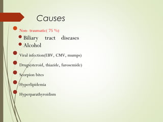
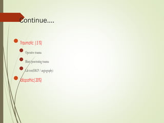
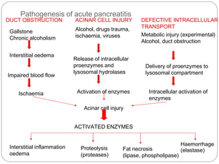
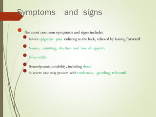
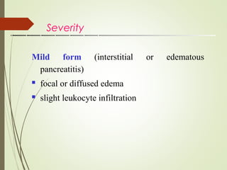
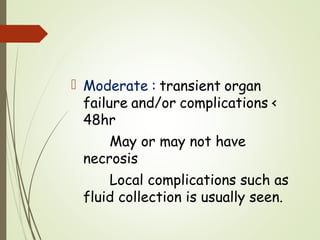
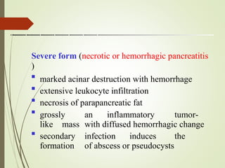
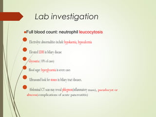
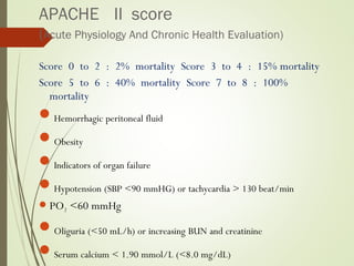
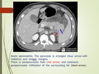
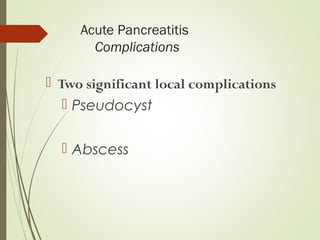
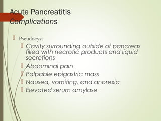
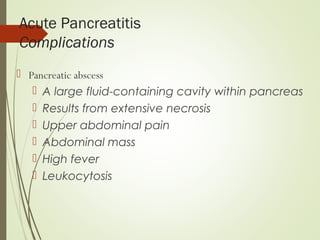
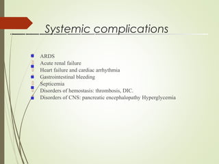
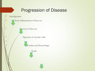
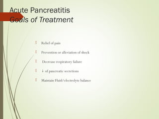
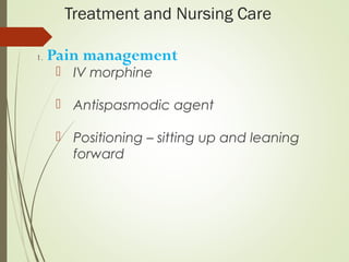
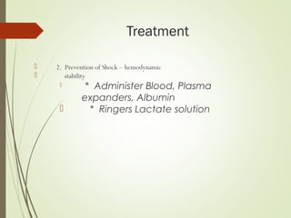
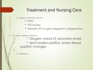
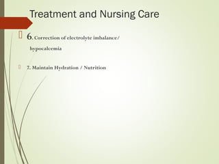
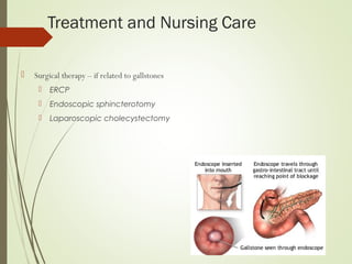
Welcome
To
Morning Session
Acute Pancreatitis
Dr.Amitav Biswas
Intern Doctor
Department of Medicine
TMMC&H
Definition
 Acute pancreatitis is a inflammation of the
pancreas
pancreatic
causes.
induced by the activation of the
enzymes derived from various
Causes
Non- traumatic( 75 %)
Biliary tract diseases
Alcohol
Viral infection(EBV, CMV, mumps)
Drugs(steroid, thiazide, furosemide)
Scorpion bites
Hyperlipidemia
Hyperparathyroidism
Continue….
Traumatic (5%)
Operativetrauma
Blunt/penetratingtrauma
Labtest(ERCP/angiography)
Idiopathic(20%)
Pathogenesis of acute pancreatitis
Interstitial oedema
Impaired blood flow
Ischaemia
Acinar cell injury
ACTIVATED ENZYMES
Interstitial inflammation
oedema
Gallstone
Chronic alcoholism
Release of intracellular
proenzymes and
lysosomal hydrolases
Activation of enzymes
Delivery of proenzymes to
lysosomal compartment
Intracellular activation of
enzymes
Proteolysis
(proteases)
Fat necrosis
(lipase, phospholipase)
Haemorrhage
(elastase)
ACINAR CELL INJURY
Alcohol, drugs trauma,
ischaemia, viruses
DEFECTIVE INTRACELLULAR
TRANSPORT
Metabolic injury (experimental)
Alcohol, duct obstruction
DUCT OBSTRUCTION
Symptoms and signs
The most common symptoms and signs include:
Severe epigastric pain radiating to the back, relieved by leaning forward
Nausea, vomiting, diarrhea and loss of appetite
Fever/chills
Hemodynamic instability, including shock
In severe case may present with tenderness, guarding, rebound.
Signs which are less common, and indicate severe disease, include:
Grey-Turner's sign (hemorrhagic discoloration of the flanks)
Cullen's sign (hemorrhagic discoloration of the umbilicus)
Cullen sign
Grey-Turner sign- discolouration in the flanks
Severity
Mild form (interstitial or edematous
pancreatitis)
focal or diffused edema
slight leukocyte infiltration


 Moderate : transient organ
failure and/or complications <
48hr
May or may not have
necrosis
Local complications such as
fluid collection is usually seen.
Severe form (necrotic or hemorrhagic pancreatitis
)

marked acinar destruction with hemorrhage
extensive leukocyte infiltration
necrosis of parapancreatic fat
grossly an inflammatory tumor-
like mass with diffused hemorrhagic change
secondary infection induces the
formation of abscess or pseudocysts




Full blood count: neutrophil leucocytosis
Lab investigation
Electrolyte abnormalities include hypokaemia, hypocalcemia
ElevatedLDHinbiliary disease
Glycosuria(10%ofcases)
Bloodsugar:hyperglycaemiainseverecases
Ultrasound look for stonesinbiliary tract diseases.
AbdominalCTscanmay revealphlegmon(inflammatory mass), pseudocyst or
abscess(complications of acute pancreatitis)
Lab investigation
Amylase and lipase
Elevated serum amylase and lipase levels, in combination with severe
abdominal pain, often trigger the initial diagnosis of acute
pancreatitis.
Serum lipase rises 4 to 8 hours from the onset of symptoms and
normalizes within 7 to 14 days after treatment.
Marked elevation of serum amylase level during first 24 hours
Reasons for false positive elevated serum amylase include salivary gland disease
(elevated salivary amylase) and macroamylasemia.
Ifthelipaselevelisabout2.5 to 3 times thatofAmylase,itis an indication of
pancreatitis due to Alcohol or gallstone
Thedegreeofamylase/lipaseelevationdoes not correlatewith severity of acute
pancreatitis.
Ranson Score
predicting the severity of acute pancreatitis
At admission
age in years > 55 years
white blood cell count > 16000 cells/mm3
blood glucose > 11 mmol/L (> 200 mg/dL)
serum ALT > 250 IU/L
serum LDH > 350 IU/L At 48 hours
Calcium(serumcalcium<2.0mmol/L(<8.0mg/dL)
Hematocritfall>10%
Oxygen(hypoxemiaPO2<60mmHg)
BUNincreasedby1.8ormoremmol/L(5ormoremg/dL)afterIVfluidhydration
Basedeficit(negativebaseexcess)>4mEq/L
Sequestrationoffluids>6L
APACHE II score
(Acute Physiology And Chronic Health Evaluation)
Score 0 to 2 : 2% mortality Score 3 to 4 : 15% mortality
Score 5 to 6 : 40% mortality Score 7 to 8 : 100%
mortality
Hemorrhagic peritoneal fluid
Obesity
Indicators of organ failure
Hypotension (SBP <90 mmHG) or tachycardia > 130 beat/min
PO2 <60 mmHg
Oliguria (<50 mL/h) or increasing BUN and creatinine
Serum calcium < 1.90 mmol/L (<8.0 mg/dL)
Acute pancreatitis. The pancreas is enlarged (blue arrow) with
indistinct and shaggy margins.
There is peripancreatic fluid (red arrow) and extensive
peripancreatic infiltration of the surrounding fat (black arrow).
Acute Pancreatitis
Complications
 Two significant local complications
 Pseudocyst
 Abscess
Acute Pancreatitis
Complications
 Pseudocyst
 Cavity surrounding outside of pancreas
filled with necrotic products and liquid
secretions
 Abdominal pain
 Palpable epigastric mass
 Nausea, vomiting, and anorexia
 Elevated serum amylase
Acute Pancreatitis
Complications
 Pancreatic abscess
 A large fluid-containing cavity within pancreas
 Results from extensive necrosis
 Upper abdominal pain
 Abdominal mass
 High fever
 Leukocytosis
Systemic complications
 ARDS
 Acute renal failure
 Heart failure and cardiac arrhythmia
 Gastrointestinal bleeding
 Septicemia
 Disorders of hemostasis: thrombosis, DIC.
 Disorders of CNS: pancreatic encephalopathy Hyperglycemia





Progression of Disease
 Autodigestion
 Acute Inflammation of Pancreas
 Necrosis of Pancreas
 Digestion of vascular walls

 Thrombus and Hemorrhage
 Death
Acute Pancreatitis
Goals of Treatment
 Relief of pain
 Prevention or alleviation of shock
 Decrease respiratory failure
 ↓ of pancreatic secretions
 Maintain Fluid/electrolyte balance
Treatment and Nursing Care
1. Pain management
 IV morphine
 Antispasmodic agent
 Positioning – sitting up and leaning
forward
Treatment
 2. Prevention of Shock – hemodynamic
 stability
 * Administer Blood, Plasma
expanders, Albumin
 * Ringers Lactate solution
Treatment and Nursing Care
 3. Suppress pancreatic enzymes
 * NPO
 * NG suction
 * Antacids, H2 receptor antagonists, antispasmotics
4. Decrease respiratory distress
 * Oxygen; check O2 saturation levels
 * Semi-fowlers position, knees flexed,
position changes

 5. Antibiotics
Treatment and Nursing Care
 6. Correction of electrolyte imbalance/
hypocalcemia
 7. Maintain Hydration / Nutrition
Treatment and Nursing Care
 Surgical therapy – if related to gallstones
 ERCP
 Endoscopic sphincterotomy
 Laparoscopic cholecystectomy
 Follow up care
 Dietary teaching
 High-carbohydrate, low-fat diet
 Abstinence from alcohol,
 Patient/family teaching
 * Signs of infection, high blood
 glucose, steatorrhea

Treatment - Home Care
Reference
• Davidson’s Principles and Practice of Medicie 22nd
edition
• Oxford Handbook of Clinical Medicine 10th
edition
• Harrison’s Principles of Internal Medicine 19th
edition
• www.Medscape.com
Thank You

Acute pancreatitis kollol

  • 1.
  • 2.
    Acute Pancreatitis Dr.Amitav Biswas InternDoctor Department of Medicine TMMC&H
  • 3.
    Definition  Acute pancreatitisis a inflammation of the pancreas pancreatic causes. induced by the activation of the enzymes derived from various
  • 4.
    Causes Non- traumatic( 75%) Biliary tract diseases Alcohol Viral infection(EBV, CMV, mumps) Drugs(steroid, thiazide, furosemide) Scorpion bites Hyperlipidemia Hyperparathyroidism
  • 5.
  • 6.
    Pathogenesis of acutepancreatitis Interstitial oedema Impaired blood flow Ischaemia Acinar cell injury ACTIVATED ENZYMES Interstitial inflammation oedema Gallstone Chronic alcoholism Release of intracellular proenzymes and lysosomal hydrolases Activation of enzymes Delivery of proenzymes to lysosomal compartment Intracellular activation of enzymes Proteolysis (proteases) Fat necrosis (lipase, phospholipase) Haemorrhage (elastase) ACINAR CELL INJURY Alcohol, drugs trauma, ischaemia, viruses DEFECTIVE INTRACELLULAR TRANSPORT Metabolic injury (experimental) Alcohol, duct obstruction DUCT OBSTRUCTION
  • 7.
    Symptoms and signs Themost common symptoms and signs include: Severe epigastric pain radiating to the back, relieved by leaning forward Nausea, vomiting, diarrhea and loss of appetite Fever/chills Hemodynamic instability, including shock In severe case may present with tenderness, guarding, rebound.
  • 8.
    Signs which areless common, and indicate severe disease, include: Grey-Turner's sign (hemorrhagic discoloration of the flanks) Cullen's sign (hemorrhagic discoloration of the umbilicus)
  • 9.
  • 10.
  • 11.
    Severity Mild form (interstitialor edematous pancreatitis) focal or diffused edema slight leukocyte infiltration  
  • 12.
     Moderate :transient organ failure and/or complications < 48hr May or may not have necrosis Local complications such as fluid collection is usually seen.
  • 13.
    Severe form (necroticor hemorrhagic pancreatitis )  marked acinar destruction with hemorrhage extensive leukocyte infiltration necrosis of parapancreatic fat grossly an inflammatory tumor- like mass with diffused hemorrhagic change secondary infection induces the formation of abscess or pseudocysts    
  • 14.
    Full blood count:neutrophil leucocytosis Lab investigation Electrolyte abnormalities include hypokaemia, hypocalcemia ElevatedLDHinbiliary disease Glycosuria(10%ofcases) Bloodsugar:hyperglycaemiainseverecases Ultrasound look for stonesinbiliary tract diseases. AbdominalCTscanmay revealphlegmon(inflammatory mass), pseudocyst or abscess(complications of acute pancreatitis)
  • 15.
    Lab investigation Amylase andlipase Elevated serum amylase and lipase levels, in combination with severe abdominal pain, often trigger the initial diagnosis of acute pancreatitis. Serum lipase rises 4 to 8 hours from the onset of symptoms and normalizes within 7 to 14 days after treatment. Marked elevation of serum amylase level during first 24 hours
  • 16.
    Reasons for falsepositive elevated serum amylase include salivary gland disease (elevated salivary amylase) and macroamylasemia. Ifthelipaselevelisabout2.5 to 3 times thatofAmylase,itis an indication of pancreatitis due to Alcohol or gallstone Thedegreeofamylase/lipaseelevationdoes not correlatewith severity of acute pancreatitis.
  • 17.
    Ranson Score predicting theseverity of acute pancreatitis At admission age in years > 55 years white blood cell count > 16000 cells/mm3 blood glucose > 11 mmol/L (> 200 mg/dL) serum ALT > 250 IU/L serum LDH > 350 IU/L At 48 hours
  • 18.
  • 19.
    APACHE II score (AcutePhysiology And Chronic Health Evaluation) Score 0 to 2 : 2% mortality Score 3 to 4 : 15% mortality Score 5 to 6 : 40% mortality Score 7 to 8 : 100% mortality Hemorrhagic peritoneal fluid Obesity Indicators of organ failure Hypotension (SBP <90 mmHG) or tachycardia > 130 beat/min PO2 <60 mmHg Oliguria (<50 mL/h) or increasing BUN and creatinine Serum calcium < 1.90 mmol/L (<8.0 mg/dL)
  • 20.
    Acute pancreatitis. Thepancreas is enlarged (blue arrow) with indistinct and shaggy margins. There is peripancreatic fluid (red arrow) and extensive peripancreatic infiltration of the surrounding fat (black arrow).
  • 21.
    Acute Pancreatitis Complications  Twosignificant local complications  Pseudocyst  Abscess
  • 22.
    Acute Pancreatitis Complications  Pseudocyst Cavity surrounding outside of pancreas filled with necrotic products and liquid secretions  Abdominal pain  Palpable epigastric mass  Nausea, vomiting, and anorexia  Elevated serum amylase
  • 23.
    Acute Pancreatitis Complications  Pancreaticabscess  A large fluid-containing cavity within pancreas  Results from extensive necrosis  Upper abdominal pain  Abdominal mass  High fever  Leukocytosis
  • 24.
    Systemic complications  ARDS Acute renal failure  Heart failure and cardiac arrhythmia  Gastrointestinal bleeding  Septicemia  Disorders of hemostasis: thrombosis, DIC.  Disorders of CNS: pancreatic encephalopathy Hyperglycemia     
  • 25.
    Progression of Disease Autodigestion  Acute Inflammation of Pancreas  Necrosis of Pancreas  Digestion of vascular walls   Thrombus and Hemorrhage  Death
  • 26.
    Acute Pancreatitis Goals ofTreatment  Relief of pain  Prevention or alleviation of shock  Decrease respiratory failure  ↓ of pancreatic secretions  Maintain Fluid/electrolyte balance
  • 27.
    Treatment and NursingCare 1. Pain management  IV morphine  Antispasmodic agent  Positioning – sitting up and leaning forward
  • 28.
    Treatment  2. Preventionof Shock – hemodynamic  stability  * Administer Blood, Plasma expanders, Albumin  * Ringers Lactate solution
  • 29.
    Treatment and NursingCare  3. Suppress pancreatic enzymes  * NPO  * NG suction  * Antacids, H2 receptor antagonists, antispasmotics 4. Decrease respiratory distress  * Oxygen; check O2 saturation levels  * Semi-fowlers position, knees flexed, position changes   5. Antibiotics
  • 30.
    Treatment and NursingCare  6. Correction of electrolyte imbalance/ hypocalcemia  7. Maintain Hydration / Nutrition
  • 31.
    Treatment and NursingCare  Surgical therapy – if related to gallstones  ERCP  Endoscopic sphincterotomy  Laparoscopic cholecystectomy
  • 32.
     Follow upcare  Dietary teaching  High-carbohydrate, low-fat diet  Abstinence from alcohol,  Patient/family teaching  * Signs of infection, high blood  glucose, steatorrhea  Treatment - Home Care
  • 33.
    Reference • Davidson’s Principlesand Practice of Medicie 22nd edition • Oxford Handbook of Clinical Medicine 10th edition • Harrison’s Principles of Internal Medicine 19th edition • www.Medscape.com
  • 34.