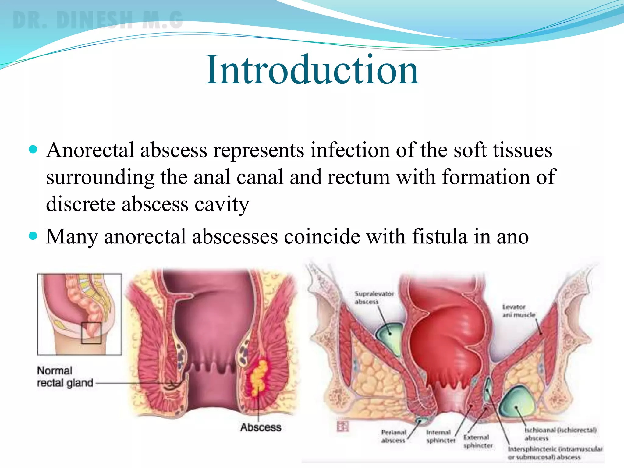
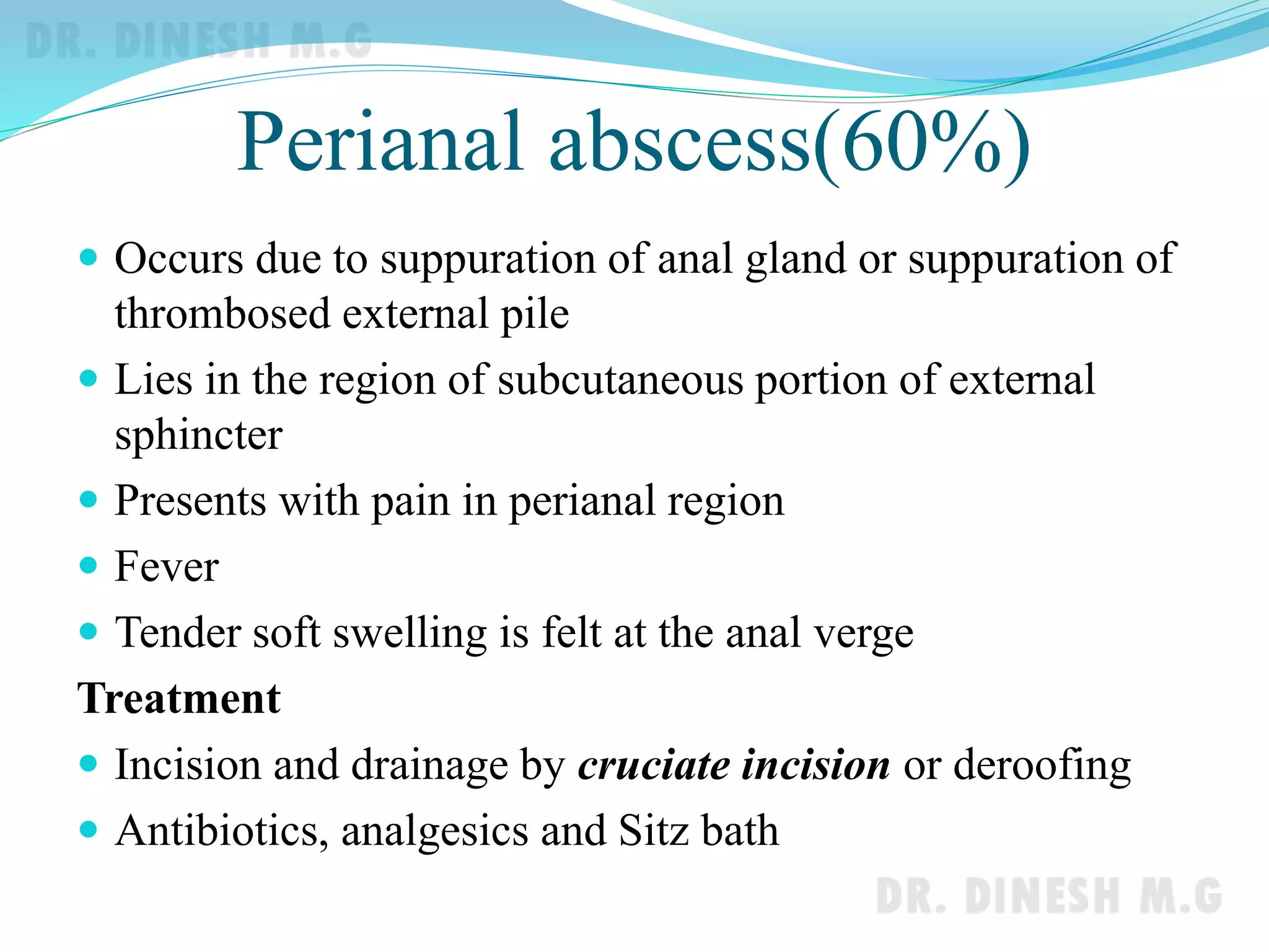
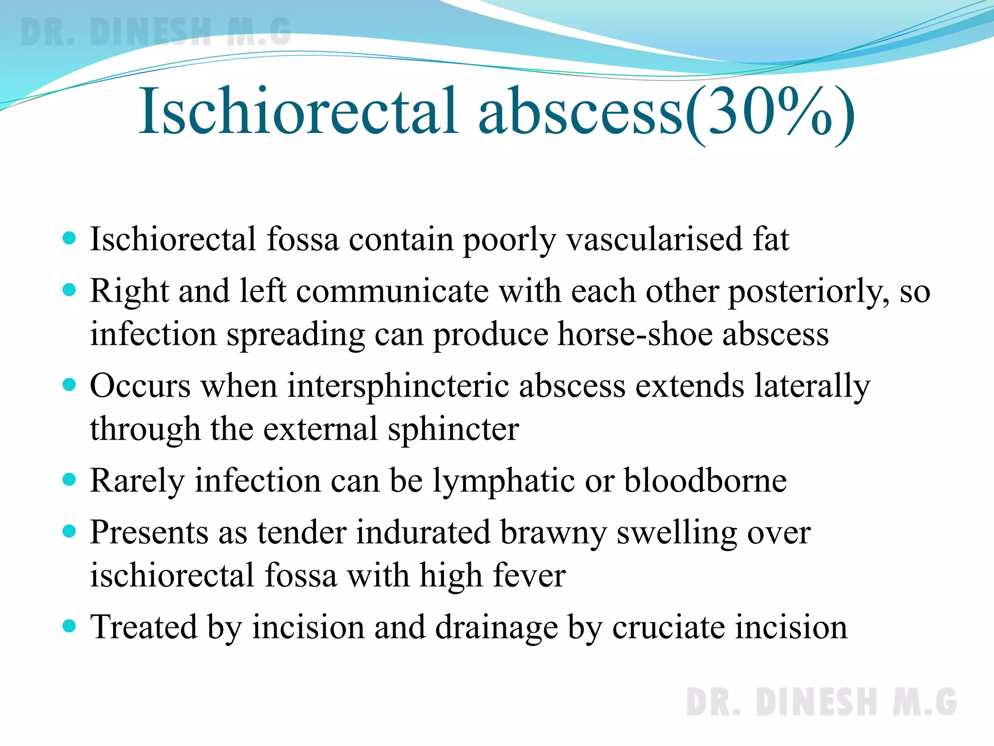
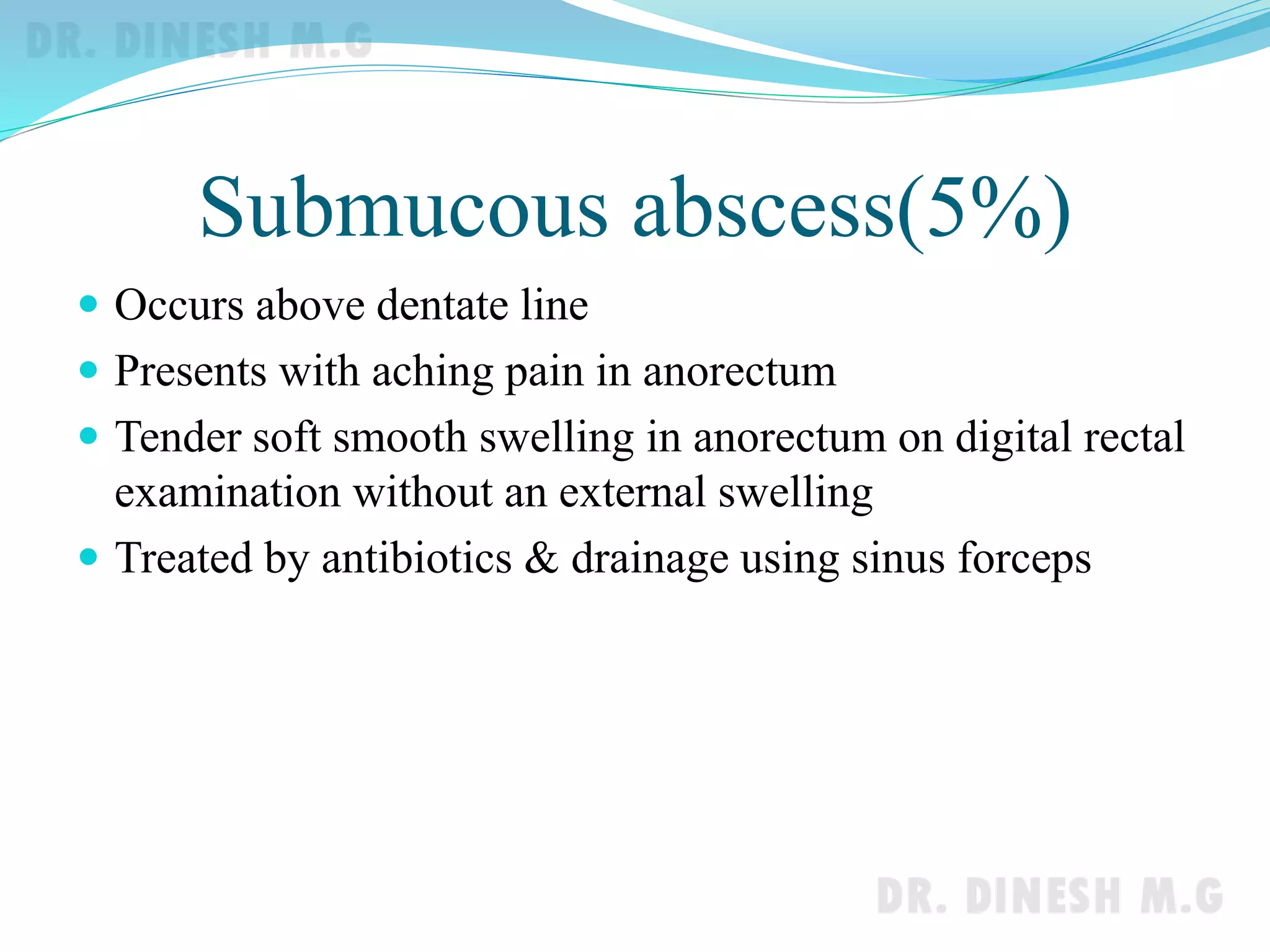
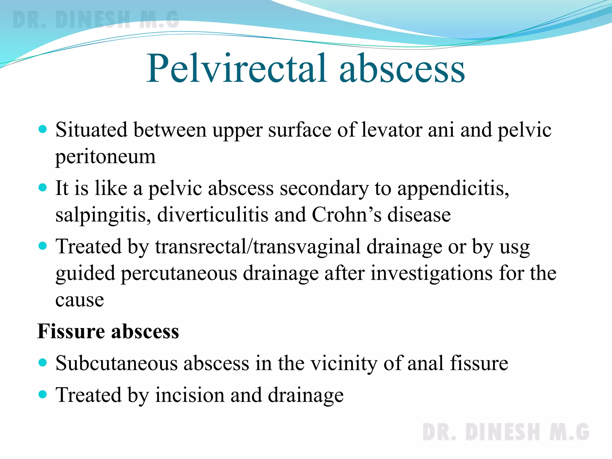
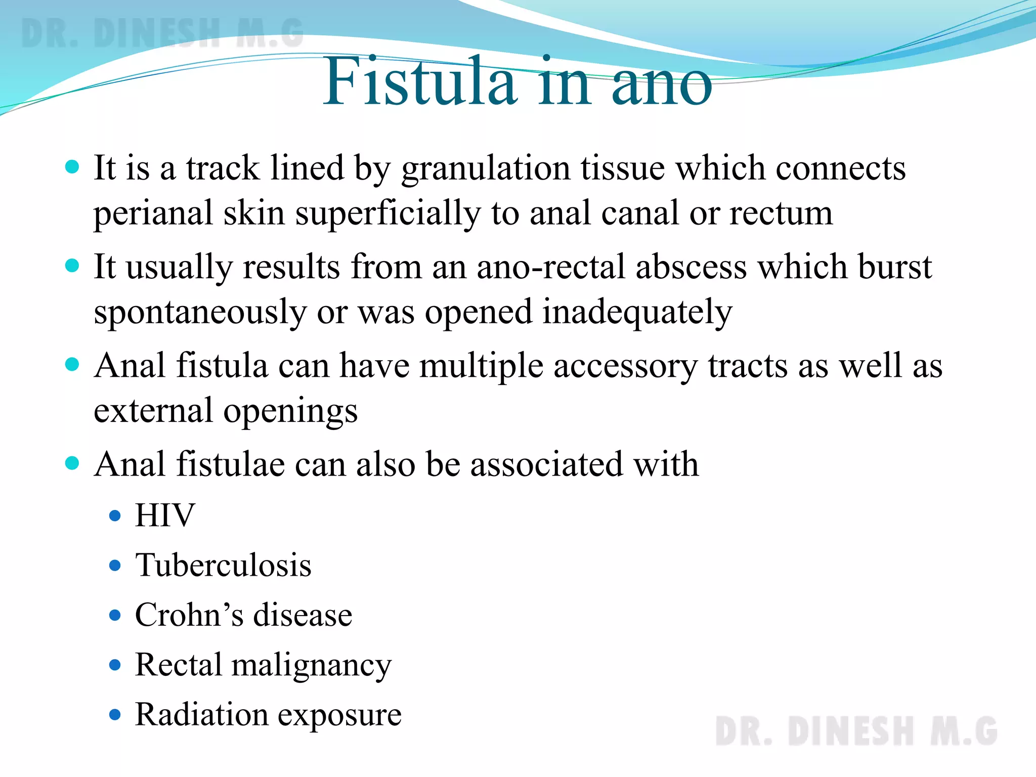
Dr. Dinesh. M.G
Professor of Surgery
J.J.M.M.C.
Davangere
Introduction
 Anorectal abscess represents infection of the soft tissues
surrounding the anal canal and rectum with formation of
discrete abscess cavity
 Many anorectal abscesses coincide with fistula in ano
Incidence
 Peak incidence in 3rd and 4th decades
 Males are more affected with male to female ratio of 2:1
Aetiology
 Infection of anal gland in relation to crypts( 90%)
 Injury to anorectum( e.g. fishbone)
 Blood borne infection
 Extension of cutaneous boil
 Fissure in ano
 Crohn’s disease
 Tuberculosis
 Rectal malignancy
Patients with diabetes and AIDS are more prone for
anorectal abscesses
Pathophysiology
 Infection of anal glands( about 4-10 near dentate line) in
anal crypts leads to suppuration and abscess typically
forms in the intersphincteric space and can spread along
various spaces
 Common causative organisms
 E.coli
 Bacteroids
 B. Proteus
 Streptococcus
 Staphylococcus aureus
 Fistula is more likely if pus culture reveals bowel
organisms rather than skin organisms
Classification
 Perianal
 Submucosal
 Ischiorectal
 Pelvirectal(supralevator)
 Fissure abscess
Perianal abscess(60%)
 Occurs due to suppuration of anal gland or suppuration of
thrombosed external pile
 Lies in the region of subcutaneous portion of external
sphincter
 Presents with pain in perianal region
 Fever
 Tender soft swelling is felt at the anal verge
Treatment
 Incision and drainage by cruciate incision or deroofing
 Antibiotics, analgesics and Sitz bath
Ischiorectal abscess(30%)
 Ischiorectal fossa contain poorly vascularised fat
 Right and left communicate with each other posteriorly, so
infection spreading can produce horse-shoe abscess
 Occurs when intersphincteric abscess extends laterally
through the external sphincter
 Rarely infection can be lymphatic or bloodborne
 Presents as tender indurated brawny swelling over
ischiorectal fossa with high fever
 Treated by incision and drainage by cruciate incision
Ischiorectal abscess
Cruciate incision & deroofing
Submucous abscess(5%)
 Occurs above dentate line
 Presents with aching pain in anorectum
 Tender soft smooth swelling in anorectum on digital rectal
examination without an external swelling
 Treated by antibiotics & drainage using sinus forceps
Pelvirectal abscess
 Situated between upper surface of levator ani and pelvic
peritoneum
 It is like a pelvic abscess secondary to appendicitis,
salpingitis, diverticulitis and Crohn’s disease
 Treated by transrectal/transvaginal drainage or by usg
guided percutaneous drainage after investigations for the
cause
Fissure abscess
 Subcutaneous abscess in the vicinity of anal fissure
 Treated by incision and drainage
Fistula in ano
 It is a track lined by granulation tissue which connects
perianal skin superficially to anal canal or rectum
 It usually results from an ano-rectal abscess which burst
spontaneously or was opened inadequately
 Anal fistula can have multiple accessory tracts as well as
external openings
 Anal fistulae can also be associated with
 HIV
 Tuberculosis
 Crohn’s disease
 Rectal malignancy
 Radiation exposure
Classification of anal fistulae
Standard classification
1. Subcutaneous
2. Submucous
3. Low anal
4. High anal
5. Pelvirectal
Classification of anal fistulae
Park’s classification
1. Intersphincteric
2. Transphincteric
3. Supralevator /Suprasphincteric
4. Extrasphincteric
Other classifications
Low fistula High fistula
Internal opening below anorectal
ring
Internal opening at or above
anorectal ring
Simple fistula Complex fistula
Do not have secondary tracts Have multiple secondary tracts
Low-level fistulae
Clinical features
 Seropurulent discharge with one or more opening on one
side of midline with induration of tract
 Horese shoe fistulae involving both ischiorectal fossae
present with openings on both sides
 Anorectal examination may reveal nodular internal
opening
Goodsall’s rule
 Anterior external opening
is of direct type
 Posterior external opening has
curved track with an internal
opening in the midline
posteriorly
Investigations
 Complete haemogram and HIV
 Chest x-ray for pulmonary TB
 Fistulogram- not popular nowadays
 Transrectal ultrasound : useful to identify internal opening
 MRI fistulogram
Endorectal ultrasound MRI of anal fistulae
Differential diagnosis
 Urethral fistula in males
 Chronic infection of Bartholin’s gland in females
 Pilonidal sinus
 Hidradenitis suppurativa
 Carcinoma
 Crohn’s,Tuberculosis and Ulcerative colitis
Treatment
 Fistulotomy
 Fistulectomy
 Seton
Newer methods of treatment
 Anal fistula plug repair
 Injection of glue into the fistulous tract
 Mucosal flap procedure
 LIFT( Ligation of intersphincteric fistula track)
 VAAFT procedure( Video assisted anal fistula track
ligation)
Treatment
 Fistulotomy is laying open of track after probing the
fistulous track
Treatment
 Fistulectomy is complete excision of fistulous tract after
probing
Treatment
 Seton is a non absorbable suture (e.g. silk) or tape that is
passed across the fistula and tied
Treatment
 Anal fistula plug repair
 Glue injection
Treatment
 Video assisted anal fistula track ligation
High-level fistula
 Internal opening is above anorectal ring
 Common causes are
 Crohn’s disease
 Ulcerative colitis
 Trauma
 Carcinoma
 Foreign body
 Investigations
 Colonoscopy/barium enema, chest x-ray & biopsy
Treatment of high-level fistulae
 Staged procedure
 Initial colostomy
 Definitive procedure with sphincter repair
 Colostomy closure
 Seton
 Loose setons
 Cutting setons
Thank you

Anorectal abscess & Anal fistulae

  • 1.
    Dr. Dinesh. M.G Professorof Surgery J.J.M.M.C. Davangere
  • 2.
    Introduction  Anorectal abscessrepresents infection of the soft tissues surrounding the anal canal and rectum with formation of discrete abscess cavity  Many anorectal abscesses coincide with fistula in ano
  • 3.
    Incidence  Peak incidencein 3rd and 4th decades  Males are more affected with male to female ratio of 2:1
  • 4.
    Aetiology  Infection ofanal gland in relation to crypts( 90%)  Injury to anorectum( e.g. fishbone)  Blood borne infection  Extension of cutaneous boil  Fissure in ano  Crohn’s disease  Tuberculosis  Rectal malignancy Patients with diabetes and AIDS are more prone for anorectal abscesses
  • 5.
    Pathophysiology  Infection ofanal glands( about 4-10 near dentate line) in anal crypts leads to suppuration and abscess typically forms in the intersphincteric space and can spread along various spaces  Common causative organisms  E.coli  Bacteroids  B. Proteus  Streptococcus  Staphylococcus aureus  Fistula is more likely if pus culture reveals bowel organisms rather than skin organisms
  • 6.
    Classification  Perianal  Submucosal Ischiorectal  Pelvirectal(supralevator)  Fissure abscess
  • 7.
    Perianal abscess(60%)  Occursdue to suppuration of anal gland or suppuration of thrombosed external pile  Lies in the region of subcutaneous portion of external sphincter  Presents with pain in perianal region  Fever  Tender soft swelling is felt at the anal verge Treatment  Incision and drainage by cruciate incision or deroofing  Antibiotics, analgesics and Sitz bath
  • 8.
    Ischiorectal abscess(30%)  Ischiorectalfossa contain poorly vascularised fat  Right and left communicate with each other posteriorly, so infection spreading can produce horse-shoe abscess  Occurs when intersphincteric abscess extends laterally through the external sphincter  Rarely infection can be lymphatic or bloodborne  Presents as tender indurated brawny swelling over ischiorectal fossa with high fever  Treated by incision and drainage by cruciate incision
  • 9.
  • 10.
  • 11.
    Submucous abscess(5%)  Occursabove dentate line  Presents with aching pain in anorectum  Tender soft smooth swelling in anorectum on digital rectal examination without an external swelling  Treated by antibiotics & drainage using sinus forceps
  • 12.
    Pelvirectal abscess  Situatedbetween upper surface of levator ani and pelvic peritoneum  It is like a pelvic abscess secondary to appendicitis, salpingitis, diverticulitis and Crohn’s disease  Treated by transrectal/transvaginal drainage or by usg guided percutaneous drainage after investigations for the cause Fissure abscess  Subcutaneous abscess in the vicinity of anal fissure  Treated by incision and drainage
  • 13.
    Fistula in ano It is a track lined by granulation tissue which connects perianal skin superficially to anal canal or rectum  It usually results from an ano-rectal abscess which burst spontaneously or was opened inadequately  Anal fistula can have multiple accessory tracts as well as external openings  Anal fistulae can also be associated with  HIV  Tuberculosis  Crohn’s disease  Rectal malignancy  Radiation exposure
  • 14.
    Classification of analfistulae Standard classification 1. Subcutaneous 2. Submucous 3. Low anal 4. High anal 5. Pelvirectal
  • 15.
    Classification of analfistulae Park’s classification 1. Intersphincteric 2. Transphincteric 3. Supralevator /Suprasphincteric 4. Extrasphincteric
  • 16.
    Other classifications Low fistulaHigh fistula Internal opening below anorectal ring Internal opening at or above anorectal ring Simple fistula Complex fistula Do not have secondary tracts Have multiple secondary tracts
  • 17.
    Low-level fistulae Clinical features Seropurulent discharge with one or more opening on one side of midline with induration of tract  Horese shoe fistulae involving both ischiorectal fossae present with openings on both sides  Anorectal examination may reveal nodular internal opening
  • 18.
    Goodsall’s rule  Anteriorexternal opening is of direct type  Posterior external opening has curved track with an internal opening in the midline posteriorly
  • 19.
    Investigations  Complete haemogramand HIV  Chest x-ray for pulmonary TB  Fistulogram- not popular nowadays  Transrectal ultrasound : useful to identify internal opening  MRI fistulogram Endorectal ultrasound MRI of anal fistulae
  • 20.
    Differential diagnosis  Urethralfistula in males  Chronic infection of Bartholin’s gland in females  Pilonidal sinus  Hidradenitis suppurativa  Carcinoma  Crohn’s,Tuberculosis and Ulcerative colitis
  • 21.
    Treatment  Fistulotomy  Fistulectomy Seton Newer methods of treatment  Anal fistula plug repair  Injection of glue into the fistulous tract  Mucosal flap procedure  LIFT( Ligation of intersphincteric fistula track)  VAAFT procedure( Video assisted anal fistula track ligation)
  • 22.
    Treatment  Fistulotomy islaying open of track after probing the fistulous track
  • 23.
    Treatment  Fistulectomy iscomplete excision of fistulous tract after probing
  • 24.
    Treatment  Seton isa non absorbable suture (e.g. silk) or tape that is passed across the fistula and tied
  • 25.
    Treatment  Anal fistulaplug repair  Glue injection
  • 26.
    Treatment  Video assistedanal fistula track ligation
  • 27.
    High-level fistula  Internalopening is above anorectal ring  Common causes are  Crohn’s disease  Ulcerative colitis  Trauma  Carcinoma  Foreign body  Investigations  Colonoscopy/barium enema, chest x-ray & biopsy
  • 28.
    Treatment of high-levelfistulae  Staged procedure  Initial colostomy  Definitive procedure with sphincter repair  Colostomy closure  Seton  Loose setons  Cutting setons
  • 29.