Acute abdomen
Dr. SUNDARPRAKASH SIVALINGAM
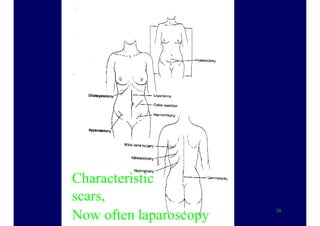
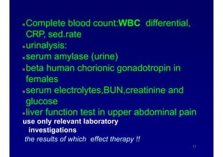
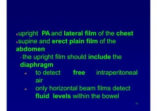
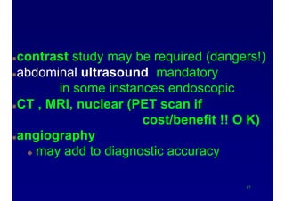
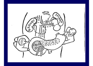
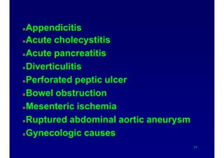
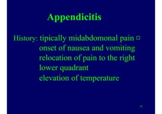
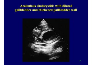
Acute abdomen is an abdominal emergency no
temporizing is ever justifiable.
Patients present more likely in the evening
hours.Never wait for your decision till the
next morning
1
Characteristics of acute
abdomen
has been present for less than 24 hours.
Sudden and unexpected onset of
abdominal pain.
associated symptoms:
nausea, vomiting, abdominal dystension,
diarrhea, constipations, anorexia
The pain may arise from intra-and extra-
abdominal structures.
2
Neural innervation of the gastrointestinal
tract
5
History
4
allow the patient to give his/her entire
current history before asking specific
questions.
the character and onset of pain are
essential.
–colicky pain: obstructive processes
–sustained pain :infectious processes
Referred pain patterns may give a clue.
7
Abdominal pain onset patterns
I. sudden(seconds)
–A. perforation or rupture of a viscus:peptic
ulcer, abdominal aortic aneurysm,
esophagus, ectopic pregnancy
–B. infarction:gut, heart, lung
Abdominal pain onset patternsII. rapid(minutes)
–A. colic syndromes: biliary, ureteral, small
bowel obstruction(high)
–B. inflammatory processes:
pancreatitis, diverticulitis,
appendicitis, penetrating ulcer,
cholecystitis
–C. ischemic processes:
strangulation, torsion
Abdominal pain onset patterns
III. Gradual(hours)
A. inflammatory :appendicitis, cholec.,
pancreat., divertic., salpingitis, ¤
prostatitis, inflamm.bowel dis., intra-
abdominal abscess
B. obstruction:distal small bowel or
colon,ectopic pregnancy,urinary retention,
incarcerated hernia
C. neoplastic:perforating or penetrating
tumors (colon, stomach, small intestine)9
Physical examination
Observation of the patients body
habitus and facial expression
–peritonitis :unwillingness to change
posture, hip flexion with the knees
drawn up, shallow breathing
–colicky pain: intense movements to
alleviate
10
Physical examination
Inspection of the abdomen:localized or
generalized dystension, visible
peristaltsis, hernial bulges, erythema
Auscultation of bowel sounds, if no
sounds are heard : paralytic ileus
 Percussion
11
absence of hepatic
dullness (!!) : perforation
13
Physical examinationPalpation : superficial, gentle of all
quadrants, first at the least painful areas,
after this deeper.
classic rebound tenderness (deep
palpation followed by rapid release) is not
specific.
have the patient laugh, cough, distend or
maximally reduce his/her abdominal girth.
A rectal digital examination is obligatory
13
16
Characteristic
scars,
Now often laparoscopy
Laboratory evaluation
Complete blood count:WBC differential,
CRP, sed.rate
urinalysis:
serum amylase (urine)
beta human chorionic gonadotropin in
females
serum electrolytes,BUN,creatinine and
glucose
liver function test in upper abdominal pain
use only relevant laboratory
investigations
the results of which effect therapy !!
15
X-ray evaluation
upright PA and lateral film of the chest
supine and erect plain film of the
abdomen
–the upright film should include the
diaphragm
 to detect free intraperitoneal
air
 only horizontal beam films detect
fluid levels within the bowel
16
X-ray evaluation
contrast study may be required (dangers!)
abdominal ultrasound mandatory
in some instances endoscopic
CT , MRI, nuclear (PET scan if
cost/benefit !! O K)
angiography
 may add to diagnostic accuracy
17
20
Causes of acute abdomen
Appendicitis
Acute cholecystitis
Acute pancreatitis
Diverticulitis
Perforated peptic ulcer
Bowel obstruction
Mesenteric ischemia
Ruptured abdominal aortic aneurysm
Gynecologic causes
19
Appendicitis
History: tipically midabdomonal pain ¤
onset of nausea and vomiting
relocation of pain to the right
lower quadrant
elevation of temperature
20
23ZETA (Sir Zachary Cope)
Appendicitis : bacterial infection with
contributory factors:intraluminal
obstruction -fecalith lymphoid
hyperplasia, parasites, carcinoid tu.
–typical symptoms:midabdominal
pain moving to the right lower
quadrant- elicited by coughing
laughing or bumping, nausea and
vomiting, anorexia,fever.
22
Differntial dg of appendicitis
Localization of the appendix
ascending:
cholec,perf
duodenal ulc
perinephr absc
hydronephr
Iliacal
penetrating duod ulc
Crohn diseas !!
Ileocecal cc.
Tbc
uretolith
23
pyonephr
pyelitis
nephrolith
omental torsion
Meckel’s diverticulum
Psoas absc
hip !!
muscle rupture
typhlitis
 Appendicitis 2 :
abdominal X-ray rarely useful,
ultrasound(periappendicular
fluid,edema,abscess,visualization of
the lumen) increasing significance
Peak incidence 15-24 years
choice of treatment ,surgery:10-20%
negative appendectomy
Keep in mind the danger of perforation
in the elderly
24
Acute cholecystitis
obstruction of the bile duct by stone
1. bacterial in 50-85% of cases
2. Chemical agents : lysolecithin,
other tissue factors
3. Inflammation from
mechanical strech
25
Acalculous cholecystitis with dilated
gallbladder and thickened gallbladder wall
26
Diagnosis of stone disease by ultrasound
shadow
27
Cholesterol stones gall bladder
28
Appearance of gallstones
29
Characteristic symptoms:
colic, localized to the right upper
quadrant
RUQ tenderness
patient suddenly stops inspiration
(Murphy‘sign)
irradiates to the right shoulder or
scapula
vomiting , exsiccosis
fever usually moderate, but also
chills
30
The „convergence projection” :
in the lateral spinothalamic tract the fiber
number is less than the sensory fibers
somatic> visceralis
the brain “learns” that on the given tract
the somatic signals are transmitted
31
32
Acute pancreatitis
Increasing incidence: 36 to 44/100 000 adults
in California (1994-2001)
200 000 hospital admission/year in the USA
Bile reflux is the trigger (1856 Claude Bernard)
2 enzymes are released from acinar cells
amylase and lipase
33
Causes
gallstones 38%
alcohol abuse 36%
pancreas divisum ( congenital abnormality of
the pancreatic duct)
intraductal papillary tumors
ERCP (increase of serum amylase
after the procedure )
Serum triglyceride >11mmol/L
some drugs
infections
34
Diagnosis
Symptoms of acute abdomen
•Constant acute pain in the epigastric area or the
right upper quadrant
•Nausea , vomiting
•Tenderness in the upper abdomen
•Cullen’s sign:
38
36
20% severe (4% die)
Early development
sequential organ failure
increased capillary permeability
decreased intravascular volume
hypovolemia
renal dysfunction
pulmonary complication
Pancreatic necrosis a very severe complication
37
Severity is assessed by CT and contrast enhanced CT
38
Treatment
Correct fluid losses
monitor respiratory, cardiovascular and
renal function. Multidisciplinary
Stop parenteral nutrition : a rule!??
Infection
antibiotic prophylaxis is debated in
proven infection: imipenem
39
Lancet 2008;371:143 ¤43
BMJ 2004;328:1407 41
42
Causes of acute abdomen
Diverticulitis
 prevalence 5% , increases with age
 the sigmoid colon is most commonly involved
 in 50% the only segment, right sided 0,1-2,5%
 signs and symptoms protean
 left lower quadrant pain, low grade fever,
 leucocytosis,nausea, vomiting, distension
 Sigmoidoscopy not indicated(perforation!!),nor
 barium enema, not in acute phase ,only later
"elective "
 X-ray or CT scanning
43
Causes of acute abdomen
Mesenteric ischemia: 0,4% of abdominal surgery
vascular disorders-usually catastrophic illness
–embolic occlusion or thrombosis:intestinal infarction--
gangrenous bowel
–mortality 40-70%
abdominal pain,vomiting diarrhea, melena ,
distension,tenderness
bowel sounds from hypoactivity to absent
Bloody peritoneal transsudate,leucocytosis 20 t
hemoconcentration
history of abdominal angina,atrial fibrillation
rapid visceral angiography
44
Causes of acute abdomen
Perforated peptic ulcer 10% ofhospital
admission for ulcer 7-10 pts/100000/year
undiagnosed pts die,duodenal 6-8x more often¤
sudden onset epigastric pain"hit with a knife"
– spreading to the entire abdomen:rigidity,diffuse
tenderness-hypovolemia, shock
upright or left lateral decubitus X-ray 55-85%
pneumoperitoneum:on physical disappearance of
hepatic dullness, X-ray
may heal spontaneously,dudenal anterior wall ¤
surgery,broad spect.antibiot,fluid
45
49
Succussion splash
Colonic perforation
50
Causes of acute abdomen
Ruptured abdominal aortic aneurysm
51
pain,sudden onset ,midabdominal,paravertebral
pulsatile abdominal mass,hypotension "triad"
risk: atheroscler.diameter and rate of increase
– 5,5 cm threshold for electivesurgery
– Abdominal ultrasound
X-ray (contrast iv.deviation of the ureters,aortic
 wall,CT,angio time consuming
MR
emergency operation-high mortality
Classification of thoraco-abdominal aortic
aneurysms
49
Atherosclerotic abdominal aortic aneurysm after
fatal rupture
50
Causes of acute abdomen
Bowel obstruction: ileus 20% of all acute
surgical hospital admissions
causes: mechanical
extrinsic: adhesions,hernias,volvulus,masses
intraluminal objects: fecal impaction,gallstone,
gastric bezoars,foreign bodies
intrinsic lesions:neoplasms,inflammation,
intussusception,hematoma
51
52
Causes of acute abdomen
Ileus 2 : adynamic(paralytic)
reflex inhibition:laparotomy,trauma
inflammation:peritonitis,toxic megacolon,
acute irradiation
infectious process:appendicitis,cholecystitis
ischemic processes:arterial insuff.
retroperitoneal :ureter,kidney
drug induced:opiates,anticholinergic drugs
metabolic:porphyria ,ketoacidosis
X-ray diagnosis: air-fluid levels -small or large bowel
53
Causes of acute abdomen
Gynecoligical:in reproductive age
 pelvic inflammatory, ectopic pregnancy, ovarian
cyst hemorrhage,adnexal or ovarian torsion
pain,delayed menstrual period,diffuse pelvic
 tenderness, acute rupture of blood filled
fallopian tube
SYNCOPE,pelvic examination,pregnancy test
54
„A good eater must be a good man,
for a good eater must have good
digestion, and good digestion
depends upon good conscience”
Benjamin Disraeli
1804-1881
Prime minister of Great-Britain: 1868, 1874-80
55
56
Some reminder of anatomy and
pathophysiology
57
58
.
The foregut,midgut and hindgut have and
retain their own innervation and blood
supply
forgut : oropharynx to the duodenum (bile
duct)
midgut: distal duodenum,jejunum,
ileum,appendix, ascending colon,
proximal 2/3 transverse colon
59
.
hindgut: distal1/3 of transverse colon to
anus
peritoneum: visceral autonomic
innervation dull,crampy or aching pain
:parietal somatic innervation
sharp, severe and persistent pain
60
Acute abdomen
Abdominal pain :visceral, somatic or referred
abdominal wall: anterior and lateral spinal T7-L1
Two types of nociceptors
–A-delta fibers rapid : sharp well localized
–C-fibers slow:dull, poorly localized
 :posterior L2-L5
pain fibers enter spinal cord ipsilaterally
 visceral pain arises in the midline
61
 fibers enter spinal cord bilaterally
“ To study the phenomenon of disease
without books is to sail an uncharted
sea,
while to study books without patients
is not to go sea atall”
William Osler
62
A University should be
a place of light,
of liberty, and of learning.
Benjamin DISRAELI, 1873
63
64
65
Diagnosis:
66

Acute abdomen

  • 1.
  • 2.
    Acute abdomen isan abdominal emergency no temporizing is ever justifiable. Patients present more likely in the evening hours.Never wait for your decision till the next morning 1
  • 3.
    Characteristics of acute abdomen hasbeen present for less than 24 hours. Sudden and unexpected onset of abdominal pain. associated symptoms: nausea, vomiting, abdominal dystension, diarrhea, constipations, anorexia The pain may arise from intra-and extra- abdominal structures. 2
  • 4.
    Neural innervation ofthe gastrointestinal tract 5
  • 5.
    History 4 allow the patientto give his/her entire current history before asking specific questions. the character and onset of pain are essential. –colicky pain: obstructive processes –sustained pain :infectious processes Referred pain patterns may give a clue.
  • 6.
  • 7.
    Abdominal pain onsetpatterns I. sudden(seconds) –A. perforation or rupture of a viscus:peptic ulcer, abdominal aortic aneurysm, esophagus, ectopic pregnancy –B. infarction:gut, heart, lung
  • 8.
    Abdominal pain onsetpatternsII. rapid(minutes) –A. colic syndromes: biliary, ureteral, small bowel obstruction(high) –B. inflammatory processes: pancreatitis, diverticulitis, appendicitis, penetrating ulcer, cholecystitis –C. ischemic processes: strangulation, torsion
  • 9.
    Abdominal pain onsetpatterns III. Gradual(hours) A. inflammatory :appendicitis, cholec., pancreat., divertic., salpingitis, ¤ prostatitis, inflamm.bowel dis., intra- abdominal abscess B. obstruction:distal small bowel or colon,ectopic pregnancy,urinary retention, incarcerated hernia C. neoplastic:perforating or penetrating tumors (colon, stomach, small intestine)9
  • 10.
    Physical examination Observation ofthe patients body habitus and facial expression –peritonitis :unwillingness to change posture, hip flexion with the knees drawn up, shallow breathing –colicky pain: intense movements to alleviate 10
  • 11.
    Physical examination Inspection ofthe abdomen:localized or generalized dystension, visible peristaltsis, hernial bulges, erythema Auscultation of bowel sounds, if no sounds are heard : paralytic ileus  Percussion 11 absence of hepatic dullness (!!) : perforation
  • 12.
  • 13.
    Physical examinationPalpation :superficial, gentle of all quadrants, first at the least painful areas, after this deeper. classic rebound tenderness (deep palpation followed by rapid release) is not specific. have the patient laugh, cough, distend or maximally reduce his/her abdominal girth. A rectal digital examination is obligatory 13
  • 14.
  • 15.
    Laboratory evaluation Complete bloodcount:WBC differential, CRP, sed.rate urinalysis: serum amylase (urine) beta human chorionic gonadotropin in females serum electrolytes,BUN,creatinine and glucose liver function test in upper abdominal pain use only relevant laboratory investigations the results of which effect therapy !! 15
  • 16.
    X-ray evaluation upright PAand lateral film of the chest supine and erect plain film of the abdomen –the upright film should include the diaphragm  to detect free intraperitoneal air  only horizontal beam films detect fluid levels within the bowel 16
  • 17.
    X-ray evaluation contrast studymay be required (dangers!) abdominal ultrasound mandatory in some instances endoscopic CT , MRI, nuclear (PET scan if cost/benefit !! O K) angiography  may add to diagnostic accuracy 17
  • 18.
  • 19.
    Causes of acuteabdomen Appendicitis Acute cholecystitis Acute pancreatitis Diverticulitis Perforated peptic ulcer Bowel obstruction Mesenteric ischemia Ruptured abdominal aortic aneurysm Gynecologic causes 19
  • 20.
    Appendicitis History: tipically midabdomonalpain ¤ onset of nausea and vomiting relocation of pain to the right lower quadrant elevation of temperature 20
  • 21.
  • 22.
    Appendicitis : bacterialinfection with contributory factors:intraluminal obstruction -fecalith lymphoid hyperplasia, parasites, carcinoid tu. –typical symptoms:midabdominal pain moving to the right lower quadrant- elicited by coughing laughing or bumping, nausea and vomiting, anorexia,fever. 22
  • 23.
    Differntial dg ofappendicitis Localization of the appendix ascending: cholec,perf duodenal ulc perinephr absc hydronephr Iliacal penetrating duod ulc Crohn diseas !! Ileocecal cc. Tbc uretolith 23 pyonephr pyelitis nephrolith omental torsion Meckel’s diverticulum Psoas absc hip !! muscle rupture typhlitis
  • 24.
     Appendicitis 2: abdominal X-ray rarely useful, ultrasound(periappendicular fluid,edema,abscess,visualization of the lumen) increasing significance Peak incidence 15-24 years choice of treatment ,surgery:10-20% negative appendectomy Keep in mind the danger of perforation in the elderly 24
  • 25.
    Acute cholecystitis obstruction ofthe bile duct by stone 1. bacterial in 50-85% of cases 2. Chemical agents : lysolecithin, other tissue factors 3. Inflammation from mechanical strech 25
  • 26.
    Acalculous cholecystitis withdilated gallbladder and thickened gallbladder wall 26
  • 27.
    Diagnosis of stonedisease by ultrasound shadow 27
  • 28.
  • 29.
  • 30.
    Characteristic symptoms: colic, localizedto the right upper quadrant RUQ tenderness patient suddenly stops inspiration (Murphy‘sign) irradiates to the right shoulder or scapula vomiting , exsiccosis fever usually moderate, but also chills 30
  • 31.
    The „convergence projection”: in the lateral spinothalamic tract the fiber number is less than the sensory fibers somatic> visceralis the brain “learns” that on the given tract the somatic signals are transmitted 31
  • 32.
  • 33.
    Acute pancreatitis Increasing incidence:36 to 44/100 000 adults in California (1994-2001) 200 000 hospital admission/year in the USA Bile reflux is the trigger (1856 Claude Bernard) 2 enzymes are released from acinar cells amylase and lipase 33
  • 34.
    Causes gallstones 38% alcohol abuse36% pancreas divisum ( congenital abnormality of the pancreatic duct) intraductal papillary tumors ERCP (increase of serum amylase after the procedure ) Serum triglyceride >11mmol/L some drugs infections 34
  • 35.
    Diagnosis Symptoms of acuteabdomen •Constant acute pain in the epigastric area or the right upper quadrant •Nausea , vomiting •Tenderness in the upper abdomen •Cullen’s sign: 38
  • 36.
  • 37.
    20% severe (4%die) Early development sequential organ failure increased capillary permeability decreased intravascular volume hypovolemia renal dysfunction pulmonary complication Pancreatic necrosis a very severe complication 37
  • 38.
    Severity is assessedby CT and contrast enhanced CT 38
  • 39.
    Treatment Correct fluid losses monitorrespiratory, cardiovascular and renal function. Multidisciplinary Stop parenteral nutrition : a rule!?? Infection antibiotic prophylaxis is debated in proven infection: imipenem 39
  • 40.
  • 41.
  • 42.
  • 43.
    Causes of acuteabdomen Diverticulitis  prevalence 5% , increases with age  the sigmoid colon is most commonly involved  in 50% the only segment, right sided 0,1-2,5%  signs and symptoms protean  left lower quadrant pain, low grade fever,  leucocytosis,nausea, vomiting, distension  Sigmoidoscopy not indicated(perforation!!),nor  barium enema, not in acute phase ,only later "elective "  X-ray or CT scanning 43
  • 44.
    Causes of acuteabdomen Mesenteric ischemia: 0,4% of abdominal surgery vascular disorders-usually catastrophic illness –embolic occlusion or thrombosis:intestinal infarction-- gangrenous bowel –mortality 40-70% abdominal pain,vomiting diarrhea, melena , distension,tenderness bowel sounds from hypoactivity to absent Bloody peritoneal transsudate,leucocytosis 20 t hemoconcentration history of abdominal angina,atrial fibrillation rapid visceral angiography 44
  • 45.
    Causes of acuteabdomen Perforated peptic ulcer 10% ofhospital admission for ulcer 7-10 pts/100000/year undiagnosed pts die,duodenal 6-8x more often¤ sudden onset epigastric pain"hit with a knife" – spreading to the entire abdomen:rigidity,diffuse tenderness-hypovolemia, shock upright or left lateral decubitus X-ray 55-85% pneumoperitoneum:on physical disappearance of hepatic dullness, X-ray may heal spontaneously,dudenal anterior wall ¤ surgery,broad spect.antibiot,fluid 45
  • 46.
  • 47.
  • 48.
    Causes of acuteabdomen Ruptured abdominal aortic aneurysm 51 pain,sudden onset ,midabdominal,paravertebral pulsatile abdominal mass,hypotension "triad" risk: atheroscler.diameter and rate of increase – 5,5 cm threshold for electivesurgery – Abdominal ultrasound X-ray (contrast iv.deviation of the ureters,aortic  wall,CT,angio time consuming MR emergency operation-high mortality
  • 49.
  • 50.
    Atherosclerotic abdominal aorticaneurysm after fatal rupture 50
  • 51.
    Causes of acuteabdomen Bowel obstruction: ileus 20% of all acute surgical hospital admissions causes: mechanical extrinsic: adhesions,hernias,volvulus,masses intraluminal objects: fecal impaction,gallstone, gastric bezoars,foreign bodies intrinsic lesions:neoplasms,inflammation, intussusception,hematoma 51
  • 52.
  • 53.
    Causes of acuteabdomen Ileus 2 : adynamic(paralytic) reflex inhibition:laparotomy,trauma inflammation:peritonitis,toxic megacolon, acute irradiation infectious process:appendicitis,cholecystitis ischemic processes:arterial insuff. retroperitoneal :ureter,kidney drug induced:opiates,anticholinergic drugs metabolic:porphyria ,ketoacidosis X-ray diagnosis: air-fluid levels -small or large bowel 53
  • 54.
    Causes of acuteabdomen Gynecoligical:in reproductive age  pelvic inflammatory, ectopic pregnancy, ovarian cyst hemorrhage,adnexal or ovarian torsion pain,delayed menstrual period,diffuse pelvic  tenderness, acute rupture of blood filled fallopian tube SYNCOPE,pelvic examination,pregnancy test 54
  • 55.
    „A good eatermust be a good man, for a good eater must have good digestion, and good digestion depends upon good conscience” Benjamin Disraeli 1804-1881 Prime minister of Great-Britain: 1868, 1874-80 55
  • 56.
  • 57.
    Some reminder ofanatomy and pathophysiology 57
  • 58.
  • 59.
    . The foregut,midgut andhindgut have and retain their own innervation and blood supply forgut : oropharynx to the duodenum (bile duct) midgut: distal duodenum,jejunum, ileum,appendix, ascending colon, proximal 2/3 transverse colon 59
  • 60.
    . hindgut: distal1/3 oftransverse colon to anus peritoneum: visceral autonomic innervation dull,crampy or aching pain :parietal somatic innervation sharp, severe and persistent pain 60
  • 61.
    Acute abdomen Abdominal pain:visceral, somatic or referred abdominal wall: anterior and lateral spinal T7-L1 Two types of nociceptors –A-delta fibers rapid : sharp well localized –C-fibers slow:dull, poorly localized  :posterior L2-L5 pain fibers enter spinal cord ipsilaterally  visceral pain arises in the midline 61  fibers enter spinal cord bilaterally
  • 62.
    “ To studythe phenomenon of disease without books is to sail an uncharted sea, while to study books without patients is not to go sea atall” William Osler 62
  • 63.
    A University shouldbe a place of light, of liberty, and of learning. Benjamin DISRAELI, 1873 63
  • 64.
  • 65.
  • 66.