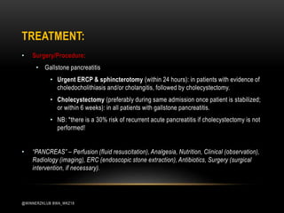
Group: WinnerzKlub
Year: 2020
ACUTE PANCREATITIS
ACUTE PANCREATITIS:
• An inflammatory condition of the pancreas.
• Most commonly caused by biliary tract disease or alcohol abuse.
• Damage to the pancreas leads to release of pancreatic enzymes (proteolytic) which
autodigests pancreatic tissue.
• Acute pancreatitis usually resolves within one-to-two weeks.
• Solid foods are generally avoided for a while in order to reduce the strain on the
pancreas.
@WINNERZKLUB BWA_WKZ18
ETIOLOGY:
• Most common causes:
i. Biliary pancreatitis (e.g. gallstones, constriction of ampulla of Vater) ~40% cases.
ii. Alcohol-induced (~30% of cases).
iii. Idiopathic (~15% - 25% of cases).
• Other causes:
• Severe hypertriglyceridemia (> 1,000 mg/dl), hypercalcemia
• Post-ERCP (upto 10% patients develop pancreatitis post-ERCP).
• Certain drugs:
• Steroids, Sulfonamides, Loop & thiazide diuretics, Estrogen, Anticonvulsants.
@WINNERZKLUB BWA_WKZ18
PATHOPHYSIOLOGY:
• Intrapancreatic activation of pancreatic enzymes: 2° to pancreatic ductal outflow
obstruction (e.g. gallstones, cystic fibrosis) or direct injury to pancreatic acinar cells (e.g.
alcohol, drugs).
• Increased proteolytic & lipolytic enzyme activity – destruction of pancreatic parenchyma.
• Attraction of pancreatic cells (neutrophils, macrophages) – release of inflammatory
cytokines – pancreatic inflammation (pancreatitis).
@WINNERZKLUB BWA_WKZ18
CLINICAL FEATURES:
• Constant, severe epigastric pain
• Classically radiating towards the back
• Worse after meals & when supine
• Improves on leaning forwards
• Nausea, vomiting
• General physical examination:
• Signs of shock: tachycardia, hypotension, oliguria/anuria.
• Possibly jaundice in patients with biliary pancreatitis.
• Abdominal examination:
• Abdominal tenderness, distension (ascites), guarding
• Ileus with reduced bowel sounds & tympany on percussion.
• Pathognomonic sign:
• Cullen sign – bluish discoloration
preumbilical area).
• Grey-Turner sign – flank ecchymosis
with discoloration.
• Fox sign – ecchymosis over the
inguinal ligament.
@WINNERZKLUB BWA_WKZ18
CLINICAL FEATURES:
@WINNERZKLUB BWA_WKZ18
DIAGNOSIS:
• Pancreatitis can be diagnosed both clinically and chemically.
• Acute pancreatitis is diagnosed based on a typical clinical presentation, with abdominal
pain radiating to the back, and either detection of highly elevated pancreatic enzymes or
characteristic findings on imaging.
• Diagnosis tests which will be discussed here includes:
• Laboratory tests
• Imaging.
@WINNERZKLUB BWA_WKZ18
DIAGNOSIS – LABORATORY TESTS:
i. Tests to confirm clinical diagnosis
• Increased serum pancreatic enzymes (Normal enzyme levels does not rule out
pancreatitis).
• Lipase: if upper reference range increases by 3 times or more.
• Amylase (nonspecific as increased level seen in may conditions).
ii. Tests to assess severity:
• Hematocrit (Hct)
• Should be conducted at presentation as well as 12hrs & 24hrs after admissions.
• Increased: (hemoconcentration) indicates 3rd space fluid loss & inadequate fluid
resuscitation.
• Decreased: indicates the rarer acute hemorrhagic pancreatitis.
@WINNERZKLUB BWA_WKZ18
• WBC count (leukocytosis is an indication of severe pancreatitis).
• Blood urea nitrogen (Rising level >48hrs of resuscitation indicates persistent
third spacing of fluid & is a predictor of severe pancreatitis.
• Increased CRP and procalcitonin levels
• Increased ALT (Alanine aminotransferase) levels
iii. Tests to determine etiology:
• Alkaline phosphatase, bilirubin levels (evidence of gallstone pancreatitis).
• Serum calcium levels
• Serum triglyceride levels (fasting).
• NB: Hypercalcemia may cause pancreatitis, which may then, in turn, cause
hypocalcemia!
DIAGNOSIS – LABORATORY TESTS:
@WINNERZKLUB BWA_WKZ18
i. Ultrasound: (most useful initial test)
• Indicated in all patients with acute pancreatitis
• Main purpose – detection of gallstones and/or dilatation of the biliary tract (indicating
biliary origin).
• Signs of pancreatitis:
• Indistinct pancreatic margins
• Peripancreatic build-up of fluid (hypoechogenic): evidence of ascites in some cases.
• Evidence of necrosis, abscesses, pancreatic pseudocysts.
ii. CT scan: not routinely indicated.
• Useful to assess severity and complications of acute pancreatitis.
DIAGNOSIS – IMAGING:
@WINNERZKLUB BWA_WKZ18
iii. MRCP and ERCP
• Indications: suspected biliary or pancreatic duct obstructions.
• MRCP is noninvasive but less sensitive than ERCP.
• ERCP can be combined with sphincterotomy & stone extraction; but may worsen pancreatitis.
iv. Conventional X-ray:
• Sentinel loop sign: dilatation of a loop of small intestine in the upper abdomen
(duodenum/jejunum)
• A specific radiological sign of acute pancreatitis.
• Caused due to a functional ileus of small intestine adjacent to the pancreas due to
pancreatic inflammation.
• Colon cut-off sign: gaseous distention of the ascending & transverse colon that abruptly
terminates at the splenic flexure.
• Evidence of possible complications: pleural effusions, pancreatic calcium stones, helps rule
out intestinal perforation with free air.
DIAGNOSIS – IMAGING:
@WINNERZKLUB BWA_WKZ18
DIFFERENTIAL DIAGNOSES:
• Intestinal manifestations:
• Acute peritonitis
• Appendicitis
• Acute mesenteric ischemia
• Acute cholecystitis
• Acute cholangitis
• Peptic ulcer disease
• Biliary colic
• Abdominal aorta aneurism
• Extra-intestinal manifestations:
• Myocardial infarction
• Bacterial pneumonia
@WINNERZKLUB BWA_WKZ18
TREATMENT:
i. General measures:
• Admission to hospital and assessment of disease severity (consider ICU admission).
• Fluid resuscitation: aggressive hydration with crystalloids (e.g., lactated Ringer’s
solution, normal saline).
• Analgesia: IV opioids (e.g. fentanyl)
• Nil-by-mouth & IV fluids are recommended until the pain subsides.
• Nasogastric tube insertion: not routinely recommended; indicated in patients with
vomiting and/or significant abdominal distention.
• Nutrition:
• Begin enteral feeding (oral/ nasogastric/ nasojejunal) as soon as pain subsides.
• Total parental nutrition: only in patients who cannot tolerate enteral feeds (e.g.
those with persistent ileus & abdominal pain).
@WINNERZKLUB BWA_WKZ18
ii. Drug Therapy:
• Analgesics: e.g. fentanyl or hydromorphone
• Antibiotics:
• Prophylactic antibiotic therapy is not recommended.
• Antibiotics should only be used in patients with evidence of infected necrosis
• (Samples from CT-guided fine needle aspiration sent for culture and Gram
staining to identify the appropriate antibiotic).
• Fenofibrates: in hyperlipidemia-induced acute pancreatitis.
• *Fenofibrates – a fibric acid derivative that functions to lower triglycerides.
• *Hyperlipidemia – condition of elevated serum concentration of both
triglycerides (>200 mg/dL) and cholesterol (>190 mg/dL).
TREATMENT:
@WINNERZKLUB BWA_WKZ18
• Surgery/Procedure:
• Gallstone pancreatitis
• Urgent ERCP & sphincterotomy (within 24 hours): in patients with evidence of
choledocholithiasis and/or cholangitis, followed by cholecystectomy.
• Cholecystectomy (preferably during same admission once patient is stabilized;
or within 6 weeks): in all patients with gallstone pancreatitis.
• NB: *there is a 30% risk of recurrent acute pancreatitis if cholecystectomy is not
performed!
• “PANCREAS” – Perfusion (fluid resuscitation), Analgesia, Nutrition, Clinical (observation),
Radiology (imaging), ERC (endoscopic stone extraction), Antibiotics, Surgery (surgical
intervention, if necessary).
TREATMENT:
@WINNERZKLUB BWA_WKZ18
• Localized:
• Bacterial superinfection of necrotic
tissue
• Pancreatic pseudocysts
• Pancreatic abscess
• Systemic:
• SIRS, sepsis, DIC
• Pneumonia, respiratory failure, ARDS
• Shock
• Prerenal failure due to volume
depletion
• Hypocalcemia
• Pleural effusion, pancreatic ascites
• Paralytic ileus
COMPLICATION(S):
@WINNERZKLUB BWA_WKZ18
THE END:
@WINNERZKLUB BWA_WKZ18

Acute pancreatitis

  • 1.
  • 2.
    ACUTE PANCREATITIS: • Aninflammatory condition of the pancreas. • Most commonly caused by biliary tract disease or alcohol abuse. • Damage to the pancreas leads to release of pancreatic enzymes (proteolytic) which autodigests pancreatic tissue. • Acute pancreatitis usually resolves within one-to-two weeks. • Solid foods are generally avoided for a while in order to reduce the strain on the pancreas. @WINNERZKLUB BWA_WKZ18
  • 3.
    ETIOLOGY: • Most commoncauses: i. Biliary pancreatitis (e.g. gallstones, constriction of ampulla of Vater) ~40% cases. ii. Alcohol-induced (~30% of cases). iii. Idiopathic (~15% - 25% of cases). • Other causes: • Severe hypertriglyceridemia (> 1,000 mg/dl), hypercalcemia • Post-ERCP (upto 10% patients develop pancreatitis post-ERCP). • Certain drugs: • Steroids, Sulfonamides, Loop & thiazide diuretics, Estrogen, Anticonvulsants. @WINNERZKLUB BWA_WKZ18
  • 4.
    PATHOPHYSIOLOGY: • Intrapancreatic activationof pancreatic enzymes: 2° to pancreatic ductal outflow obstruction (e.g. gallstones, cystic fibrosis) or direct injury to pancreatic acinar cells (e.g. alcohol, drugs). • Increased proteolytic & lipolytic enzyme activity – destruction of pancreatic parenchyma. • Attraction of pancreatic cells (neutrophils, macrophages) – release of inflammatory cytokines – pancreatic inflammation (pancreatitis). @WINNERZKLUB BWA_WKZ18
  • 5.
    CLINICAL FEATURES: • Constant,severe epigastric pain • Classically radiating towards the back • Worse after meals & when supine • Improves on leaning forwards • Nausea, vomiting • General physical examination: • Signs of shock: tachycardia, hypotension, oliguria/anuria. • Possibly jaundice in patients with biliary pancreatitis. • Abdominal examination: • Abdominal tenderness, distension (ascites), guarding • Ileus with reduced bowel sounds & tympany on percussion. • Pathognomonic sign: • Cullen sign – bluish discoloration preumbilical area). • Grey-Turner sign – flank ecchymosis with discoloration. • Fox sign – ecchymosis over the inguinal ligament. @WINNERZKLUB BWA_WKZ18
  • 6.
  • 7.
    DIAGNOSIS: • Pancreatitis canbe diagnosed both clinically and chemically. • Acute pancreatitis is diagnosed based on a typical clinical presentation, with abdominal pain radiating to the back, and either detection of highly elevated pancreatic enzymes or characteristic findings on imaging. • Diagnosis tests which will be discussed here includes: • Laboratory tests • Imaging. @WINNERZKLUB BWA_WKZ18
  • 8.
    DIAGNOSIS – LABORATORYTESTS: i. Tests to confirm clinical diagnosis • Increased serum pancreatic enzymes (Normal enzyme levels does not rule out pancreatitis). • Lipase: if upper reference range increases by 3 times or more. • Amylase (nonspecific as increased level seen in may conditions). ii. Tests to assess severity: • Hematocrit (Hct) • Should be conducted at presentation as well as 12hrs & 24hrs after admissions. • Increased: (hemoconcentration) indicates 3rd space fluid loss & inadequate fluid resuscitation. • Decreased: indicates the rarer acute hemorrhagic pancreatitis. @WINNERZKLUB BWA_WKZ18
  • 9.
    • WBC count(leukocytosis is an indication of severe pancreatitis). • Blood urea nitrogen (Rising level >48hrs of resuscitation indicates persistent third spacing of fluid & is a predictor of severe pancreatitis. • Increased CRP and procalcitonin levels • Increased ALT (Alanine aminotransferase) levels iii. Tests to determine etiology: • Alkaline phosphatase, bilirubin levels (evidence of gallstone pancreatitis). • Serum calcium levels • Serum triglyceride levels (fasting). • NB: Hypercalcemia may cause pancreatitis, which may then, in turn, cause hypocalcemia! DIAGNOSIS – LABORATORY TESTS: @WINNERZKLUB BWA_WKZ18
  • 10.
    i. Ultrasound: (mostuseful initial test) • Indicated in all patients with acute pancreatitis • Main purpose – detection of gallstones and/or dilatation of the biliary tract (indicating biliary origin). • Signs of pancreatitis: • Indistinct pancreatic margins • Peripancreatic build-up of fluid (hypoechogenic): evidence of ascites in some cases. • Evidence of necrosis, abscesses, pancreatic pseudocysts. ii. CT scan: not routinely indicated. • Useful to assess severity and complications of acute pancreatitis. DIAGNOSIS – IMAGING: @WINNERZKLUB BWA_WKZ18
  • 11.
    iii. MRCP andERCP • Indications: suspected biliary or pancreatic duct obstructions. • MRCP is noninvasive but less sensitive than ERCP. • ERCP can be combined with sphincterotomy & stone extraction; but may worsen pancreatitis. iv. Conventional X-ray: • Sentinel loop sign: dilatation of a loop of small intestine in the upper abdomen (duodenum/jejunum) • A specific radiological sign of acute pancreatitis. • Caused due to a functional ileus of small intestine adjacent to the pancreas due to pancreatic inflammation. • Colon cut-off sign: gaseous distention of the ascending & transverse colon that abruptly terminates at the splenic flexure. • Evidence of possible complications: pleural effusions, pancreatic calcium stones, helps rule out intestinal perforation with free air. DIAGNOSIS – IMAGING: @WINNERZKLUB BWA_WKZ18
  • 12.
    DIFFERENTIAL DIAGNOSES: • Intestinalmanifestations: • Acute peritonitis • Appendicitis • Acute mesenteric ischemia • Acute cholecystitis • Acute cholangitis • Peptic ulcer disease • Biliary colic • Abdominal aorta aneurism • Extra-intestinal manifestations: • Myocardial infarction • Bacterial pneumonia @WINNERZKLUB BWA_WKZ18
  • 13.
    TREATMENT: i. General measures: •Admission to hospital and assessment of disease severity (consider ICU admission). • Fluid resuscitation: aggressive hydration with crystalloids (e.g., lactated Ringer’s solution, normal saline). • Analgesia: IV opioids (e.g. fentanyl) • Nil-by-mouth & IV fluids are recommended until the pain subsides. • Nasogastric tube insertion: not routinely recommended; indicated in patients with vomiting and/or significant abdominal distention. • Nutrition: • Begin enteral feeding (oral/ nasogastric/ nasojejunal) as soon as pain subsides. • Total parental nutrition: only in patients who cannot tolerate enteral feeds (e.g. those with persistent ileus & abdominal pain). @WINNERZKLUB BWA_WKZ18
  • 14.
    ii. Drug Therapy: •Analgesics: e.g. fentanyl or hydromorphone • Antibiotics: • Prophylactic antibiotic therapy is not recommended. • Antibiotics should only be used in patients with evidence of infected necrosis • (Samples from CT-guided fine needle aspiration sent for culture and Gram staining to identify the appropriate antibiotic). • Fenofibrates: in hyperlipidemia-induced acute pancreatitis. • *Fenofibrates – a fibric acid derivative that functions to lower triglycerides. • *Hyperlipidemia – condition of elevated serum concentration of both triglycerides (>200 mg/dL) and cholesterol (>190 mg/dL). TREATMENT: @WINNERZKLUB BWA_WKZ18
  • 15.
    • Surgery/Procedure: • Gallstonepancreatitis • Urgent ERCP & sphincterotomy (within 24 hours): in patients with evidence of choledocholithiasis and/or cholangitis, followed by cholecystectomy. • Cholecystectomy (preferably during same admission once patient is stabilized; or within 6 weeks): in all patients with gallstone pancreatitis. • NB: *there is a 30% risk of recurrent acute pancreatitis if cholecystectomy is not performed! • “PANCREAS” – Perfusion (fluid resuscitation), Analgesia, Nutrition, Clinical (observation), Radiology (imaging), ERC (endoscopic stone extraction), Antibiotics, Surgery (surgical intervention, if necessary). TREATMENT: @WINNERZKLUB BWA_WKZ18
  • 16.
    • Localized: • Bacterialsuperinfection of necrotic tissue • Pancreatic pseudocysts • Pancreatic abscess • Systemic: • SIRS, sepsis, DIC • Pneumonia, respiratory failure, ARDS • Shock • Prerenal failure due to volume depletion • Hypocalcemia • Pleural effusion, pancreatic ascites • Paralytic ileus COMPLICATION(S): @WINNERZKLUB BWA_WKZ18
  • 17.