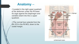
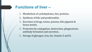
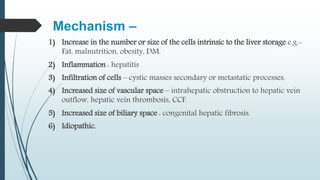
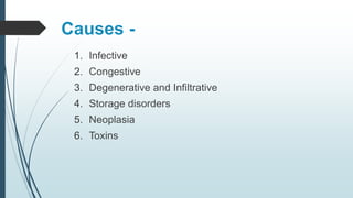
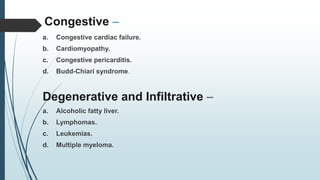
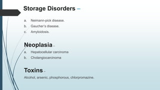
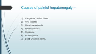
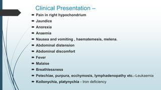
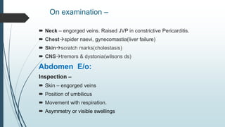
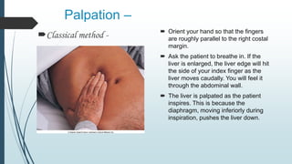
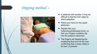
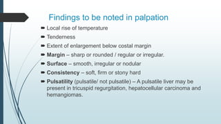
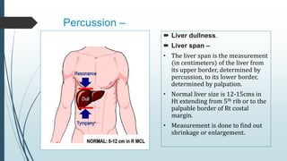
Enlargement of liver.
Presence of a palpable liver does not always represent
hepatomegaly.
Anatomy –
Located in the right upper quadrant
of the abdomen under the Rt lower
rib cage against the diaphragm and
variable extent into the Lt upper
quadrant.
The normal liver extends from the
5th ICS in the Rt MCL down to the
costal margin.
Functions of liver –
1. Metabolism of carbohydrates, fats, proteins.
2. Synthesis of bile and prothrombin.
3. Excretion of drugs, toxins, poisons, bile pigment &
heavy metals.
4. Protective by conjugation, destruction, phagocytosis,
antibody formation and excretion.
5. Storage of glycogen, iron, fat, vitamin A and D.
Mechanism –
1) Increase in the number or size of the cells intrinsic to the liver storage e.g.-
Fat, malnutrition, obesity, DM.
2) Inflammation : hepatitis
3) Infiltration of cells – cystic masses secondary or metastatic processes.
4) Increased size of vascular space – intrahepatic obstruction to hepatic vein
outflow, hepatic vein thrombosis, CCF.
5) Increased size of biliary space : congenital hepatic fibrosis.
6) Idiopathic.
Causes -
1. Infective
2. Congestive
3. Degenerative and Infiltrative
4. Storage disorders
5. Neoplasia
6. Toxins
Infective –
a. Along the biliary tree – Cholangitis
b. Along Portal vein – Amoebiasis, schistosomiasis, bacterial infections.
c. Along hepatic artery –
 Bacterial – typhoid, brucellosis, Tuberculosis, Syphilis, weil’s disease.
 Viral – Infective hepatitis, infectious mononucleosis.
 Protozoal – Malaria, kala-azar.
 Fungal – Actinomycosis, histoplasmosis.
 Parasitic – Echinococcosis[hydatid cyst]
Congestive –
a. Congestive cardiac failure.
b. Cardiomyopathy.
c. Congestive pericarditis.
d. Budd-Chiari syndrome.
Degenerative and Infiltrative –
a. Alcoholic fatty liver.
b. Lymphomas.
c. Leukemias.
d. Multiple myeloma.
Storage Disorders –
a. Neimann-pick disease.
b. Gaucher’s disease.
c. Amyloidosis.
Neoplasia–
a. Hepatocellular carcinoma
b. Cholangiocarcinoma
Toxins–
Alcohol, arsenic, phosphorous, chlorpromazine.
Causes of painful hepatomegaly –
1) Congestive cardiac failure.
2) Viral hepatitis
3) Hepatic Amoebiasis
4) Pyemic abscess
5) Hepatoma
6) Actinomycosis
7) Budd-Chiari syndrome.
Clinical Presentation –
 Pain in right hypochondrium
 Jaundice
 Anorexia
 Anaemia
 Nausea and vomiting , haematemesis, melena.
 Abdominal distension
 Abdominal discomfort
 Fever
 Malaise
 Breathlessness
 Petechiae, purpura, ecchymosis, lymphadenopathy etc.–Leukaemia
 Koilonychia, platynychia - Iron deficiency
On examination –
 Neck – engorged veins. Raised JVP in constrictive Pericarditis.
 Chestspider naevi, gynecomastia(liver failure)
 Skinscratch marks(cholestasis)
 CNStremors & dystonia(wilsons ds)
Abdomen E/o:
Inspection –
 Skin – engorged veins
 Position of umbilicus
 Movement with respiration.
 Asymmetry or visible swellings
Palpation –
Classical method -  Orient your hand so that the fingers
are roughly parallel to the right costal
margin.
 Ask the patient to breathe in. If the
liver is enlarged, the liver edge will hit
the side of your index finger as the
liver moves caudally. You will feel it
through the abdominal wall.
 The liver is palpated as the patient
inspires. This is because the
diaphragm, moving inferiorly during
inspiration, pushes the liver down.
Dipping method –
 In patients with ascites, it may be
difficult to feel the liver edge by
direct palpation.
 Place your hand on the upper
abdomen.
 Rapidly flex your
metacarpophalangeal joints, so
that your fingers suddenly dip
into the patient’s abdomen.
 The fingers will displacing the
underlying fluid and hit the liver,
confirming that a mass, likely to
be liver, is present
Findings to be noted in palpation
 Local rise of temperature
 Tenderness
 Extent of enlargement below costal margin
 Margin – sharp or rounded / regular or irregular.
 Surface – smooth, irregular or nodular
 Consistency – soft, firm or stony hard
 Pulsatility (pulsatile/ not pulsatile) – A pulsatile liver may be
present in tricuspid regurgitation, hepatocellular carcinoma and
hemangiomas.
Percussion –
 Liver dullness.
 Liver span –
• The liver span is the measurement
(in centimeters) of the liver from
its upper border, determined by
percussion, to its lower border,
determined by palpation.
• Normal liver size is 12-15cms in
Ht extending from 5th rib or to the
palpable border of Rt costal
margin.
• Measurement is done to find out
shrinkage or enlargement.
 The following are screenings and tests performed to determine the
cause of hepatomegaly:
 Abdominal CT scans
 Abdominal X-rays
 Abdominal MRI
 Abdominal Ultrasound
 Liver function test
 Blood test for Hepatitis
 Biopsy to rule out cancer
Hepatomegaly

Hepatomegaly

  • 1.
    Enlargement of liver. Presenceof a palpable liver does not always represent hepatomegaly.
  • 2.
    Anatomy – Located inthe right upper quadrant of the abdomen under the Rt lower rib cage against the diaphragm and variable extent into the Lt upper quadrant. The normal liver extends from the 5th ICS in the Rt MCL down to the costal margin.
  • 3.
    Functions of liver– 1. Metabolism of carbohydrates, fats, proteins. 2. Synthesis of bile and prothrombin. 3. Excretion of drugs, toxins, poisons, bile pigment & heavy metals. 4. Protective by conjugation, destruction, phagocytosis, antibody formation and excretion. 5. Storage of glycogen, iron, fat, vitamin A and D.
  • 4.
    Mechanism – 1) Increasein the number or size of the cells intrinsic to the liver storage e.g.- Fat, malnutrition, obesity, DM. 2) Inflammation : hepatitis 3) Infiltration of cells – cystic masses secondary or metastatic processes. 4) Increased size of vascular space – intrahepatic obstruction to hepatic vein outflow, hepatic vein thrombosis, CCF. 5) Increased size of biliary space : congenital hepatic fibrosis. 6) Idiopathic.
  • 5.
    Causes - 1. Infective 2.Congestive 3. Degenerative and Infiltrative 4. Storage disorders 5. Neoplasia 6. Toxins
  • 6.
    Infective – a. Alongthe biliary tree – Cholangitis b. Along Portal vein – Amoebiasis, schistosomiasis, bacterial infections. c. Along hepatic artery –  Bacterial – typhoid, brucellosis, Tuberculosis, Syphilis, weil’s disease.  Viral – Infective hepatitis, infectious mononucleosis.  Protozoal – Malaria, kala-azar.  Fungal – Actinomycosis, histoplasmosis.  Parasitic – Echinococcosis[hydatid cyst]
  • 7.
    Congestive – a. Congestivecardiac failure. b. Cardiomyopathy. c. Congestive pericarditis. d. Budd-Chiari syndrome. Degenerative and Infiltrative – a. Alcoholic fatty liver. b. Lymphomas. c. Leukemias. d. Multiple myeloma.
  • 8.
    Storage Disorders – a.Neimann-pick disease. b. Gaucher’s disease. c. Amyloidosis. Neoplasia– a. Hepatocellular carcinoma b. Cholangiocarcinoma Toxins– Alcohol, arsenic, phosphorous, chlorpromazine.
  • 9.
    Causes of painfulhepatomegaly – 1) Congestive cardiac failure. 2) Viral hepatitis 3) Hepatic Amoebiasis 4) Pyemic abscess 5) Hepatoma 6) Actinomycosis 7) Budd-Chiari syndrome.
  • 10.
    Clinical Presentation – Pain in right hypochondrium  Jaundice  Anorexia  Anaemia  Nausea and vomiting , haematemesis, melena.  Abdominal distension  Abdominal discomfort  Fever  Malaise  Breathlessness  Petechiae, purpura, ecchymosis, lymphadenopathy etc.–Leukaemia  Koilonychia, platynychia - Iron deficiency
  • 11.
    On examination – Neck – engorged veins. Raised JVP in constrictive Pericarditis.  Chestspider naevi, gynecomastia(liver failure)  Skinscratch marks(cholestasis)  CNStremors & dystonia(wilsons ds) Abdomen E/o: Inspection –  Skin – engorged veins  Position of umbilicus  Movement with respiration.  Asymmetry or visible swellings
  • 12.
    Palpation – Classical method-  Orient your hand so that the fingers are roughly parallel to the right costal margin.  Ask the patient to breathe in. If the liver is enlarged, the liver edge will hit the side of your index finger as the liver moves caudally. You will feel it through the abdominal wall.  The liver is palpated as the patient inspires. This is because the diaphragm, moving inferiorly during inspiration, pushes the liver down.
  • 13.
    Dipping method – In patients with ascites, it may be difficult to feel the liver edge by direct palpation.  Place your hand on the upper abdomen.  Rapidly flex your metacarpophalangeal joints, so that your fingers suddenly dip into the patient’s abdomen.  The fingers will displacing the underlying fluid and hit the liver, confirming that a mass, likely to be liver, is present
  • 14.
    Findings to benoted in palpation  Local rise of temperature  Tenderness  Extent of enlargement below costal margin  Margin – sharp or rounded / regular or irregular.  Surface – smooth, irregular or nodular  Consistency – soft, firm or stony hard  Pulsatility (pulsatile/ not pulsatile) – A pulsatile liver may be present in tricuspid regurgitation, hepatocellular carcinoma and hemangiomas.
  • 15.
    Percussion –  Liverdullness.  Liver span – • The liver span is the measurement (in centimeters) of the liver from its upper border, determined by percussion, to its lower border, determined by palpation. • Normal liver size is 12-15cms in Ht extending from 5th rib or to the palpable border of Rt costal margin. • Measurement is done to find out shrinkage or enlargement.
  • 16.
     The followingare screenings and tests performed to determine the cause of hepatomegaly:  Abdominal CT scans  Abdominal X-rays  Abdominal MRI  Abdominal Ultrasound  Liver function test  Blood test for Hepatitis  Biopsy to rule out cancer