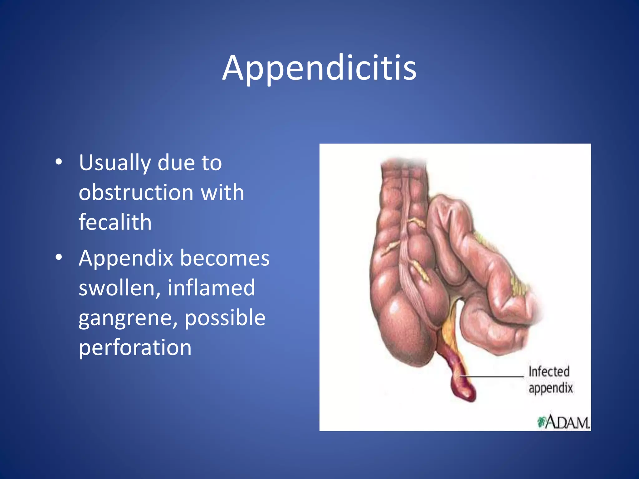
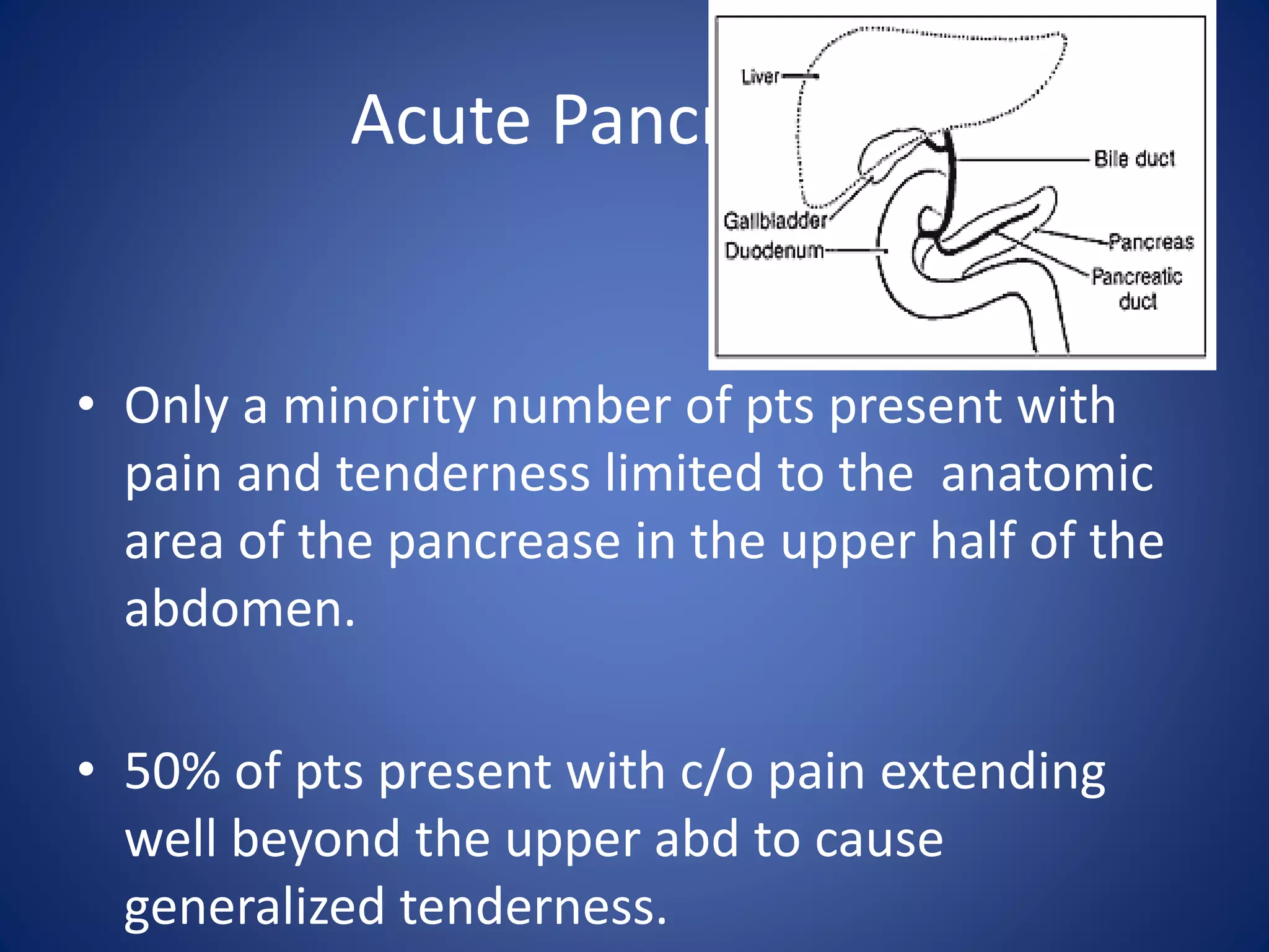
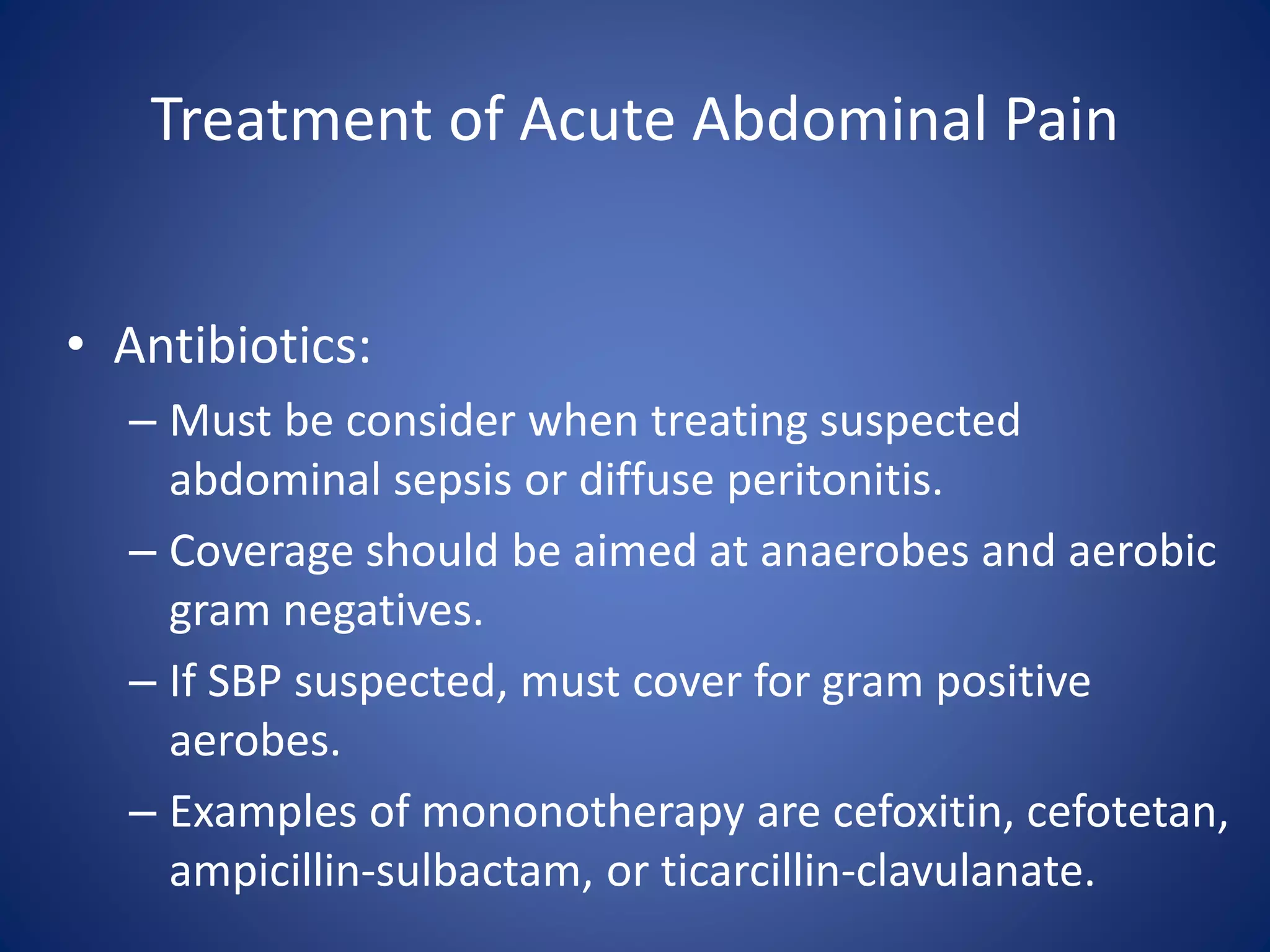
SlideShare a Scribd company logo
Acute Abdomen
The “Black Hole” of Medicine
Acute Abdomen
• Challenge to Surgeons
• Most common cause of surgical emergency
admission
• Encompass various conditions ranging from
the trivial to the life-threatening
• Clinical course can vary from minutes to hours,
to weeks
• It can be an acute exacerbation of a chronic
problem e.g. Chronic Pancreatitis, Vascular
Insufficiency
Differential Diagnosis
The primary symptom of the "acute
abdomen" is –
Abdominal pain.
ASSESMENT
• A Full history
• Thorough physical examination
Diagnosis can be made most of the time by
a good history and a proper physical
examination.
- An exact diagnosis often impossible to make after the initial
assessment, and often relying on further investigation
Types of Abdominal Pain:
• Three types of pain exist:
1. Visceral
2. Parietal
3. Referred
1. Visceral Pain
• Due to stretching of fibers innervating the
walls of hollow or solid organs.
• It occurs early and poorly localized
• It can be due to early ischemia or
inflammation.
2. Parietal Pain
• Caused by irritation of parietal peritoneum
fibers.
• It occurs late and better localized.
• Can be localized to a dermatome superficial to
site of the painful stimulus.
3. Referred Pain
• Pain is felt at a site away from the pathological
organ.
• Pain is usually ipsilateral to the involved organ
and is felt midline if pathology is midline.
• Pattern based on developmental embryology.
Referred Pain Locations
Acute Abdominal Pain
• Two approaches to evaluate pts with acute
abdominal pain:
1. Classification of abd pain into systems
2. Abdominal Topography (4 quadrants)
Classification on Abdominal Pain
• Three main categories of abdominal pain:
1. Intra-abdominal (arising from within the abd
cavity / retroperitoneum) involves:
• GI (Appendicitis, Diverticulitis, etc, etc, etc)
• GU (Renal Colic, etc, etc, etc)
• Gyn (Acute PID, Pregnancy, etc)
• Vascular systems (AAA, Mesenteric Ischemia, etc)
Classification on Abdominal Pain
2. Extra-abdominal (less common) involves:
• Cardiopulmonary (AMI, etc)
• Abdominal wall (Hernia, Zoster etc)
• Toxic-metabolic (DKA, OD, lead, etc)
• Neurogenic pain (Zoster, etc)
• Psychic (Anxiety, Depression, etc)
3. Nonspecific Abd pain – not well explained or
described.
Abdominal Topography
History (S)
• Duration?
• Nausea, vomiting? Bloody? (Coffee grounds emesis?)
• Change in urinary habits? Urine appearance?
• Change in bowel habits? Melena (Dark, tarry stools?)
• Regular food/water intake?
History (S)
• Females
–Last menstrual period?
–Abnormal bleeding?
In females, abdominal pain =
GYN problem until proven otherwise
Historical features of Abd Pain
• Location, quality, severity, onset, and duration
of pain, aggravating and alleviating factors
• GI symptoms (N/V/D)
• GU symptoms
• Vascular symptoms (A. fib / AMI / AAA)
• Can overlap i.e. Nausea seen in both GI / GU
pathologies.
Historical features of Abd Pain
• PMH
– Recent / current medications
– Past hospitalizations
– Past surgery
– Chronic disease
– Social history
– Occupation / Toxic exposure (CO / lead)
Physical Exam (O)
• Palpate each quadrant
– Work toward area
of pain
– Warm hands
– Patient on back,
knee bent (if
possible)
– Note tenderness,
rigidity, guarding,
masses
Physical Examination of the Abdomen
• Note pt’s general appearance. Realize that the
intensity of the abdominal pain may have no
relationship to severity of illness.
• One of the initial steps of the PE should be
obtaining and interpreting the vitals.
• Pts with visceral pain are unable to lie still.
• Pts with peritonitis like to stay immobile.
Physical Examination of the Abdomen
• INSPECT for distention, scars, masses, rash.
• AUSCULATE for hyperactive, obstructive,
absent, or normal bowel sounds.
• PALPATION to look for guarding, rigidity,
rebound tenderness, organomegally, or
hernias.
• Women should have pelvic exam (check FHR if
pregnant).
• Anyone with a rectum should have rectal
exam.
Laboratory Test
• CBC (limited clinical utility)
• UA / Urine culture
• Lactic acid
• LFT / Amylase / Lipase
• CE / Troponin
• HCG (quant / qual)
• Stool Culture
Radiographic Test
• Plain abdominal radiographs or abdominal
series has several limitations and is subject to
reader interpretation.
• CT scan in conjunction with ultrasound is
superior in identifying any abnormality seen
on plain film.
Specific Diagnoses
• In patients above fifty years of age the top four reasons for
acute abdominal pain are: Biliary Tract Disease (21%,) NSAP
(16%), Appendicitis(15%), and Bowel Obstruction (12%).
• In patients under fifty years of age the top three reasons for
acute abdominal pain are: NSAP (40%,) Appendicitis (32%,)
and Other (13%.)
Appendicitis
• Usually due to
obstruction with
fecalith
• Appendix becomes
swollen, inflamed
gangrene, possible
perforation

Appendicitis
• Pain begins periumbilical; moves to RLQ
• Nausea, vomiting, anorexia, fever
• Patient lies on side; right hip, knee flexed
• Pain may not localize to RLQ if appendix in odd
location
• Sudden relief of pain = possible perforation
Acute Appendicitis
• “In spite of a large number of algorithms and
decision rules incorporating many different
clinical and laboratory features, an accurate
preoperative diagnosis of appendicitis has
remain elusive for more than a century.”
Acute Appendicitis
• Clinical features with some predictive value
include:
• Pain located in the RLQ
• Pain migration from the periumbilical area to the RLQ
• Rigidity
• Pain before vomiting
• Positive psoas sign
• Note: Anorexia is not a useful symptom (33% pts not
anorectic preoperatively.)
Acute Appendicitis
• Ultrasound can be used for detection, but CT
is preferred in adults and non-pregnant
women.
• The CT scan can be with and without contrast
(oral & IV.)
• A neg. CT does not exclude diagnosis, but a
positive scan confirms it.
Peptic Ulcer Disease
• Steady, well-localized
epigastric or LUQ pain
• Described as a “burning”,
“gnawing”, “aching”
• Increased by coffee,
stress, spicy food,
smoking
• Decreased by alkaline
food, antacids
Peptic Ulcer Disease
• Erosion of the lining of the stomach,
duodenum, or esophagus
• May cause massive GI bleed
• Patient lies very still with complaint of intense,
steady pain, rigid abdomen with exam,
suspect perforation
Gastroesophageal Reflux
• Also known as GERD
• Signs and symptoms
can mimic cardiac
pain.
• Usually onset after
eating.
• Typically resolved with
medication.
Cholecystitis
• Inflammation of gall
bladder
• Commonly associated
with gall stones
• More common in 30 to 50
year old females
• Nausea, vomiting; RUQ
pain, tenderness; fever
• Attacks triggered by
ingestion of fatty foods
Biliary Tract Disease
• Most common diagnosis in ED of pts > 50.
– Composed of:
• Acute Cholecystitis (acalculus / calculus)
• Biliary Colic
• Common Duct Obstruction (Ascending Cholangitis –
painful jundice / fever / MSΔ).
• Of those patients found to have acute
cholecystitis, the majority lack fever and 40%
lack leukocytosis.
Biliary Tract Disease
• Patients may complain of:
– Diffuse pain in upper half of abdomen
– Generalized tenderness throughout belly
– RUQ or RLQ pain.
Biliary Tract Disease
• Sonography (US) is the initial test of choice for patients with
suspected biliary tract disease. More sensitive than CT scan to
detect CBD obstruction.
• CT scan is better in the identification of cholecystitis than in
the detection of CBD obstruction.
• Cholescintigraphy (radionclide / HIDA scan) of the biliary tree
is a more sensitive test than US for the diagnosis of both of
these conditions.
Biliary Tract Disease
• MR cholangiography (MRCP)
– Has good specificity and sensitivity in picking up
stones and common duct obstructions.
– Less invasive / less complications than ERCP
(ERCP can induce GI perforation, pancreatitis, biliary duct injury)
Bowel Obstruction
• Blockage of inside of intestine
• Interrupts normal flow of contents
• Causes include adhesions, hernias, fecal
impactions, tumors
• Cramping abdominal pain, nausea,
vomiting (often of fecal matter),
abdominal distension
Small Bowel Obstruction
• SBO may result from previous abdominal surgeries.
• Patient may present with intermittent, colicky pain,
abdominal distention, and abnormal BS.
• Only 2 historical features (previous abd surgery
and intermittent / colicky pain) and 2 physical
findings (abd distention and abn BS) appear to
have predictive value in diagnosing SBO.
Small Bowel Obstruction
• Plain abd films has a large number of
indeterminate readings and can be very
limited due to the following:
• Pt is obese
• Pt is bedridden / contracted (limited lateral decub /
upright view)
• Technical limitations
ABDOMINAL XR
Bowel Obstruction
• Small Bowel Large Bowel
– Central Peripheral
– Valvulae conniventes Haustrae
– Dia > 5cm > 10cm
SBO LBO
Small Bowel Obstruction
• CT scan is better than plain film in detecting
high grade SBO.
• CT scan can also give more info that might not
be seen on plain film (i.e. ischemic bowel)
• Low grade SBO may require small bowel
follow through.
Pancreatitis
• Inflammation of pancreas
• Triggered by ingestion of
EtOH; large amounts of
fatty foods
• Nausea, vomiting;
abdominal tenderness;
pain radiating from upper
abdomen straight through
to back
• Signs, symptoms of
hypovolemic shock
Acute Pancreatitis
• 80% of cases are due to ETOH abuse or gallstones.
• Other common causes:
– Drugs ( Valproic acid, Tetracycline, Hydrochlorothiazide, Furosemide)
– Pancreatic cancer
– Abdominal trauma/surgery
– Ulcer with pancreatic involvement
– Familial pancreatitis (Hypertriglycerides / Hypercalcemia)
– Iatrogenic (ERCP)
– In Trinidad, the sting of the scorpion Tityus trinitatis is the most common cause of acute
pancreatitis
• Definition :
– Inflammation of the pancreas
– Associated with edema, pancreatic autodigestion, necrosis and possible
hemorrhage
Acute Pancreatitis
• Only a minority number of pts present with
pain and tenderness limited to the anatomic
area of the pancrease in the upper half of the
abdomen.
• 50% of pts present with c/o pain extending
well beyond the upper abd to cause
generalized tenderness.
Acute Pancreatitis
• The inflammatory process around the
pancreas may cause other signs and
symptoms such as:
– Pleural effusion
– Grey Turner's sign ( flank discoloration )
– Cullen's sign ( discoloration around the umbilicus )
– Ascites
– Jaundice
Acute Pancreatitis
• Lipase testing is preferred in ED.
• Other test to consider: (CBC, Amylase, UA and
CE/trop)
• The height of the pancreatic enzyme
elevations do not have prognostic value
• A double contrast helical CT scan stages
severity and predicts mortality sooner than
Ranson’s Criteria.
Acute Pancreatitis
• Should consider ICU admission for pts with
high Ranson’s Criteria.
• When making the diagnosis of Acute
Pancreatitis, it maybe necessary to assess
the pt for the following:
1. Biliary pancreatitis
2. Peripancreatic complications
Acute Pancreatitis
Biliary pancreatitis
-Due to CBD obstruction.
-Can lead to Ascending Cholangitis
Clinical findings: May have a fever, jaundice / icterus
Lab findings: ↑AST / ALT, ↑Total Bilirubin
Radiological std:
MRCP - Test of choice to get clear images of the pancrease and CBD.
Double contrast CT - can also be use, may have limited view of the CBD – 2nd most
common test to be ordered in ED
Ultrasound – 1st most common test to be order in ED to evaluate for CBD obstruction.
More sensitive than CT scan to evaluate the CBD. Its use is safer in pregnancy.
Acute Pancreatitis
Peripancreatic complications:
• Necrosis (Necrotizing Pancreatitis)
• Hemorrhage (Hemorrhagic Pancreatitis)
• Drainable fluid collections (Ruptured Pancreatic Pseudocyst)
– Clinical findings: May have a distended Abd, appear septic,
Cullen’s sign, and / or Grey Turner’s Sign.
– Lab findings: No definite lab test will help in the diagnosis. May
see decrease Hg or ↑Lactic Acid level.
– Radiological test: of choice to evaluate for the above
complications is a double contrast CT scan.
Diverticulitis
• Pouches become
blocked and infected
with fecal matter
causing inflammation.
• Pain, perforation,
severe peritonitis.
Acute Diverticulitis
• Less than ¼ of pts present with LLQ pain.
• 1/3 of pts present with pain to the lower half of
the abdomen.
• 20% of elderly pts with operatively confirmed
diverticulitis lacked abdominal tenderness.
• Elderly pts are at risk for a severe and often fatal
complication of diverticulitis.
(Free perforation of the colon)
Acute Diverticulitis
• CT with contrast:
– Test of choice for Acute Diverticulitis.
– Can identify abscesses, other complications, and
inform surgical management strategies.
• US:
– Relies on identification of an inflamed
diverticulum to make the diagnosis which is often
obscured in pts with complicated diverticulitis.
Esophageal Varices
• Dilated veins in
lower part of
esophagus
• Common in EtOH
abusers, patients
with liver disease
• Produce massive
upper GI bleeds
Renal Colic
• Pts may present with abrupt, colicky, unilateral flank pain that
radiates to the groin, testicle, or labia.
• Hematuria and plain abd films can be helpful however do not
provide a strong support in the diagnostic evaluation of
suspected renal colic.
• Noncontrast helical CT is standard for the diagnosis. IVP has poor
sensitivity and time consuming in ED setting.
• Must rule out AAA.
Kidney Stone
• Mineral deposits form in
kidney, move to ureter
• Often associated with
history of recent UTI
• Severe flank pain
radiates to groin, scrotum
• Nausea, vomiting,
hematuria
• Extreme restlessness
Acute Pelvic Inflammatory Disease
• Patient may complain of pain / tenderness in
lower abdomen, adnexal or cervix.
• Most importantly patient may complain of
abnormal vaginal discharge (most common
finding).
• Fever, palpable mass, ↑WBC have been
inconsistently associated with PID.
• The best noninvasive test is transvaginal
ultrasound.
Ectopic Pregnancy
• Fertilized egg is
implanted outside the
uterus.
• Growth causes
rupture and can lead
to massive bleeding.
• Patient c/o of severe
RLQ or LLQ pain with
radiation.
Ectopic Pregnancy
• Symptoms include abdominal pain (most
common) and vaginal bleeding (maybe the
only complaint).
• Female pts (child bearing age) that present
with these symptoms automatically get a
pregnancy test and HCG quantitative level.
Ectopic Pregnancy
• If the pt is pregnant, then order a transvaginal
US to evaluate for ectopic pregnancy.
• Clear view of an IUP in 2 perpendicular views
essentially excludes an ectopic pregnancy.
• If an IUP is not seen, this must be interpreted
in the context of the discriminatory zone (DZ)
of the quantitative HCG.
Ectopic Pregnancy
• The DZ (1500 mlU/ml) is the threshold level of
serum HCG, above which a normal IUP should be
seen on sonography.
• Although there is a broad range of normal
variation in HCG, failure of levels to increase by
about 66% within 48 h in 1st trim pregnancy
suggests an abnormal gestation (either a
threatened miscarriage or blighted pregnancy
from an ectopic.)
• If the diagnosis is not made with US and there is
still a high suspicion for ectopic than laparoscopy
is indicated.
Pelvic Inflammatory Disease
• Inflammation of the
fallopian tubes and
tissues of the pelvis
• Typically lower
abdominal or pelvic
pain, nausea, vomiting
Abdominal Aortic Aneurysm
• Localized weakness of
blood vessel wall with
dilation (like bubble on
tire)
• Pulsating mass in
abdomen
• Can cause lower back pain
• Rupture shock,
exsanguination

Abdominal Aortic Aneurysm
• Dissections produce chest or upper back pain
that can migrates to abdomen as the dissection
extend distally.
• AAA rather than dissect, it enlarge, leak, and
rupture.
• <50% of pts with AAA present with hypotension,
abdominal/back pain, and/or pulsatile abd mass.
Can present similar to renal colic.
• Neither the presence or the absence of femoral
pulse or an abdominal bruit are helpful clinically.
Abdominal Aortic Aneurysm
• Palpation is an important part of physical exam. Maybe
able to detect an enlarged aorta.
• Any stable pt > 50 yrs old presenting with recent onset of
abd / flank / low back pain should have a CT scan to
exclude AAA from the differential diagnosis.
• Can use bedside ultrasound FAST scan, but this will not
provide information about leakage or rupture.
• MRI is limited in its ability to identify fresh bleeding. It is
not an appropriate emergency procedure.
Mesenteric Ischemia (MI)
• Diagnosis can be divided into the following:
1. Arterial insufficiency
• Occlusive – Embolic (A. Fib) / Thrombotic
– Embolic MI has the most abrupt onset.
• Nonocclusive – Low flow state (AMI / Shock)
– Usually has clinical evidence of a low flow state ( acute cardiac
disease)
Mesenteric Ischemia (MI)
2. Venous – Mesenteric Venous Thrombosis
• Occurs in hypercoagulable states.
• Usually is found in younger pts.
• Has a lower mortality.
• Can be treated with immediate anticoagulation.
Mesenteric Ischemia
• Pt is usually older, has significant co-morbidity,
and with visceral type abdominal pain poorly
localized without tenderness.
• Pt may have a diversion for food or weight loss.
• Elevated Lactate level may help in the diagnosis.
• Abd films may have findings of perforated viscus
and / or obstruction.
• May find pneumotosis intestinalis, free fluid,
dilated bowel consistent with an ileus and / or
obstructive pattern on CT scan.
• Angiography is the diagnostic and initial
therapeutic procedure of choice.
Ischemic Colitis
• It is a diagnosis of an older patient.
• Pain described as diffuse, lower abdominal pain
in 80% of pts.
• Can be accompanied by diarrhea often mixed
with blood in 60% of patients.
• Compares to mesenteric ischemia, this is not due
to large vessel occlusive disease.
• Angiography is not indicated. If it is performed it
is often normal.
Ischemic Colitis
• Can be seen post – Abd Aorta surgery
• The diagnosis is made by colonoscopy.
• A color doppler ultrasound can also be used.
• In most cases only segmental areas of the
mucosa and submucosa are affected.
• Chronic cases can lead to colonic stricture.
• Treatment may include conservative
management or if bowel necrosis occurs surgery
may be needed for colectomy.
Extrabdominal Diagnoses of Acute Abdominal
Pain: Cardiopulmonary
• Pain is usually in upper half of abdomen.
• A chest film should be done to look for pneumonia,
pulmonary infarction, pleura effusion, and / or
pnemothorax.
• A neg. film plus pleuritic pain could mean PE.
• If epigastric pain is present one should inquire about
cardiac history, get and ECG, and consider further
cardiac evaluation .
Extrabdominal Diagnoses of Acute Abdominal
Pain: Abdominal Wall
• Carnett’s sign: The examiner finds point of
maximum abdominal tenderness on patient.
Patient asked to sit up half way, and if
palpation produces same or increased
tenderness than test is positive for an
abdominal wall syndrome.
• Abd wall syndrome overlaps with hernia,
neuropathic causes of acute abdominal pain
Extrabdominal Diagnoses of Acute Abdominal
Pain: Hernias
• Characterized by a defect through which
intraabdominal contents protrude during
increases in the intraabdominal pressure
• Several types exist: inguinal, incisional,
periumbilical, and femoral (common in Female).
• Uncomplicated hernias can be asymptomatic,
aching / uncomfortable, and reducible on exam.
• Significant pain could mean strangulation (blood
supply is compromised) / incarceration (not
reducible).
Inguinal Hernia
• Protrusion of the
intestine through a
tear in the inguinal
canal.
• Usually identified by
abnormal mass in
lower quadrant, with
or without pain.
• Strangulation can lead
to necrosis.
Toxic causes for
Acute Abdominal Pain
• Pt may present with symptoms of N/V/D and/or +/- fever to
suggest a gastroenteritis or enterocolitis.
• Most of these infections are confine to the mucosa of the GI
tract, therefore, pts may not present with significant tenderness.
• Other Infectious etiology that can cause abd pain includes: Gp A
Beta Hem. Strep Pharyngitis, Henoch-Schonlein purpura, Rocky
Mountain spotted fever, Scarlet fever, early toxic shock
syndrome.
Other Toxic causes for
Acute Abdominal Pain
• Other toxic cause includes poisoning and OD
– Black Widow Spider  Abd muscle spasm
– Cocaine induced intestinal ischemia
– Iron poisoning
– Lead toxicity
– Mercury salts
– Electrical injury
– Opoid withdrawal
– Mushroom toxicity
– Isopropranol induced hemorrhagic gastritis
Metabolic causes for
Acute Abdominal Pain
• DKA
• AKA (ETOH)
– Note both AKA / DKA can be a cause or a
consequence of acute pancreatitis.
• Adrenal crisis
• Thyroid storm
• Hypo / hypercalcemia
• Sickle cell crisis – consider these causes for pain
splenomegaly / heptomegaly, splenic infarct,
cholecystitis, pancreatitis, Salmonella infect, or
mesenteric venous thrombosis.
Neurogenic causes for
Acute Abdominal Pain
• “Hover Sign” – the pt show signs of discomfort
when the examining hand is hovering just
above or is passed very lightly over the area of
dysesthesia.
• Zosteriform Radiculopathy- follows
dermatome distribution and is characterized
by shooting or continuous burning sensation.
• May be due to diabetic neuropathic
involvement of root, plexus, or nerve.
NSAP causes for
Acute Abdominal Pain
• A good portion of ER patients will have
nonspecific abdominal pain.
• Patients may have nausea, midepigastric pain,
or RLQ tenderness.
• The lab workup is usually normal.
• WBC may be elevated.
• Diagnosis should be confirm with repeated
exam.
Special Considerations
• In pts >50 you must consider mesenteric
ischemia, ischemic colitis, and AAA.
• In an elderly patient symptoms do not manifest
in the same manner as those younger.
• Compared to young pts, only 20% of elderly pts
with abdominal pain will be diagnose with NSAP
• Assume an elderly patient has a surgical cause of
pain unless proven otherwise.
• 40% of those > 65 yrs old that present to ED with
abdominal pain need surgery.
HIV/AIDS
• Enterocolitis with diarrhea and dehydration is
most common cause of abdominal pain.
• CMV related large bowel perforation is
possible.
• Watch for obstruction due to Kaposi Sarcoma,
lymphoma, or atypical mycobacteria.
• Watch for biliary tract disease (CMV,
Cryptosporidium.)
Treatment of Acute Abdominal Pain
• Hypotension:
– In younger pts probably due to volume depletion
from vomiting, diarrhea, decreased oral intake or
third spacing.
• Treatment would be isotonic crystalloid.
– Younger patients may also have abdominal sepsis
(septic shock).
• Treatment would include isotonic crystalloid,
antibiotics, and vasopressors (levophed or dopamine).
Treatment of Acute Abdominal Pain
• Hypotension:
– In older patients CV disease should be added to
the differential.
• If AMI is the diagnosis, a aortic balloon pump may be
needed until angioplasty or bypass is done. If CHF is
diagnosed than dobutamine with isotonic crystalloid
may be used
– Must also consider hemorrhage as a cause:
• Initiate treatment with isotonic crystalloid then
consider blood transfusion
Treatment of Acute Abdominal Pain
• Analgesics:
– Though in past ER physicians did not treat acute
abdominal pain with analgesics for fear of altering
or obscuring the diagnosis, current literature
favors the use of opoids judiciously in such
patients.
Treatment of Acute Abdominal Pain
• Antibiotics:
– Must be consider when treating suspected
abdominal sepsis or diffuse peritonitis.
– Coverage should be aimed at anaerobes and aerobic
gram negatives.
– If SBP suspected, must cover for gram positive
aerobes.
– Examples of mononotherapy are cefoxitin, cefotetan,
ampicillin-sulbactam, or ticarcillin-clavulanate.
Disposition of Acute Abdominal Pain
• Indications for admissions:
– Pts who appear ill.
– Very young / Elderly
– Immunocompromised
– Unclear diagnosis
– Intractable pain, nausea, or vomiting
– Altered mental status
– Those using drugs, alcohol, or that lack social
support.
– Pts with poor follow-up and/or noncompliant.
Disposition of Acute Abdominal Pain
• Non-specific abdominal pain
– If this is the working diagnosis, patients must be
re-examined in 24 hours. This may be done in the
outpatient setting.
??? QUESTION #1 ???
• A 45 year-old male patient presents with severe abdominal pain which is
worse with movement. He has fever, tachycardia, tachypnea and a
narrow pulse pressure. There is guarding, and rebound tenderness in the
right lower quadrant. Which of the following is the most likely diagnosis?
A. Perforated appendicitis
B. Acute unperforated appendicitis
C. Perforated gallbladder
D. Ruptured diverticulum
E. Acute cholecystitis
??? QUESTION #2 ???
• A 45 year-old male with peptic ulcer disease (PUD) presents to the ED
with an abrupt onset of severe epigastric pain 1 hour prior to arrival. Abd
exam leads you to suspect an early acute surgical abdomen. Describe the
findings and treatment with this complication of PUD. Physical
examination findings suggestive of perforation include all of the following
except?
A. A reactive pleural effusion is frequent seen with gastric perforation.
B. Tympany may indicate free air, confirmed by upright chest x-ray or
lateral decubitus film
C. Acute pancreatitis may result from posterior perforation.
D. Chemical peritonitis progresses to abdominal rigidity, bacterial
peritonitis and sepsis.
??? QUESTION #3 ???
• Acute pancreatitis may range from mild inflammation to severe
hemorrhagic pancreatitis with extensive necrosis of the gland. Serum
amylase and lipase are elevated. Laboratory findings suggesting a poor
prognosis include all of the following except:
A. Elevated blood glucose
B. Elevated hematocrit (due to dehydration)
C. Elevated LDH
D. Elevated WBC
E. Elevated AST
??? QUESTION #4 ???
• Most hernias are asymptomatic, but signs and symptoms may include all
of the following except:
A. Chronic postprandial pain and belching.
B. Nausea and vomiting with pain, inflammation and toxicity, progressing to
perforation, peritonitis and sepsis with strangulated hernias.
C. Abdominal or focal pain and tenderness, possibly with signs of
obstruction with incarceration. Possibly tachycardia and fever,
leukocytosis and left shift.
D. Local swelling; intermittent "dragging" sensation or minor aching
discomfort.
??? QUESTION #5 ???
• All of the following are true regarding the plain radiographic evaluation of
bowel obstruction except:
A. A stepladder pattern of air-fluid levels suggests obstruction.
B. Gas in the rectum or sigmoid excludes obstruction.
C. A dilated loop may terminate abruptly at the site of obstruction.
D. Obtain an upright chest x-ray to exclude free air in the abdomen.
E. Obtain flat and upright abdominal films or decubitus films to look for
air fluid levels.
F. Dilated loops without stepladder air-fluid levels may be due to ileus.
ANSWERS
1. A -These findings are highly suggestive of bacterial peritonitis and
sepsis.
2. A
3. B
4. A
5. A -With complete obstruction, distal gas will usually be absent. Gas may
still be present early in obstruction, however, or may be introduced
during the rectal examination.
THANK YOU 

More Related Content

What's hot

Anal Fissure
Anal FissureAnal Fissure
Intestinal obstruction
Intestinal obstructionIntestinal obstruction
Intestinal obstruction
syed ubaid
 
Rectal bleeding
Rectal bleedingRectal bleeding
Rectal bleeding
Ruwandika Jayawickrama
 
acute pancreatitis
acute pancreatitisacute pancreatitis
acute pancreatitis
ssn zhd
 
Anorectal fistula
Anorectal fistula Anorectal fistula
Anorectal fistula vidyaveer
 
Pancreatic pseudocyst
Pancreatic pseudocystPancreatic pseudocyst
Pancreatic pseudocystdraakif
 
Haematemesis and malena
Haematemesis and malenaHaematemesis and malena
Haematemesis and malena
Mohammed Musa
 
Acute cholecystitis..
Acute cholecystitis..Acute cholecystitis..
Acute cholecystitis..Sarif Raza
 
Cholelithiasis
CholelithiasisCholelithiasis
Cholelithiasis
Nikhil Gupta
 
Inguinal hernia
Inguinal herniaInguinal hernia
Inguinal hernia
fathimma sahir
 
chronic liver disease (CLD)
chronic liver disease (CLD)chronic liver disease (CLD)
chronic liver disease (CLD)
Kashif Hussain
 
PERIANAL ABSCESS & ISCHIORECTAL ABSCESS
PERIANAL ABSCESS & ISCHIORECTAL ABSCESSPERIANAL ABSCESS & ISCHIORECTAL ABSCESS
PERIANAL ABSCESS & ISCHIORECTAL ABSCESS
Navya Teja Malla
 
Intussusception
IntussusceptionIntussusception
Intussusception
Uma Chidiebere
 
Approach to Management of Upper Gastrointestinal (GI) Bleeding
Approach to Management of Upper Gastrointestinal (GI) BleedingApproach to Management of Upper Gastrointestinal (GI) Bleeding
Approach to Management of Upper Gastrointestinal (GI) Bleeding
Arun Vasireddy
 
Intestinal perforation
Intestinal perforationIntestinal perforation
Intestinal perforation
Sara Memon
 
Abdiminal tuberculosis
Abdiminal tuberculosisAbdiminal tuberculosis
Abdiminal tuberculosis
Thorlikonda Sasidhar
 
RECTAL PROLAPSE
RECTAL PROLAPSE RECTAL PROLAPSE
RECTAL PROLAPSE
Kushal kumar
 
ascites
 ascites ascites
ascites
Sumer Yadav
 

What's hot (20)

Anal Fissure
Anal FissureAnal Fissure
Anal Fissure
 
Intestinal obstruction
Intestinal obstructionIntestinal obstruction
Intestinal obstruction
 
Rectal bleeding
Rectal bleedingRectal bleeding
Rectal bleeding
 
acute pancreatitis
acute pancreatitisacute pancreatitis
acute pancreatitis
 
Anorectal fistula
Anorectal fistula Anorectal fistula
Anorectal fistula
 
Pancreatic pseudocyst
Pancreatic pseudocystPancreatic pseudocyst
Pancreatic pseudocyst
 
Haematemesis and malena
Haematemesis and malenaHaematemesis and malena
Haematemesis and malena
 
Acute cholecystitis..
Acute cholecystitis..Acute cholecystitis..
Acute cholecystitis..
 
Cholelithiasis
CholelithiasisCholelithiasis
Cholelithiasis
 
Inguinal hernia
Inguinal herniaInguinal hernia
Inguinal hernia
 
chronic liver disease (CLD)
chronic liver disease (CLD)chronic liver disease (CLD)
chronic liver disease (CLD)
 
PERIANAL ABSCESS & ISCHIORECTAL ABSCESS
PERIANAL ABSCESS & ISCHIORECTAL ABSCESSPERIANAL ABSCESS & ISCHIORECTAL ABSCESS
PERIANAL ABSCESS & ISCHIORECTAL ABSCESS
 
Intestinal obstruction
Intestinal obstructionIntestinal obstruction
Intestinal obstruction
 
Intussusception
IntussusceptionIntussusception
Intussusception
 
Abdominal Exam
Abdominal ExamAbdominal Exam
Abdominal Exam
 
Approach to Management of Upper Gastrointestinal (GI) Bleeding
Approach to Management of Upper Gastrointestinal (GI) BleedingApproach to Management of Upper Gastrointestinal (GI) Bleeding
Approach to Management of Upper Gastrointestinal (GI) Bleeding
 
Intestinal perforation
Intestinal perforationIntestinal perforation
Intestinal perforation
 
Abdiminal tuberculosis
Abdiminal tuberculosisAbdiminal tuberculosis
Abdiminal tuberculosis
 
RECTAL PROLAPSE
RECTAL PROLAPSE RECTAL PROLAPSE
RECTAL PROLAPSE
 
ascites
 ascites ascites
ascites
 

Viewers also liked

Acute appendicitis &amp;lump
Acute appendicitis &amp;lumpAcute appendicitis &amp;lump
Acute appendicitis &amp;lump
syed ubaid
 
Crohns disease
Crohns diseaseCrohns disease
Crohns disease
syed ubaid
 
Paralytic ileus
Paralytic ileusParalytic ileus
Paralytic ileus
syed ubaid
 
Ulcerative colitis
Ulcerative colitisUlcerative colitis
Ulcerative colitis
syed ubaid
 
Tumours of large intestine
Tumours of large intestineTumours of large intestine
Tumours of large intestine
surgerymgmcri
 
Neoplasm of Large Intestine
Neoplasm of Large IntestineNeoplasm of Large Intestine
Neoplasm of Large Intestine
Abdullah Mamun
 
Thyroid anatomy and physiology
Thyroid anatomy and physiologyThyroid anatomy and physiology
Thyroid anatomy and physiology
syed ubaid
 
Goiter
Goiter Goiter
Goiter
syed ubaid
 
benign tumor of large intestine
benign tumor of large intestinebenign tumor of large intestine
benign tumor of large intestine
snich
 
Intra abdominal abscess
Intra abdominal abscessIntra abdominal abscess
Intra abdominal abscess
Abdul Rahim Shaan
 
Austin Journal of Gastroenterology
Austin Journal of GastroenterologyAustin Journal of Gastroenterology
Austin Journal of Gastroenterology
Austin Publishing Group
 
Marketing and the Muslim women community in Asia
Marketing and the Muslim women community in AsiaMarketing and the Muslim women community in Asia
Marketing and the Muslim women community in Asia
InsightAsia
 
Stonehaven 2020 Strategic Plan
Stonehaven 2020 Strategic PlanStonehaven 2020 Strategic Plan
Stonehaven 2020 Strategic Plan
accaknights
 
14th century food and cookery
14th century food and cookery14th century food and cookery
14th century food and cookeryLauren Flagg
 
Familial Polyposis and Lynch syndrome review March 2014
Familial Polyposis and Lynch syndrome review March 2014Familial Polyposis and Lynch syndrome review March 2014
Familial Polyposis and Lynch syndrome review March 2014
Douglas Riegert-Johnson
 
TAEM10: Acute Abdomen - Nurse
TAEM10: Acute Abdomen - NurseTAEM10: Acute Abdomen - Nurse
TAEM10: Acute Abdomen - Nurse
taem
 
Food Habits In Different Countries by Nikita Jain.R
Food Habits In Different Countries by Nikita Jain.RFood Habits In Different Countries by Nikita Jain.R
Food Habits In Different Countries by Nikita Jain.R
nikitan9
 

Viewers also liked (20)

Acute appendicitis &amp;lump
Acute appendicitis &amp;lumpAcute appendicitis &amp;lump
Acute appendicitis &amp;lump
 
Crohns disease
Crohns diseaseCrohns disease
Crohns disease
 
Paralytic ileus
Paralytic ileusParalytic ileus
Paralytic ileus
 
Ulcerative colitis
Ulcerative colitisUlcerative colitis
Ulcerative colitis
 
Tumours of large intestine
Tumours of large intestineTumours of large intestine
Tumours of large intestine
 
Neoplasm of Large Intestine
Neoplasm of Large IntestineNeoplasm of Large Intestine
Neoplasm of Large Intestine
 
Thyroid anatomy and physiology
Thyroid anatomy and physiologyThyroid anatomy and physiology
Thyroid anatomy and physiology
 
Abdominal Abcess
Abdominal AbcessAbdominal Abcess
Abdominal Abcess
 
Goiter
Goiter Goiter
Goiter
 
benign tumor of large intestine
benign tumor of large intestinebenign tumor of large intestine
benign tumor of large intestine
 
Intra abdominal abscess
Intra abdominal abscessIntra abdominal abscess
Intra abdominal abscess
 
Austin Journal of Gastroenterology
Austin Journal of GastroenterologyAustin Journal of Gastroenterology
Austin Journal of Gastroenterology
 
Marketing and the Muslim women community in Asia
Marketing and the Muslim women community in AsiaMarketing and the Muslim women community in Asia
Marketing and the Muslim women community in Asia
 
Stonehaven 2020 Strategic Plan
Stonehaven 2020 Strategic PlanStonehaven 2020 Strategic Plan
Stonehaven 2020 Strategic Plan
 
14th century food and cookery
14th century food and cookery14th century food and cookery
14th century food and cookery
 
Familial Polyposis and Lynch syndrome review March 2014
Familial Polyposis and Lynch syndrome review March 2014Familial Polyposis and Lynch syndrome review March 2014
Familial Polyposis and Lynch syndrome review March 2014
 
Case discussions in polyposis
Case discussions in polyposisCase discussions in polyposis
Case discussions in polyposis
 
Colonoscopy
ColonoscopyColonoscopy
Colonoscopy
 
TAEM10: Acute Abdomen - Nurse
TAEM10: Acute Abdomen - NurseTAEM10: Acute Abdomen - Nurse
TAEM10: Acute Abdomen - Nurse
 
Food Habits In Different Countries by Nikita Jain.R
Food Habits In Different Countries by Nikita Jain.RFood Habits In Different Countries by Nikita Jain.R
Food Habits In Different Countries by Nikita Jain.R
 

Similar to Acute abdomen

TheacuteabdomenageneralsurgerySurgeonsapproach
TheacuteabdomenageneralsurgerySurgeonsapproachTheacuteabdomenageneralsurgerySurgeonsapproach
TheacuteabdomenageneralsurgerySurgeonsapproach
vishalvaishnavi2
 
DR. LUCIA Presentation on Acute abdomen.pptx
DR. LUCIA Presentation on Acute abdomen.pptxDR. LUCIA Presentation on Acute abdomen.pptx
DR. LUCIA Presentation on Acute abdomen.pptx
JustineNDeodatus
 
Acute abdomen
Acute abdomenAcute abdomen
Acute abdomen
Dr. Kiran Pandey
 
Abdominal Pain
Abdominal PainAbdominal Pain
Abdominal Pain
Pro Faather
 
ACUTE_ABDOMEN-1.ppt
ACUTE_ABDOMEN-1.pptACUTE_ABDOMEN-1.ppt
ACUTE_ABDOMEN-1.ppt
kwasikyie
 
Acute Abdomen by Dr KD DELE
Acute Abdomen by Dr KD DELEAcute Abdomen by Dr KD DELE
Acute Abdomen by Dr KD DELE
Kemi Dele-Ijagbulu
 
CPC-06-2003.ppt
CPC-06-2003.pptCPC-06-2003.ppt
CPC-06-2003.ppt
sridharamurthy8
 
I.o Intestinal
I.o IntestinalI.o Intestinal
I.o Intestinal
MochiManja
 
Intestinal obstruction, Ileus, and volvulus
Intestinal obstruction, Ileus, and volvulusIntestinal obstruction, Ileus, and volvulus
Intestinal obstruction, Ileus, and volvulus
Khaled AlKhodari
 
Abdominal Pain
Abdominal PainAbdominal Pain
Abdominal Pain
Pallaavi Goel
 
Gi emergenceis.pptx
Gi emergenceis.pptxGi emergenceis.pptx
Gi emergenceis.pptx
EidleMohamedsaed
 
Intestinal obstruction
Intestinal obstructionIntestinal obstruction
Intestinal obstruction
BISHAL SAPKOTA
 
ED abdominal pain lecture
ED abdominal pain lectureED abdominal pain lecture
ED abdominal pain lecture
Marion Sills
 
ACUTE ABDOMEN-CLINICAL PRESENTATION AND MANAGEMENT.pptx
ACUTE ABDOMEN-CLINICAL PRESENTATION AND MANAGEMENT.pptxACUTE ABDOMEN-CLINICAL PRESENTATION AND MANAGEMENT.pptx
ACUTE ABDOMEN-CLINICAL PRESENTATION AND MANAGEMENT.pptx
AjilAntony10
 
ACUTE ABDOMEN pptx
ACUTE ABDOMEN pptxACUTE ABDOMEN pptx
ACUTE ABDOMEN pptx
HarizIkhwan10
 
26-Central_Abd_Pain_and_mass__final.pptx
26-Central_Abd_Pain_and_mass__final.pptx26-Central_Abd_Pain_and_mass__final.pptx
26-Central_Abd_Pain_and_mass__final.pptx
ReshopNanda1
 
APPROACH TO PATIENT WITH GI DISEASE (2)-3.ppt
APPROACH TO PATIENT WITH GI DISEASE (2)-3.pptAPPROACH TO PATIENT WITH GI DISEASE (2)-3.ppt
APPROACH TO PATIENT WITH GI DISEASE (2)-3.ppt
DR.Mtonda
 
Acute abdomen Dr Komolafe.pptx
Acute abdomen Dr Komolafe.pptxAcute abdomen Dr Komolafe.pptx
Acute abdomen Dr Komolafe.pptx
AyodeleKomolafe2
 
4522504.ppt
4522504.ppt4522504.ppt
4522504.ppt
KHForhadRumi
 
Epigastric pain differential diagnosis
Epigastric pain differential diagnosisEpigastric pain differential diagnosis
Epigastric pain differential diagnosis
abdelrazekdawod
 

Similar to Acute abdomen (20)

TheacuteabdomenageneralsurgerySurgeonsapproach
TheacuteabdomenageneralsurgerySurgeonsapproachTheacuteabdomenageneralsurgerySurgeonsapproach
TheacuteabdomenageneralsurgerySurgeonsapproach
 
DR. LUCIA Presentation on Acute abdomen.pptx
DR. LUCIA Presentation on Acute abdomen.pptxDR. LUCIA Presentation on Acute abdomen.pptx
DR. LUCIA Presentation on Acute abdomen.pptx
 
Acute abdomen
Acute abdomenAcute abdomen
Acute abdomen
 
Abdominal Pain
Abdominal PainAbdominal Pain
Abdominal Pain
 
ACUTE_ABDOMEN-1.ppt
ACUTE_ABDOMEN-1.pptACUTE_ABDOMEN-1.ppt
ACUTE_ABDOMEN-1.ppt
 
Acute Abdomen by Dr KD DELE
Acute Abdomen by Dr KD DELEAcute Abdomen by Dr KD DELE
Acute Abdomen by Dr KD DELE
 
CPC-06-2003.ppt
CPC-06-2003.pptCPC-06-2003.ppt
CPC-06-2003.ppt
 
I.o Intestinal
I.o IntestinalI.o Intestinal
I.o Intestinal
 
Intestinal obstruction, Ileus, and volvulus
Intestinal obstruction, Ileus, and volvulusIntestinal obstruction, Ileus, and volvulus
Intestinal obstruction, Ileus, and volvulus
 
Abdominal Pain
Abdominal PainAbdominal Pain
Abdominal Pain
 
Gi emergenceis.pptx
Gi emergenceis.pptxGi emergenceis.pptx
Gi emergenceis.pptx
 
Intestinal obstruction
Intestinal obstructionIntestinal obstruction
Intestinal obstruction
 
ED abdominal pain lecture
ED abdominal pain lectureED abdominal pain lecture
ED abdominal pain lecture
 
ACUTE ABDOMEN-CLINICAL PRESENTATION AND MANAGEMENT.pptx
ACUTE ABDOMEN-CLINICAL PRESENTATION AND MANAGEMENT.pptxACUTE ABDOMEN-CLINICAL PRESENTATION AND MANAGEMENT.pptx
ACUTE ABDOMEN-CLINICAL PRESENTATION AND MANAGEMENT.pptx
 
ACUTE ABDOMEN pptx
ACUTE ABDOMEN pptxACUTE ABDOMEN pptx
ACUTE ABDOMEN pptx
 
26-Central_Abd_Pain_and_mass__final.pptx
26-Central_Abd_Pain_and_mass__final.pptx26-Central_Abd_Pain_and_mass__final.pptx
26-Central_Abd_Pain_and_mass__final.pptx
 
APPROACH TO PATIENT WITH GI DISEASE (2)-3.ppt
APPROACH TO PATIENT WITH GI DISEASE (2)-3.pptAPPROACH TO PATIENT WITH GI DISEASE (2)-3.ppt
APPROACH TO PATIENT WITH GI DISEASE (2)-3.ppt
 
Acute abdomen Dr Komolafe.pptx
Acute abdomen Dr Komolafe.pptxAcute abdomen Dr Komolafe.pptx
Acute abdomen Dr Komolafe.pptx
 
4522504.ppt
4522504.ppt4522504.ppt
4522504.ppt
 
Epigastric pain differential diagnosis
Epigastric pain differential diagnosisEpigastric pain differential diagnosis
Epigastric pain differential diagnosis
 

More from syed ubaid

Fissure and fistula
Fissure and fistulaFissure and fistula
Fissure and fistula
syed ubaid
 
Ca rectum
Ca rectumCa rectum
Ca rectum
syed ubaid
 
Acute pancreatitis
Acute pancreatitisAcute pancreatitis
Acute pancreatitis
syed ubaid
 
Abdominal tuberculosis dr syed obaid
Abdominal tuberculosis dr syed obaidAbdominal tuberculosis dr syed obaid
Abdominal tuberculosis dr syed obaid
syed ubaid
 
Liver trauma
Liver traumaLiver trauma
Liver trauma
syed ubaid
 
Liver tomour
Liver tomourLiver tomour
Liver tomour
syed ubaid
 
Liver abcess
Liver abcessLiver abcess
Liver abcess
syed ubaid
 
Hydatid cyst
Hydatid cystHydatid cyst
Hydatid cyst
syed ubaid
 

More from syed ubaid (8)

Fissure and fistula
Fissure and fistulaFissure and fistula
Fissure and fistula
 
Ca rectum
Ca rectumCa rectum
Ca rectum
 
Acute pancreatitis
Acute pancreatitisAcute pancreatitis
Acute pancreatitis
 
Abdominal tuberculosis dr syed obaid
Abdominal tuberculosis dr syed obaidAbdominal tuberculosis dr syed obaid
Abdominal tuberculosis dr syed obaid
 
Liver trauma
Liver traumaLiver trauma
Liver trauma
 
Liver tomour
Liver tomourLiver tomour
Liver tomour
 
Liver abcess
Liver abcessLiver abcess
Liver abcess
 
Hydatid cyst
Hydatid cystHydatid cyst
Hydatid cyst
 

Recently uploaded

Triangles of Neck and Clinical Correlation by Dr. RIG.pptx
Triangles of Neck and Clinical Correlation by Dr. RIG.pptxTriangles of Neck and Clinical Correlation by Dr. RIG.pptx
Triangles of Neck and Clinical Correlation by Dr. RIG.pptx
Dr. Rabia Inam Gandapore
 
Prix Galien International 2024 Forum Program
Prix Galien International 2024 Forum ProgramPrix Galien International 2024 Forum Program
Prix Galien International 2024 Forum Program
Levi Shapiro
 
Hemodialysis: Chapter 3, Dialysis Water Unit - Dr.Gawad
Hemodialysis: Chapter 3, Dialysis Water Unit - Dr.GawadHemodialysis: Chapter 3, Dialysis Water Unit - Dr.Gawad
Hemodialysis: Chapter 3, Dialysis Water Unit - Dr.Gawad
NephroTube - Dr.Gawad
 
24 Upakrama.pptx class ppt useful in all
24 Upakrama.pptx class ppt useful in all24 Upakrama.pptx class ppt useful in all
24 Upakrama.pptx class ppt useful in all
DrSathishMS1
 
New Directions in Targeted Therapeutic Approaches for Older Adults With Mantl...
New Directions in Targeted Therapeutic Approaches for Older Adults With Mantl...New Directions in Targeted Therapeutic Approaches for Older Adults With Mantl...
New Directions in Targeted Therapeutic Approaches for Older Adults With Mantl...
i3 Health
 
Lung Cancer: Artificial Intelligence, Synergetics, Complex System Analysis, S...
Lung Cancer: Artificial Intelligence, Synergetics, Complex System Analysis, S...Lung Cancer: Artificial Intelligence, Synergetics, Complex System Analysis, S...
Lung Cancer: Artificial Intelligence, Synergetics, Complex System Analysis, S...
Oleg Kshivets
 
Charaka Samhita Sutra Sthana 9 Chapter khuddakachatuspadadhyaya
Charaka Samhita Sutra Sthana 9 Chapter khuddakachatuspadadhyayaCharaka Samhita Sutra Sthana 9 Chapter khuddakachatuspadadhyaya
Charaka Samhita Sutra Sthana 9 Chapter khuddakachatuspadadhyaya
Dr KHALID B.M
 
Flu Vaccine Alert in Bangalore Karnataka
Flu Vaccine Alert in Bangalore KarnatakaFlu Vaccine Alert in Bangalore Karnataka
Flu Vaccine Alert in Bangalore Karnataka
addon Scans
 
ANATOMY AND PHYSIOLOGY OF URINARY SYSTEM.pptx
ANATOMY AND PHYSIOLOGY OF URINARY SYSTEM.pptxANATOMY AND PHYSIOLOGY OF URINARY SYSTEM.pptx
ANATOMY AND PHYSIOLOGY OF URINARY SYSTEM.pptx
Swetaba Besh
 
Surat @ℂall @Girls ꧁❤8527049040❤꧂@ℂall @Girls Service Vip Top Model Safe
Surat @ℂall @Girls ꧁❤8527049040❤꧂@ℂall @Girls Service Vip Top Model SafeSurat @ℂall @Girls ꧁❤8527049040❤꧂@ℂall @Girls Service Vip Top Model Safe
Surat @ℂall @Girls ꧁❤8527049040❤꧂@ℂall @Girls Service Vip Top Model Safe
Savita Shen $i11
 
Superficial & Deep Fascia of the NECK.pptx
Superficial & Deep Fascia of the NECK.pptxSuperficial & Deep Fascia of the NECK.pptx
Superficial & Deep Fascia of the NECK.pptx
Dr. Rabia Inam Gandapore
 
Non-respiratory Functions of the Lungs.pdf
Non-respiratory Functions of the Lungs.pdfNon-respiratory Functions of the Lungs.pdf
Non-respiratory Functions of the Lungs.pdf
MedicoseAcademics
 
Report Back from SGO 2024: What’s the Latest in Cervical Cancer?
Report Back from SGO 2024: What’s the Latest in Cervical Cancer?Report Back from SGO 2024: What’s the Latest in Cervical Cancer?
Report Back from SGO 2024: What’s the Latest in Cervical Cancer?
bkling
 
Physiology of Special Chemical Sensation of Taste
Physiology of Special Chemical Sensation of TastePhysiology of Special Chemical Sensation of Taste
Physiology of Special Chemical Sensation of Taste
MedicoseAcademics
 
Maxilla, Mandible & Hyoid Bone & Clinical Correlations by Dr. RIG.pptx
Maxilla, Mandible & Hyoid Bone & Clinical Correlations by Dr. RIG.pptxMaxilla, Mandible & Hyoid Bone & Clinical Correlations by Dr. RIG.pptx
Maxilla, Mandible & Hyoid Bone & Clinical Correlations by Dr. RIG.pptx
Dr. Rabia Inam Gandapore
 
Phone Us ❤85270-49040❤ #ℂall #gIRLS In Surat By Surat @ℂall @Girls Hotel With...
Phone Us ❤85270-49040❤ #ℂall #gIRLS In Surat By Surat @ℂall @Girls Hotel With...Phone Us ❤85270-49040❤ #ℂall #gIRLS In Surat By Surat @ℂall @Girls Hotel With...
Phone Us ❤85270-49040❤ #ℂall #gIRLS In Surat By Surat @ℂall @Girls Hotel With...
Savita Shen $i11
 
ARTIFICIAL INTELLIGENCE IN HEALTHCARE.pdf
ARTIFICIAL INTELLIGENCE IN  HEALTHCARE.pdfARTIFICIAL INTELLIGENCE IN  HEALTHCARE.pdf
ARTIFICIAL INTELLIGENCE IN HEALTHCARE.pdf
Anujkumaranit
 
Alcohol_Dr. Jeenal Mistry MD Pharmacology.pdf
Alcohol_Dr. Jeenal Mistry MD Pharmacology.pdfAlcohol_Dr. Jeenal Mistry MD Pharmacology.pdf
Alcohol_Dr. Jeenal Mistry MD Pharmacology.pdf
Dr Jeenal Mistry
 
Ophthalmology Clinical Tests for OSCE exam
Ophthalmology Clinical Tests for OSCE examOphthalmology Clinical Tests for OSCE exam
Ophthalmology Clinical Tests for OSCE exam
KafrELShiekh University
 
Are There Any Natural Remedies To Treat Syphilis.pdf
Are There Any Natural Remedies To Treat Syphilis.pdfAre There Any Natural Remedies To Treat Syphilis.pdf
Are There Any Natural Remedies To Treat Syphilis.pdf
Little Cross Family Clinic
 

Recently uploaded (20)

Triangles of Neck and Clinical Correlation by Dr. RIG.pptx
Triangles of Neck and Clinical Correlation by Dr. RIG.pptxTriangles of Neck and Clinical Correlation by Dr. RIG.pptx
Triangles of Neck and Clinical Correlation by Dr. RIG.pptx
 
Prix Galien International 2024 Forum Program
Prix Galien International 2024 Forum ProgramPrix Galien International 2024 Forum Program
Prix Galien International 2024 Forum Program
 
Hemodialysis: Chapter 3, Dialysis Water Unit - Dr.Gawad
Hemodialysis: Chapter 3, Dialysis Water Unit - Dr.GawadHemodialysis: Chapter 3, Dialysis Water Unit - Dr.Gawad
Hemodialysis: Chapter 3, Dialysis Water Unit - Dr.Gawad
 
24 Upakrama.pptx class ppt useful in all
24 Upakrama.pptx class ppt useful in all24 Upakrama.pptx class ppt useful in all
24 Upakrama.pptx class ppt useful in all
 
New Directions in Targeted Therapeutic Approaches for Older Adults With Mantl...
New Directions in Targeted Therapeutic Approaches for Older Adults With Mantl...New Directions in Targeted Therapeutic Approaches for Older Adults With Mantl...
New Directions in Targeted Therapeutic Approaches for Older Adults With Mantl...
 
Lung Cancer: Artificial Intelligence, Synergetics, Complex System Analysis, S...
Lung Cancer: Artificial Intelligence, Synergetics, Complex System Analysis, S...Lung Cancer: Artificial Intelligence, Synergetics, Complex System Analysis, S...
Lung Cancer: Artificial Intelligence, Synergetics, Complex System Analysis, S...
 
Charaka Samhita Sutra Sthana 9 Chapter khuddakachatuspadadhyaya
Charaka Samhita Sutra Sthana 9 Chapter khuddakachatuspadadhyayaCharaka Samhita Sutra Sthana 9 Chapter khuddakachatuspadadhyaya
Charaka Samhita Sutra Sthana 9 Chapter khuddakachatuspadadhyaya
 
Flu Vaccine Alert in Bangalore Karnataka
Flu Vaccine Alert in Bangalore KarnatakaFlu Vaccine Alert in Bangalore Karnataka
Flu Vaccine Alert in Bangalore Karnataka
 
ANATOMY AND PHYSIOLOGY OF URINARY SYSTEM.pptx
ANATOMY AND PHYSIOLOGY OF URINARY SYSTEM.pptxANATOMY AND PHYSIOLOGY OF URINARY SYSTEM.pptx
ANATOMY AND PHYSIOLOGY OF URINARY SYSTEM.pptx
 
Surat @ℂall @Girls ꧁❤8527049040❤꧂@ℂall @Girls Service Vip Top Model Safe
Surat @ℂall @Girls ꧁❤8527049040❤꧂@ℂall @Girls Service Vip Top Model SafeSurat @ℂall @Girls ꧁❤8527049040❤꧂@ℂall @Girls Service Vip Top Model Safe
Surat @ℂall @Girls ꧁❤8527049040❤꧂@ℂall @Girls Service Vip Top Model Safe
 
Superficial & Deep Fascia of the NECK.pptx
Superficial & Deep Fascia of the NECK.pptxSuperficial & Deep Fascia of the NECK.pptx
Superficial & Deep Fascia of the NECK.pptx
 
Non-respiratory Functions of the Lungs.pdf
Non-respiratory Functions of the Lungs.pdfNon-respiratory Functions of the Lungs.pdf
Non-respiratory Functions of the Lungs.pdf
 
Report Back from SGO 2024: What’s the Latest in Cervical Cancer?
Report Back from SGO 2024: What’s the Latest in Cervical Cancer?Report Back from SGO 2024: What’s the Latest in Cervical Cancer?
Report Back from SGO 2024: What’s the Latest in Cervical Cancer?
 
Physiology of Special Chemical Sensation of Taste
Physiology of Special Chemical Sensation of TastePhysiology of Special Chemical Sensation of Taste
Physiology of Special Chemical Sensation of Taste
 
Maxilla, Mandible & Hyoid Bone & Clinical Correlations by Dr. RIG.pptx
Maxilla, Mandible & Hyoid Bone & Clinical Correlations by Dr. RIG.pptxMaxilla, Mandible & Hyoid Bone & Clinical Correlations by Dr. RIG.pptx
Maxilla, Mandible & Hyoid Bone & Clinical Correlations by Dr. RIG.pptx
 
Phone Us ❤85270-49040❤ #ℂall #gIRLS In Surat By Surat @ℂall @Girls Hotel With...
Phone Us ❤85270-49040❤ #ℂall #gIRLS In Surat By Surat @ℂall @Girls Hotel With...Phone Us ❤85270-49040❤ #ℂall #gIRLS In Surat By Surat @ℂall @Girls Hotel With...
Phone Us ❤85270-49040❤ #ℂall #gIRLS In Surat By Surat @ℂall @Girls Hotel With...
 
ARTIFICIAL INTELLIGENCE IN HEALTHCARE.pdf
ARTIFICIAL INTELLIGENCE IN  HEALTHCARE.pdfARTIFICIAL INTELLIGENCE IN  HEALTHCARE.pdf
ARTIFICIAL INTELLIGENCE IN HEALTHCARE.pdf
 
Alcohol_Dr. Jeenal Mistry MD Pharmacology.pdf
Alcohol_Dr. Jeenal Mistry MD Pharmacology.pdfAlcohol_Dr. Jeenal Mistry MD Pharmacology.pdf
Alcohol_Dr. Jeenal Mistry MD Pharmacology.pdf
 
Ophthalmology Clinical Tests for OSCE exam
Ophthalmology Clinical Tests for OSCE examOphthalmology Clinical Tests for OSCE exam
Ophthalmology Clinical Tests for OSCE exam
 
Are There Any Natural Remedies To Treat Syphilis.pdf
Are There Any Natural Remedies To Treat Syphilis.pdfAre There Any Natural Remedies To Treat Syphilis.pdf
Are There Any Natural Remedies To Treat Syphilis.pdf
 

Acute abdomen

  • 1. Acute Abdomen The “Black Hole” of Medicine
  • 2. Acute Abdomen • Challenge to Surgeons • Most common cause of surgical emergency admission • Encompass various conditions ranging from the trivial to the life-threatening • Clinical course can vary from minutes to hours, to weeks • It can be an acute exacerbation of a chronic problem e.g. Chronic Pancreatitis, Vascular Insufficiency
  • 4. The primary symptom of the "acute abdomen" is – Abdominal pain.
  • 5. ASSESMENT • A Full history • Thorough physical examination Diagnosis can be made most of the time by a good history and a proper physical examination. - An exact diagnosis often impossible to make after the initial assessment, and often relying on further investigation
  • 6. Types of Abdominal Pain: • Three types of pain exist: 1. Visceral 2. Parietal 3. Referred
  • 7. 1. Visceral Pain • Due to stretching of fibers innervating the walls of hollow or solid organs. • It occurs early and poorly localized • It can be due to early ischemia or inflammation.
  • 8. 2. Parietal Pain • Caused by irritation of parietal peritoneum fibers. • It occurs late and better localized. • Can be localized to a dermatome superficial to site of the painful stimulus.
  • 9. 3. Referred Pain • Pain is felt at a site away from the pathological organ. • Pain is usually ipsilateral to the involved organ and is felt midline if pathology is midline. • Pattern based on developmental embryology.
  • 10.
  • 12. Acute Abdominal Pain • Two approaches to evaluate pts with acute abdominal pain: 1. Classification of abd pain into systems 2. Abdominal Topography (4 quadrants)
  • 13. Classification on Abdominal Pain • Three main categories of abdominal pain: 1. Intra-abdominal (arising from within the abd cavity / retroperitoneum) involves: • GI (Appendicitis, Diverticulitis, etc, etc, etc) • GU (Renal Colic, etc, etc, etc) • Gyn (Acute PID, Pregnancy, etc) • Vascular systems (AAA, Mesenteric Ischemia, etc)
  • 14. Classification on Abdominal Pain 2. Extra-abdominal (less common) involves: • Cardiopulmonary (AMI, etc) • Abdominal wall (Hernia, Zoster etc) • Toxic-metabolic (DKA, OD, lead, etc) • Neurogenic pain (Zoster, etc) • Psychic (Anxiety, Depression, etc) 3. Nonspecific Abd pain – not well explained or described.
  • 16. History (S) • Duration? • Nausea, vomiting? Bloody? (Coffee grounds emesis?) • Change in urinary habits? Urine appearance? • Change in bowel habits? Melena (Dark, tarry stools?) • Regular food/water intake?
  • 17. History (S) • Females –Last menstrual period? –Abnormal bleeding? In females, abdominal pain = GYN problem until proven otherwise
  • 18. Historical features of Abd Pain • Location, quality, severity, onset, and duration of pain, aggravating and alleviating factors • GI symptoms (N/V/D) • GU symptoms • Vascular symptoms (A. fib / AMI / AAA) • Can overlap i.e. Nausea seen in both GI / GU pathologies.
  • 19. Historical features of Abd Pain • PMH – Recent / current medications – Past hospitalizations – Past surgery – Chronic disease – Social history – Occupation / Toxic exposure (CO / lead)
  • 20. Physical Exam (O) • Palpate each quadrant – Work toward area of pain – Warm hands – Patient on back, knee bent (if possible) – Note tenderness, rigidity, guarding, masses
  • 21. Physical Examination of the Abdomen • Note pt’s general appearance. Realize that the intensity of the abdominal pain may have no relationship to severity of illness. • One of the initial steps of the PE should be obtaining and interpreting the vitals. • Pts with visceral pain are unable to lie still. • Pts with peritonitis like to stay immobile.
  • 22. Physical Examination of the Abdomen • INSPECT for distention, scars, masses, rash. • AUSCULATE for hyperactive, obstructive, absent, or normal bowel sounds. • PALPATION to look for guarding, rigidity, rebound tenderness, organomegally, or hernias. • Women should have pelvic exam (check FHR if pregnant). • Anyone with a rectum should have rectal exam.
  • 23. Laboratory Test • CBC (limited clinical utility) • UA / Urine culture • Lactic acid • LFT / Amylase / Lipase • CE / Troponin • HCG (quant / qual) • Stool Culture
  • 24. Radiographic Test • Plain abdominal radiographs or abdominal series has several limitations and is subject to reader interpretation. • CT scan in conjunction with ultrasound is superior in identifying any abnormality seen on plain film.
  • 25. Specific Diagnoses • In patients above fifty years of age the top four reasons for acute abdominal pain are: Biliary Tract Disease (21%,) NSAP (16%), Appendicitis(15%), and Bowel Obstruction (12%). • In patients under fifty years of age the top three reasons for acute abdominal pain are: NSAP (40%,) Appendicitis (32%,) and Other (13%.)
  • 26. Appendicitis • Usually due to obstruction with fecalith • Appendix becomes swollen, inflamed gangrene, possible perforation 
  • 27. Appendicitis • Pain begins periumbilical; moves to RLQ • Nausea, vomiting, anorexia, fever • Patient lies on side; right hip, knee flexed • Pain may not localize to RLQ if appendix in odd location • Sudden relief of pain = possible perforation
  • 28. Acute Appendicitis • “In spite of a large number of algorithms and decision rules incorporating many different clinical and laboratory features, an accurate preoperative diagnosis of appendicitis has remain elusive for more than a century.”
  • 29. Acute Appendicitis • Clinical features with some predictive value include: • Pain located in the RLQ • Pain migration from the periumbilical area to the RLQ • Rigidity • Pain before vomiting • Positive psoas sign • Note: Anorexia is not a useful symptom (33% pts not anorectic preoperatively.)
  • 30. Acute Appendicitis • Ultrasound can be used for detection, but CT is preferred in adults and non-pregnant women. • The CT scan can be with and without contrast (oral & IV.) • A neg. CT does not exclude diagnosis, but a positive scan confirms it.
  • 31. Peptic Ulcer Disease • Steady, well-localized epigastric or LUQ pain • Described as a “burning”, “gnawing”, “aching” • Increased by coffee, stress, spicy food, smoking • Decreased by alkaline food, antacids
  • 32. Peptic Ulcer Disease • Erosion of the lining of the stomach, duodenum, or esophagus • May cause massive GI bleed • Patient lies very still with complaint of intense, steady pain, rigid abdomen with exam, suspect perforation
  • 33. Gastroesophageal Reflux • Also known as GERD • Signs and symptoms can mimic cardiac pain. • Usually onset after eating. • Typically resolved with medication.
  • 34. Cholecystitis • Inflammation of gall bladder • Commonly associated with gall stones • More common in 30 to 50 year old females • Nausea, vomiting; RUQ pain, tenderness; fever • Attacks triggered by ingestion of fatty foods
  • 35. Biliary Tract Disease • Most common diagnosis in ED of pts > 50. – Composed of: • Acute Cholecystitis (acalculus / calculus) • Biliary Colic • Common Duct Obstruction (Ascending Cholangitis – painful jundice / fever / MSΔ). • Of those patients found to have acute cholecystitis, the majority lack fever and 40% lack leukocytosis.
  • 36. Biliary Tract Disease • Patients may complain of: – Diffuse pain in upper half of abdomen – Generalized tenderness throughout belly – RUQ or RLQ pain.
  • 37. Biliary Tract Disease • Sonography (US) is the initial test of choice for patients with suspected biliary tract disease. More sensitive than CT scan to detect CBD obstruction. • CT scan is better in the identification of cholecystitis than in the detection of CBD obstruction. • Cholescintigraphy (radionclide / HIDA scan) of the biliary tree is a more sensitive test than US for the diagnosis of both of these conditions.
  • 38. Biliary Tract Disease • MR cholangiography (MRCP) – Has good specificity and sensitivity in picking up stones and common duct obstructions. – Less invasive / less complications than ERCP (ERCP can induce GI perforation, pancreatitis, biliary duct injury)
  • 39. Bowel Obstruction • Blockage of inside of intestine • Interrupts normal flow of contents • Causes include adhesions, hernias, fecal impactions, tumors • Cramping abdominal pain, nausea, vomiting (often of fecal matter), abdominal distension
  • 40. Small Bowel Obstruction • SBO may result from previous abdominal surgeries. • Patient may present with intermittent, colicky pain, abdominal distention, and abnormal BS. • Only 2 historical features (previous abd surgery and intermittent / colicky pain) and 2 physical findings (abd distention and abn BS) appear to have predictive value in diagnosing SBO.
  • 41. Small Bowel Obstruction • Plain abd films has a large number of indeterminate readings and can be very limited due to the following: • Pt is obese • Pt is bedridden / contracted (limited lateral decub / upright view) • Technical limitations
  • 43. Bowel Obstruction • Small Bowel Large Bowel – Central Peripheral – Valvulae conniventes Haustrae – Dia > 5cm > 10cm
  • 45. Small Bowel Obstruction • CT scan is better than plain film in detecting high grade SBO. • CT scan can also give more info that might not be seen on plain film (i.e. ischemic bowel) • Low grade SBO may require small bowel follow through.
  • 46. Pancreatitis • Inflammation of pancreas • Triggered by ingestion of EtOH; large amounts of fatty foods • Nausea, vomiting; abdominal tenderness; pain radiating from upper abdomen straight through to back • Signs, symptoms of hypovolemic shock
  • 47. Acute Pancreatitis • 80% of cases are due to ETOH abuse or gallstones. • Other common causes: – Drugs ( Valproic acid, Tetracycline, Hydrochlorothiazide, Furosemide) – Pancreatic cancer – Abdominal trauma/surgery – Ulcer with pancreatic involvement – Familial pancreatitis (Hypertriglycerides / Hypercalcemia) – Iatrogenic (ERCP) – In Trinidad, the sting of the scorpion Tityus trinitatis is the most common cause of acute pancreatitis • Definition : – Inflammation of the pancreas – Associated with edema, pancreatic autodigestion, necrosis and possible hemorrhage
  • 48. Acute Pancreatitis • Only a minority number of pts present with pain and tenderness limited to the anatomic area of the pancrease in the upper half of the abdomen. • 50% of pts present with c/o pain extending well beyond the upper abd to cause generalized tenderness.
  • 49. Acute Pancreatitis • The inflammatory process around the pancreas may cause other signs and symptoms such as: – Pleural effusion – Grey Turner's sign ( flank discoloration ) – Cullen's sign ( discoloration around the umbilicus ) – Ascites – Jaundice
  • 50. Acute Pancreatitis • Lipase testing is preferred in ED. • Other test to consider: (CBC, Amylase, UA and CE/trop) • The height of the pancreatic enzyme elevations do not have prognostic value • A double contrast helical CT scan stages severity and predicts mortality sooner than Ranson’s Criteria.
  • 51. Acute Pancreatitis • Should consider ICU admission for pts with high Ranson’s Criteria. • When making the diagnosis of Acute Pancreatitis, it maybe necessary to assess the pt for the following: 1. Biliary pancreatitis 2. Peripancreatic complications
  • 52. Acute Pancreatitis Biliary pancreatitis -Due to CBD obstruction. -Can lead to Ascending Cholangitis Clinical findings: May have a fever, jaundice / icterus Lab findings: ↑AST / ALT, ↑Total Bilirubin Radiological std: MRCP - Test of choice to get clear images of the pancrease and CBD. Double contrast CT - can also be use, may have limited view of the CBD – 2nd most common test to be ordered in ED Ultrasound – 1st most common test to be order in ED to evaluate for CBD obstruction. More sensitive than CT scan to evaluate the CBD. Its use is safer in pregnancy.
  • 53. Acute Pancreatitis Peripancreatic complications: • Necrosis (Necrotizing Pancreatitis) • Hemorrhage (Hemorrhagic Pancreatitis) • Drainable fluid collections (Ruptured Pancreatic Pseudocyst) – Clinical findings: May have a distended Abd, appear septic, Cullen’s sign, and / or Grey Turner’s Sign. – Lab findings: No definite lab test will help in the diagnosis. May see decrease Hg or ↑Lactic Acid level. – Radiological test: of choice to evaluate for the above complications is a double contrast CT scan.
  • 54. Diverticulitis • Pouches become blocked and infected with fecal matter causing inflammation. • Pain, perforation, severe peritonitis.
  • 55. Acute Diverticulitis • Less than ¼ of pts present with LLQ pain. • 1/3 of pts present with pain to the lower half of the abdomen. • 20% of elderly pts with operatively confirmed diverticulitis lacked abdominal tenderness. • Elderly pts are at risk for a severe and often fatal complication of diverticulitis. (Free perforation of the colon)
  • 56. Acute Diverticulitis • CT with contrast: – Test of choice for Acute Diverticulitis. – Can identify abscesses, other complications, and inform surgical management strategies. • US: – Relies on identification of an inflamed diverticulum to make the diagnosis which is often obscured in pts with complicated diverticulitis.
  • 57. Esophageal Varices • Dilated veins in lower part of esophagus • Common in EtOH abusers, patients with liver disease • Produce massive upper GI bleeds
  • 58. Renal Colic • Pts may present with abrupt, colicky, unilateral flank pain that radiates to the groin, testicle, or labia. • Hematuria and plain abd films can be helpful however do not provide a strong support in the diagnostic evaluation of suspected renal colic. • Noncontrast helical CT is standard for the diagnosis. IVP has poor sensitivity and time consuming in ED setting. • Must rule out AAA.
  • 59. Kidney Stone • Mineral deposits form in kidney, move to ureter • Often associated with history of recent UTI • Severe flank pain radiates to groin, scrotum • Nausea, vomiting, hematuria • Extreme restlessness
  • 60. Acute Pelvic Inflammatory Disease • Patient may complain of pain / tenderness in lower abdomen, adnexal or cervix. • Most importantly patient may complain of abnormal vaginal discharge (most common finding). • Fever, palpable mass, ↑WBC have been inconsistently associated with PID. • The best noninvasive test is transvaginal ultrasound.
  • 61. Ectopic Pregnancy • Fertilized egg is implanted outside the uterus. • Growth causes rupture and can lead to massive bleeding. • Patient c/o of severe RLQ or LLQ pain with radiation.
  • 62. Ectopic Pregnancy • Symptoms include abdominal pain (most common) and vaginal bleeding (maybe the only complaint). • Female pts (child bearing age) that present with these symptoms automatically get a pregnancy test and HCG quantitative level.
  • 63. Ectopic Pregnancy • If the pt is pregnant, then order a transvaginal US to evaluate for ectopic pregnancy. • Clear view of an IUP in 2 perpendicular views essentially excludes an ectopic pregnancy. • If an IUP is not seen, this must be interpreted in the context of the discriminatory zone (DZ) of the quantitative HCG.
  • 64. Ectopic Pregnancy • The DZ (1500 mlU/ml) is the threshold level of serum HCG, above which a normal IUP should be seen on sonography. • Although there is a broad range of normal variation in HCG, failure of levels to increase by about 66% within 48 h in 1st trim pregnancy suggests an abnormal gestation (either a threatened miscarriage or blighted pregnancy from an ectopic.) • If the diagnosis is not made with US and there is still a high suspicion for ectopic than laparoscopy is indicated.
  • 65. Pelvic Inflammatory Disease • Inflammation of the fallopian tubes and tissues of the pelvis • Typically lower abdominal or pelvic pain, nausea, vomiting
  • 66. Abdominal Aortic Aneurysm • Localized weakness of blood vessel wall with dilation (like bubble on tire) • Pulsating mass in abdomen • Can cause lower back pain • Rupture shock, exsanguination 
  • 67. Abdominal Aortic Aneurysm • Dissections produce chest or upper back pain that can migrates to abdomen as the dissection extend distally. • AAA rather than dissect, it enlarge, leak, and rupture. • <50% of pts with AAA present with hypotension, abdominal/back pain, and/or pulsatile abd mass. Can present similar to renal colic. • Neither the presence or the absence of femoral pulse or an abdominal bruit are helpful clinically.
  • 68. Abdominal Aortic Aneurysm • Palpation is an important part of physical exam. Maybe able to detect an enlarged aorta. • Any stable pt > 50 yrs old presenting with recent onset of abd / flank / low back pain should have a CT scan to exclude AAA from the differential diagnosis. • Can use bedside ultrasound FAST scan, but this will not provide information about leakage or rupture. • MRI is limited in its ability to identify fresh bleeding. It is not an appropriate emergency procedure.
  • 69. Mesenteric Ischemia (MI) • Diagnosis can be divided into the following: 1. Arterial insufficiency • Occlusive – Embolic (A. Fib) / Thrombotic – Embolic MI has the most abrupt onset. • Nonocclusive – Low flow state (AMI / Shock) – Usually has clinical evidence of a low flow state ( acute cardiac disease)
  • 70. Mesenteric Ischemia (MI) 2. Venous – Mesenteric Venous Thrombosis • Occurs in hypercoagulable states. • Usually is found in younger pts. • Has a lower mortality. • Can be treated with immediate anticoagulation.
  • 71. Mesenteric Ischemia • Pt is usually older, has significant co-morbidity, and with visceral type abdominal pain poorly localized without tenderness. • Pt may have a diversion for food or weight loss. • Elevated Lactate level may help in the diagnosis. • Abd films may have findings of perforated viscus and / or obstruction. • May find pneumotosis intestinalis, free fluid, dilated bowel consistent with an ileus and / or obstructive pattern on CT scan. • Angiography is the diagnostic and initial therapeutic procedure of choice.
  • 72. Ischemic Colitis • It is a diagnosis of an older patient. • Pain described as diffuse, lower abdominal pain in 80% of pts. • Can be accompanied by diarrhea often mixed with blood in 60% of patients. • Compares to mesenteric ischemia, this is not due to large vessel occlusive disease. • Angiography is not indicated. If it is performed it is often normal.
  • 73. Ischemic Colitis • Can be seen post – Abd Aorta surgery • The diagnosis is made by colonoscopy. • A color doppler ultrasound can also be used. • In most cases only segmental areas of the mucosa and submucosa are affected. • Chronic cases can lead to colonic stricture. • Treatment may include conservative management or if bowel necrosis occurs surgery may be needed for colectomy.
  • 74. Extrabdominal Diagnoses of Acute Abdominal Pain: Cardiopulmonary • Pain is usually in upper half of abdomen. • A chest film should be done to look for pneumonia, pulmonary infarction, pleura effusion, and / or pnemothorax. • A neg. film plus pleuritic pain could mean PE. • If epigastric pain is present one should inquire about cardiac history, get and ECG, and consider further cardiac evaluation .
  • 75. Extrabdominal Diagnoses of Acute Abdominal Pain: Abdominal Wall • Carnett’s sign: The examiner finds point of maximum abdominal tenderness on patient. Patient asked to sit up half way, and if palpation produces same or increased tenderness than test is positive for an abdominal wall syndrome. • Abd wall syndrome overlaps with hernia, neuropathic causes of acute abdominal pain
  • 76. Extrabdominal Diagnoses of Acute Abdominal Pain: Hernias • Characterized by a defect through which intraabdominal contents protrude during increases in the intraabdominal pressure • Several types exist: inguinal, incisional, periumbilical, and femoral (common in Female). • Uncomplicated hernias can be asymptomatic, aching / uncomfortable, and reducible on exam. • Significant pain could mean strangulation (blood supply is compromised) / incarceration (not reducible).
  • 77. Inguinal Hernia • Protrusion of the intestine through a tear in the inguinal canal. • Usually identified by abnormal mass in lower quadrant, with or without pain. • Strangulation can lead to necrosis.
  • 78. Toxic causes for Acute Abdominal Pain • Pt may present with symptoms of N/V/D and/or +/- fever to suggest a gastroenteritis or enterocolitis. • Most of these infections are confine to the mucosa of the GI tract, therefore, pts may not present with significant tenderness. • Other Infectious etiology that can cause abd pain includes: Gp A Beta Hem. Strep Pharyngitis, Henoch-Schonlein purpura, Rocky Mountain spotted fever, Scarlet fever, early toxic shock syndrome.
  • 79. Other Toxic causes for Acute Abdominal Pain • Other toxic cause includes poisoning and OD – Black Widow Spider  Abd muscle spasm – Cocaine induced intestinal ischemia – Iron poisoning – Lead toxicity – Mercury salts – Electrical injury – Opoid withdrawal – Mushroom toxicity – Isopropranol induced hemorrhagic gastritis
  • 80. Metabolic causes for Acute Abdominal Pain • DKA • AKA (ETOH) – Note both AKA / DKA can be a cause or a consequence of acute pancreatitis. • Adrenal crisis • Thyroid storm • Hypo / hypercalcemia • Sickle cell crisis – consider these causes for pain splenomegaly / heptomegaly, splenic infarct, cholecystitis, pancreatitis, Salmonella infect, or mesenteric venous thrombosis.
  • 81. Neurogenic causes for Acute Abdominal Pain • “Hover Sign” – the pt show signs of discomfort when the examining hand is hovering just above or is passed very lightly over the area of dysesthesia. • Zosteriform Radiculopathy- follows dermatome distribution and is characterized by shooting or continuous burning sensation. • May be due to diabetic neuropathic involvement of root, plexus, or nerve.
  • 82. NSAP causes for Acute Abdominal Pain • A good portion of ER patients will have nonspecific abdominal pain. • Patients may have nausea, midepigastric pain, or RLQ tenderness. • The lab workup is usually normal. • WBC may be elevated. • Diagnosis should be confirm with repeated exam.
  • 83. Special Considerations • In pts >50 you must consider mesenteric ischemia, ischemic colitis, and AAA. • In an elderly patient symptoms do not manifest in the same manner as those younger. • Compared to young pts, only 20% of elderly pts with abdominal pain will be diagnose with NSAP • Assume an elderly patient has a surgical cause of pain unless proven otherwise. • 40% of those > 65 yrs old that present to ED with abdominal pain need surgery.
  • 84. HIV/AIDS • Enterocolitis with diarrhea and dehydration is most common cause of abdominal pain. • CMV related large bowel perforation is possible. • Watch for obstruction due to Kaposi Sarcoma, lymphoma, or atypical mycobacteria. • Watch for biliary tract disease (CMV, Cryptosporidium.)
  • 85. Treatment of Acute Abdominal Pain • Hypotension: – In younger pts probably due to volume depletion from vomiting, diarrhea, decreased oral intake or third spacing. • Treatment would be isotonic crystalloid. – Younger patients may also have abdominal sepsis (septic shock). • Treatment would include isotonic crystalloid, antibiotics, and vasopressors (levophed or dopamine).
  • 86. Treatment of Acute Abdominal Pain • Hypotension: – In older patients CV disease should be added to the differential. • If AMI is the diagnosis, a aortic balloon pump may be needed until angioplasty or bypass is done. If CHF is diagnosed than dobutamine with isotonic crystalloid may be used – Must also consider hemorrhage as a cause: • Initiate treatment with isotonic crystalloid then consider blood transfusion
  • 87. Treatment of Acute Abdominal Pain • Analgesics: – Though in past ER physicians did not treat acute abdominal pain with analgesics for fear of altering or obscuring the diagnosis, current literature favors the use of opoids judiciously in such patients.
  • 88. Treatment of Acute Abdominal Pain • Antibiotics: – Must be consider when treating suspected abdominal sepsis or diffuse peritonitis. – Coverage should be aimed at anaerobes and aerobic gram negatives. – If SBP suspected, must cover for gram positive aerobes. – Examples of mononotherapy are cefoxitin, cefotetan, ampicillin-sulbactam, or ticarcillin-clavulanate.
  • 89. Disposition of Acute Abdominal Pain • Indications for admissions: – Pts who appear ill. – Very young / Elderly – Immunocompromised – Unclear diagnosis – Intractable pain, nausea, or vomiting – Altered mental status – Those using drugs, alcohol, or that lack social support. – Pts with poor follow-up and/or noncompliant.
  • 90. Disposition of Acute Abdominal Pain • Non-specific abdominal pain – If this is the working diagnosis, patients must be re-examined in 24 hours. This may be done in the outpatient setting.
  • 91. ??? QUESTION #1 ??? • A 45 year-old male patient presents with severe abdominal pain which is worse with movement. He has fever, tachycardia, tachypnea and a narrow pulse pressure. There is guarding, and rebound tenderness in the right lower quadrant. Which of the following is the most likely diagnosis? A. Perforated appendicitis B. Acute unperforated appendicitis C. Perforated gallbladder D. Ruptured diverticulum E. Acute cholecystitis
  • 92. ??? QUESTION #2 ??? • A 45 year-old male with peptic ulcer disease (PUD) presents to the ED with an abrupt onset of severe epigastric pain 1 hour prior to arrival. Abd exam leads you to suspect an early acute surgical abdomen. Describe the findings and treatment with this complication of PUD. Physical examination findings suggestive of perforation include all of the following except? A. A reactive pleural effusion is frequent seen with gastric perforation. B. Tympany may indicate free air, confirmed by upright chest x-ray or lateral decubitus film C. Acute pancreatitis may result from posterior perforation. D. Chemical peritonitis progresses to abdominal rigidity, bacterial peritonitis and sepsis.
  • 93. ??? QUESTION #3 ??? • Acute pancreatitis may range from mild inflammation to severe hemorrhagic pancreatitis with extensive necrosis of the gland. Serum amylase and lipase are elevated. Laboratory findings suggesting a poor prognosis include all of the following except: A. Elevated blood glucose B. Elevated hematocrit (due to dehydration) C. Elevated LDH D. Elevated WBC E. Elevated AST
  • 94. ??? QUESTION #4 ??? • Most hernias are asymptomatic, but signs and symptoms may include all of the following except: A. Chronic postprandial pain and belching. B. Nausea and vomiting with pain, inflammation and toxicity, progressing to perforation, peritonitis and sepsis with strangulated hernias. C. Abdominal or focal pain and tenderness, possibly with signs of obstruction with incarceration. Possibly tachycardia and fever, leukocytosis and left shift. D. Local swelling; intermittent "dragging" sensation or minor aching discomfort.
  • 95. ??? QUESTION #5 ??? • All of the following are true regarding the plain radiographic evaluation of bowel obstruction except: A. A stepladder pattern of air-fluid levels suggests obstruction. B. Gas in the rectum or sigmoid excludes obstruction. C. A dilated loop may terminate abruptly at the site of obstruction. D. Obtain an upright chest x-ray to exclude free air in the abdomen. E. Obtain flat and upright abdominal films or decubitus films to look for air fluid levels. F. Dilated loops without stepladder air-fluid levels may be due to ileus.
  • 96. ANSWERS 1. A -These findings are highly suggestive of bacterial peritonitis and sepsis. 2. A 3. B 4. A 5. A -With complete obstruction, distal gas will usually be absent. Gas may still be present early in obstruction, however, or may be introduced during the rectal examination.

Editor's Notes

  1. Temple College EMS Professions