Downloaded 799 times


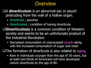


Diverticular disease is a common condition where pouches called diverticula bulge out from the colon wall, usually where blood vessels penetrate the colon. Diverticulosis is the presence of diverticula without inflammation, while diverticulitis occurs when diverticula become inflamed or infected, usually due to hard stool getting stuck in a diverticulum. Diverticulitis ranges from uncomplicated cases treated with antibiotics to complicated cases involving abscesses, fistulas, or perforation requiring surgery. Risk factors include low-fiber diet, aging, and high blood pressure.


