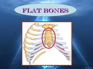
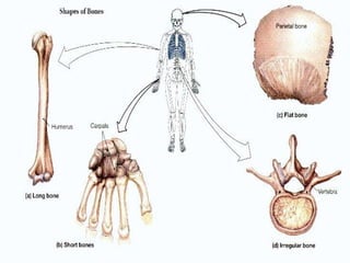
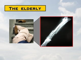
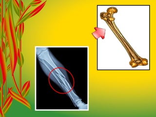
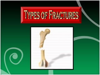
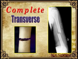
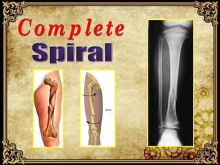
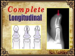
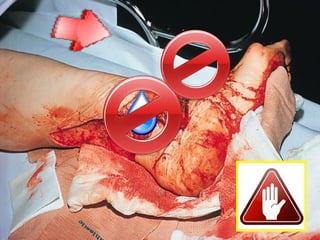
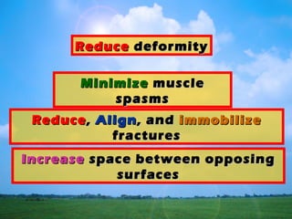
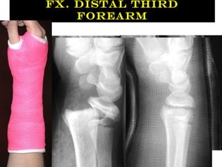
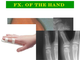
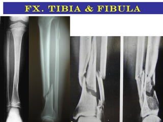
ORTHOPAEDICORTHOPAEDIC
DEPARTMENT AL-DEPARTMENT AL-
BUKAYRIA GENERALBUKAYRIA GENERAL
HOSPITALHOSPITAL
Flat bonesFlat bones
Irregular BonesIrregular Bones
People who arePeople who are
activeactive
The elderlyThe elderly
work-relatedwork-related lose inlose in
bone massbone mass
It is a break in the bone
continuity
CloseClose OpenOpen
DisplacedDisplaced
Non-DisplacedNon-Displaced
SplintinSplintin
gg
How to treat a fracture
•Principles of Fracture Management
–Splint joint above, below
–Splint bone ends
–Loosely cover open fracture sites
–Neurovascular assessment
•before and after splinting
–Gentle in-line traction of long bone
•maintain normal alignment if possible
•reduction of angulated fracture site
Fracture Management
Key Points
With few exceptions orthopedic injuries are not
life threatening.
Do not let drama of grossly deformed fracture
distract you from more serious problems involving
ABC’s.
The problem is not the damage to the bone.
The problem is the damage the bone does to the
surrounding soft tissues.
Evaluate Neurovascular Function Distally
is the applicationis the application
of a pulling forceof a pulling force
to a part of theto a part of the
bodybody
MinimizeMinimize musclemuscle
spasmsspasms
ReduceReduce,, AlignAlign, and, and immobilizeimmobilize
fracturesfractures
ReduceReduce deformitydeformity
IncreaseIncrease space between opposingspace between opposing
surfacessurfaces
MaintainingMaintaining
EffectiveEffective
TractionTraction
MaintainingMaintaining
PositioningPositioning
PreventingPreventing
Skin breakdownSkin breakdown
MonitoringMonitoring
NeurologicalNeurological
StatusStatus
ProvidingProviding
Pin Site CarePin Site Care
Early Management
•Principles:
•Splint joint above and below.
•Loosely cover open fracture sites.
•Neurovascular assessment (before
and after(.
•Gentle in-line traction of long bone:
•maintain normal alignment if possible.
•reduction of angulated fracture site.
Fracture clavicle
Fx. Proximal
Humerus
Fx. Shaft of
Humerus
Higher risk of radial nerve injury.
Fx. Supracondylar
Humerus
Absolute
Emergency.
Mostly treated by
closed reduction
and percutaneous
K-wire fixation.
Complications include:Complications include:
Volkmann's Ischemic
contracture.
Mal-union (cubitus
varus).
Fx. Distal Third
Forearm
Fx. Of The Hand
Fx. femur
Fx. Around the knee
Fx. Tibia & fibula
Fx. Of the foot
Conclusion
Evaluate Neurovascular Function Distally
Splint all fractures and when in doubt splint
ThankThank
youyou

Fracture management

Editor's Notes

  • #3 Fracture Classification and Management By DR. BASSAM YOUNIS HEAD OF ORTHOPAEDIC DEPARTMENT AL-BUKAYRIA GENERAL HOSPITAL
  • #4 Classification of Bones
  • #5 Axial skeleton: skull, vertebral column, ribs & sternum.
  • #6 Appendicular skeleton: limbs, pectoral girdle, & pelvic girdle.
  • #8 Long bones
  • #9 Short bones Cube-shaped carpals, tarsals, & patella
  • #10 Flat bones sternum, & most skull bones
  • #11 Irregular bones Vertebrae & pelvic bones
  • #14 Risk Factors for Fractures
  • #15 Anyone can fracture a bone. People who are active are more likely to break a bone, for example, people who participate in contact sports, like football and basketball, are at higher risk for fractures.
  • #16 The elderly are more likely to break bones than younger people as bones become more brittle as one grows older.
  • #17 Before the age of fifty, more men than women suffer fractures. These fractures are often caused in work-related injuries. After the age of fifty, more women than men suffer fractures. Women tend to lose bone mass more quickly than do men as they grow older.
  • #18 It is break in the bone continuity.
  • #19 It is break in the bone continuity.
  • #20 It is either:
  • #21 Closed: Overlying skin intact. Open: Wound extends from body surface to fracture site. Produced either by bones or object that caused the fracture. Danger of infection.
  • #22 Displaced and Non-displaced Fractures Displaced fracture in which the fractured bone is no longer aligned.
  • #23 Non-displaced fracture, in which the fractured bone remains aligned.
  • #24 Incomplete: Hairline. The break is not reaching all through the bone. It is usually the result of a relatively minor injury.
  • #25 Incomplete: Greenstick. Incomplete break in soft bone Similar to a green twig bending but not breaking
  • #26 complete: Transverse. Occurs in a straight line, usually at a 90-degree angle to the bone shaft. Usually caused by a direct outside blow.
  • #27 complete: Oblique. Occur when one end receives a sudden twisting while the opposite end is fix or stabilized. Fracture occurs down the length and across the width of the bone.
  • #28 complete: Spiral. Result in an S-shaped separation. Similar to an oblique fracture, except the fracture wraps around the length of the bone.
  • #29 complete: Impacted. It is a closed fracture that occurs when pressure is applied to both ends of the bone, causing it to split into two fragments that jam into each other. This type of fracture is common in falls and car accidents.
  • #30 complete: Avulsion. An avulsion fracture is an injury to the bone in a place where a tendon or ligament attaches to the bone. When it occurs, the tendon or ligament pulls off a piece of the bone.
  • #31 complete: Comminuted. Consists of three or more displaced fragments at the site of injury. Caused by a hard blow or a blow in an awkward position
  • #32 complete: Compression. It occurs when the normal vertebral body of the spine is squished, or compressed, to a smaller height. This type of fracture is common in falls and car accidents.
  • #33 complete: Longitudinal. Injury in which the bone splits along its length. Often the result of jumping from a height and landing in such a way as to transmit force to the long axis of the bone.
  • #34 complete: Pathological. It occurs where the bone becomes weakened because of a bone pathology as bone cyst or tumor. It usually occur due to minor trauma.
  • #35 complete: Stress. A stress fracture is a common overuse injury most often seen in athletes. It can occur in any bone, but is commonly seen in the foot and shin bones. They rarely occur in the upper extremity.
  • #38 SPLINT TRACTION
  • #39 Immobilize the injury and joints above and below the injury site. If questionable, treat as a fracture.
  • #40 Guidelines for Splinting Support the injured area above and below the site of the injury, including the joints. If possible, splint the injury in the position that you find it. Don’t try to realign bones or joints. After splinting, check for proper circulation (warmth, feeling, and color). Immobilize above and below the injury.
  • #44 Do not: Do not draw exposed bones back into tissue. Do not irrigate wound.
  • #45 DO: Cover the wound. Splint fracture without disturbing the wound. Place a moist dressing over bone end to prevent drying.
  • #46 is the application of a pulling force to a part of the body
  • #48 -Minimize muscle spasms. -Reduce, align, and immobilize fractures. - Reduce deformity. -Increase space between opposing surfaces.
  • #49 Maintaining Effective Traction. Maintaining Positioning. Preventing Skin breakdown. Monitoring Neurological Status. Providing Pin Site Care.