Acute Pancreatitis
•Download as PPTX, PDF•
49 likes•2,698 views

The document provides an overview of acute pancreatitis including: - It begins with anatomy and blood supply of the pancreas. - Pathophysiology involves autodigestion from inappropriately activated pancreatic enzymes. - Etiology is commonly gallstones, alcohol, or idiopathic. - Clinical presentation includes severe abdominal pain and systemic complications can involve multiple organ systems.

Recommended
More Related Content
What's hot
What's hot (20)
Similar to Acute Pancreatitis
Similar to Acute Pancreatitis (20)
Recently uploaded
Recently uploaded (20)
Acute Pancreatitis
- 2. Outline Anatomy Epidemiology Pathophysiology Etiology Clinical Presentation Workup Severity Scoring System Treatment Prognosis Complications
- 3. PANCREAS PANCREAS Greek word with literal meaning ‘pan’ (all/whole) , ‘creas’ (flesh)
- 4. Anatomy Pancreas is an elongated, accessory digestive gland that lies retroperitoneally Transversely it lies across the posterior abdominal wall posterior to the stomach between duodenum on the right and the spleen on the left.
- 5. Anatomy Parts of pancreas • Head • Neck • Body • Tail
- 6. HEAD • The expanded part of the gland that is embraced by the C shaped curve of the duodenum to the right of the superior mesenteric vessels. • The head of the pancreas rests posteriorly on the IVC Anatomy
- 7. Anatomy NECK • It is short and overlies the superior mesenteric vessels, which form a groove in its posterior aspect. • The anterior surface of neck, covered with peritoneum, is adjacent to the pylorus of the stomach.
- 8. BODY • Continues from the neck and lies to the left of the superior mesenteric vessels, • Passing over the aorta and L2 vertebra • The posterior surface of the body is devoid of peritoneum and is in contact with the aorta, SMA, left suprarenal gland and left kidney and renal vessels
- 9. TAIL • Lies anterior to the left kidney, where it is closely related to the splenic hilum and the left colic flexure. • The tip of the tail is usually blunted and turned superiorly
- 10. Main Pancreatic Duct (WIRSUNG) opens at major duodenal papilla. Accessory pancreatic duct (SANTORINI) usually (60%) communicates with the main pancreatic duct opens into the duodenum at the summit of the minor duodenal papilla
- 11. • Smooth muscle sphincter that control the flow of bile & pancreatic juice into duodenum : - Sphincter of the pancreatic duct - Sphincter of the bile duct Sphincter of ampulla - sphincter of Oddi
- 12. Blood Supply - Arterial -The pancreatic arteries derive mainly from the branches of the SPLENIC ARTERY -The anterior and posterior superior pancreaticoduodenal arteries, branches of the gastroduodenal artery -The anterior and posterior inferior pancreaticoduodenal arteries, branches of the SMA.
- 14. Venous Drainage
- 15. Lymphatics The pancreatic lymphatic vessels follow the blood vessel. Most of them end in the pancreaticosplenic nodes that lie along the splenic artery, but some vessels end in the pyloric lymph nodes. Efferent vessels from these nodes drain to the superior mesenteric lymph nodes or to the celiac lymph nodes via the hepatic lymph nodes.
- 16. Nerve Supply The nerves of the pancreas are derived from the vagus and abdominopelvic splanchnic nerves.
- 17. Pancreas - Physiology FUNCTIONS: neutralize chyme digestive enzymes hormones
- 18. Physiology The pancreas produces : • An exocrine secretion ( pancreatic juice from the acinar cells) that enter the duodenum through the main and accessory pancreatic ducts. • Endocrine secretion (glucagon & insulin) from the pancreatic islets (of langerhans) that enter blood.
- 19. PANCREATITIS Aseptic inflammation of the pancreas. PANCREAS Stroma Parenchyma Exocrine Primary injury causing PANCREATITIS Endocrine Involved secondarily or as a complication
- 21. ACUTE PANCREATITIS CLINICAL DEFINITION An acute condition presenting with abdominal pain-usually associated with raised blood/urine pancreatic enzymes as a result of pancreatic inflammation
- 22. CLASSIFICATION OF ACUTE PANCREATITIS Atlanta* criteria (1992) Revised Atlanta criteria (2012) Mild acute pancreatitis (80% cases) (Acute Interstitial/edematous pancreatitis) Acute Absence of organ failure Absence of local complications Severe acute pancreatitis(20 % cases) (Acute Hemorrhagic Necrotizing (fulminant) pancreatitis) Local complications +/- Organ failure defined as SBP < 90 mm Hg PaO2 ≤ 60 mm Hg GI bleed ≥ 500 ml/24 hrs Cret ≥ 2 mg/dL after rehydration Ranson score ≥ 3 or APACHE ≥ 8 Mild acute pancreatitis Absence of organ failure Absence of local complications Moderately severe acute pancreatitis Local complications +/- Transient organ failure(<48 h) Severe acute pancreatitis Persistent organ failure**(>48 h) and/or death *defined as a score of 2 or more for one of these(CVS, Renal, Resp) organ systems using the modified Marshall scoring system
- 23. Two Distinct phases of Acute Pancreatitis Early Phase(within 1 week) Late Phase (>1 week) Characterized by SIRS +/- organ failure Severity assessed by functional/clinical Severity Scoring System (Ranson/Galsgow etc) Characterized by local complication Severity assessed by morphological scoring system(Balthazar Scoring)
- 24. Mortality of acute pancreatitis The overall mortality remains approximately 5% to 10% Rises to >40% if sterile necrosis becomes infected
- 25. Epidemiology Gender Predilection Generally M>F In males more often related to alcohol In females more often related to biliary tract disease Idiopathic pancreatitis no clear gender predilection
- 26. Epidemiology RACE Hospitalization rates3 times higher for blacks than whites Risk for African Americans aged 35-64 years10 times higher than for any other group
- 27. PATHOPHYSIOLOGY Autodigestion of pancreatic substance by inappropriately activated pancreatic enzymes (especially trypsinogen)
- 28. Acute Pancreatitis Pathophysiology Pancreatic Ducts become obstructed Hypersecretion of the exocrine enzymes of pancreas These enzymes enter the bile duct, where they are activated and with bile back up into the pancreatic duct Pancreatitis
- 29. Acute Pancreatitis Pathophysiology autodigestion of pancreatic tissue release of enzymes into the circulation activation of white blood cells local complications local vascular insufficiency distant organ failure
- 30. MICROBIOLOGY-in infected necrosis Commonly polymicrobial infection-60% Source: gall bladder, colon, small bowel(Transmural) or hematogenous spread INCIDENCE: 24%(1 week) and 70 %( 3 weeks) ORGANISMS: E.Coli(35%), klebsiella( 25%), Enterococcus (25%) Others….. Staphlyococci, pseudomonas, proteus, enterobacter, anaerobes, candida(10%)
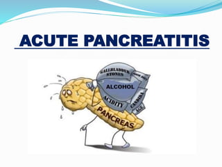
- 32. MECHANICAL CAUSES OF ACUTE PANCREATITIS 1. GALL STONES CHOLEDOCHOLITHIASIS(40-70 %) • Most common biliary tract disease leading to pancreatitis. • MOA: Ductal obstruction causing ductal hypertension and acinar cell injury • 5 % of gall stones causes pancreatitis • M:F=1:3 2. - Ampullary tumor 3. - Sphincter of Oddi dysfunction 4. - Pancreatic head CA(1-2 % ) • 5 % present as AP 5. - Choledochoceles, biliary sludge 6. - Abdominal trauma(1.5 %cases) • 17 % cases have high enzymes and 5 % have clinical AP • Penetrating>Blunt 7. - Iatrogenic Injury • Operative injury - Endoscopic procedures with dye injection
- 33. METABOLIC CAUSES OF ACUTE PANCREATITIS 1.- ALCOHOLISM (25-35%) 2.- Hyperlipoproteinemia (1-4 %) • Type I and V hyperlipidemia • >1000 mg/dL(diagnostic criteria) 3.- Malnutrition 4.- Diabetes, Azotemia,Porphyria 5.- Hypercalcemia/hyperparathyroidism • Hyper secretion and formation of calcified stones intraductallyAcinar injury 6.- Drugs (2 % cases)(usually mild) Definite association ( azathioprine, sulfonamaides, sulindac, tetracycline, valproic acid, Didanosine, Methyldopa,estrogens, furosemide, 6-Mercaptopurine, pentamidine, 5-ASA, steriods,octreotide) Probable association (thiazides, Flagyl, Methandienone, Nitrofurantoin, phenformin,piroxicam,procainamide, Colaspase, chlorthalidone, asparagine,cimetidine,cisplatin, cytosine arabinoside, diphenoxylate, ethacrynic acid)
- 34. INFECTIVE CAUSES OF ACUTE PANCREATITIS VIRUSES • MMR, Dengue virus(complication of dengue hemorrhagic fever, Coxsackie B, Hepatitis virus, CMV, EBV, Echovirus, VZV, HSV BACTERIA • M. tuberculosis, M. avium complex, Mycoplasma, Legionella, Leptospira, Salmonella, Campylobacter, Yersinia, Brucella, Nocarbia FUNGI • Aspergillus PARASITES • Clonorchis sinensis, Toxoplasma, Cryptosporidium, Ascaris, Echinococcus granulosus, • Fasciola Hepatica, Opistorhcis sp and Dicrocoelium dendriticym SCORPION AND SNAKE BITE • Trinidalian scorpion* (Tityus trinitatis) • MOA: Neurotransmitter discharge from cholinergic nerve terminals, leading to massive production of pancreatic juice(same MOA in antiacetylcholinesterase/OPC insecticide)
- 35. GENETIC CAUSES OF AUTE PANCREATITIS 1. PANCREAS DIVISUM (unfused ducts of Wirsung and Santorini) • Seen in 20-45 % cases • MOA(controversial): stenotic minor papillae and atretic duct of santorini 2. ANOMALOUS UNION OF PANCREATICOBILIARY DUCT(annular pancreas) 3. AUTOIMMUNE PANCREATITIs-associated with IBD 4. HEREDITARY PANCREATITIS- • Autosomal dominant, premature activation of trypsinogen • Acute pancreatitis in teens • Chronic in next 2 decades • 40 % risk of pancreatic cancer by 70 year 5. Celiac disease 6. Cystic fibrosis-CFTR mutation abnormality of ductal secretion
- 36. Idiopathic Acute Pancreatitis Defined as pancreatitis with no etiology established after initial laboratory (including lipid and calcium level,autoimmune markers, viral titers) and imaging tests (USG and CT in the appropriate patient) In some patients an etiology may eventually be found(70 % of IAP due to microlithiasis), yet in others no definite cause is ever established. ____________________________________________________ Careful search for the etiology must be made in all cases, and no more than 20 % of cases should fall into the idiopathic category
- 37. Post-ERCP Pancreatitis 3rd Most common cause of AP(after gallstone and alcohol) i.e. 4% Most common complication of ERCP INCIDENCE 2-4 % patients undergoing ERCP develop acute pancreatitis Risk of severe AP < 1/500 PREDISPOSING FACTORS Sphincter of Oddi dysfunction(risk increases to 30 %) H/O recurrent pancreatitis Sphincterotomy Balloon dilation of sphincter Inexperienced endoscopist
- 39. ABDOMINAL PAIN-CARDINAL SYMPTOM SITE: usually experienced first in the epigastrium but may be localized to either upper quadrant or felt diffusely throughout the abdomen or lower chest ONSET: characteristically develops quickly, generally following substantial meal SEVERITY: frequently severe, reaching max. intensity within minutes NATURE: “boring through”, “knifing” (illimitable agony) DURATION: hours-days COURSE: constant (refractory to usual doses of analgesics, not relieved by vomiting) RADIATION: directly to back(50%), chest or flanks AGGRAVATING FACTOR: food/alcohol intake, walking, lying supine. RELEIVING FACTOR: sitting or leaning/stooping forward (MUHAMMEDAN PRAYER SIGN)
- 40. OTHER MANIFESTATIONS Nausea, frequent and effortless vomiting, anorexia,diarrhea Due to reflex pylorospasm More intense in necrotizing than in edematous pancreatitis Persistent retching despite empty stomach Hiccups Due to gastric distension/diaphragmatic irritation Fever Low grade, seen in infective pancreatitis Weakness, Anxiety, Sweating Indicates severe attack
- 41. ABDOMEN EXAMINATION Tenderness + Rebound tenderness: epigastrium/upper abdomen Guarding(also called “defense musculaire” )-upper abdomen Rigidity(involuntary stiffness)-unusual
- 42. Cutaneous Ecchymosis(1 % cases)* Acute Hemorrhagic Necrotizing/fulminant Pancreatitis Periperitoneal/retroperitoneal Hemorrhage Methemalbumin formed from digested blood tracks around Fascial planes hemorrhagic spots and ecchymosis in flanks (GREY TURNER’S SIGN)** FALCIFORM LIGAMENT Bluish Discoloration around umbilicus (CULLEN’S SIGN) Below inguinal ligament (FOX SIGN)
- 43. GREY TURNER1 SIGN CULLEN2 SIGN FOX3 SIGN 1. Named after British surgeon George Grey Turner(1877-1951) 2. Named for Thomas Stephen Cullen (1869-1953), Canadian gynecologist who first described the sign in ruptured ectopic pregnancy in 1916 3.Named after George Henry Fox(1846-1937), American dermatologist
- 44. RESPIRATORY MANIFESTATIONS Left sided* Pleural effusion(10-20%) + pulmonary edema + pneumonitis *Due to close approximation of body and tail of pancreas to the left sided diaphragm
- 45. SYSTEMIC COMPLICATIONS CARDIOVASCULAR Shock- hypovolemic and septic Arrhythmias/pericardial effusion/sudden death ST-T nonspecific changes Pulmonary Respiratory failure/pneumonia/atelectasis/pleural effusion Acute Respiratory Distress Syndrome (ARDS) Renal Failure Oliguria Azotemia Renal artery/vein thrombosis Hematological Hemoconcentation Disseminated Intravascular Coagulopathy (DIC)
- 46. SYSTEMIC COMPLICATIONS Metabolic Hypocalcemia Hyperglycemia Hyperlipidemia Gastrointestinal Peptic Ulcer/Erosive gastritis Ileus Portal vein or splenic vein thrombosis with varices Neurological Visual disturbances-Sudden blindness(Purtscher’s retinopathy) Confusion,irritability,psychosis Fat emboli Alcohol withdrawal syndrome Encephalopathy Miscellaneous Subcutaneous fat necrosis Intra-abdominal saponification Arthralgia
- 47. LOCAL COMPLICATIONS Peripancreatic fluid collections Pancreatic necrosis( sterile + infected) Pancreatic abscess(Phlegmon) Pseudocyst Pancreatic ascites Pseudoaneurysm
- 48. MANIFESTAIONS OF COMPLICATIONS Hypocalcaemia circumoral numbness or paresthesia (1st symtpom to develop) /tingling of distal extremities carpopedal spasm (=main d'accoucheur- French "hand of the obstetrician”) laryngospasm generalized seizures Chvostek*(-Weiss) sign Trousseau** sign of latent tetany
- 50. Diagnostic criteria Most often established by the presence of two of the three following criteria: (i) abdominal pain consistent with the disease, (ii) serum amylase and/or lipase greater than three times the upper limit of normal, and/or (iii) characteristic findings from abdominal imaging. *CT and/or MRI of the pancreas should be reserved for patients in whom the diagnosis is unclear(typical pain with normal enzymes) who fail to improve clinically within the first 48–72 h after hospital admission (e.g., persistent pain, fever, nausea, unable to begin oral feeding) to evaluate complications
- 51. HEMATOLOGICAL BASELINES CBC: Low Hb: prolonged hemetemesis/melena, internal hemorrhage Leucocytosis (10,000-30,000/mcL)-infection, non infectious inflammation Low platelets-DIC LFT’s: raised bilirubin, AST/ALT/LDH, ALP, GGTP- gall stone pancreatitis RFT’s: raised BUN/cretainine- ATN ARF Coagulation profile: increased INR-DIC BSR: > 180 mg/dl-diabetes as a sequelae or cause Serum electrolytes: Serum Protein: low protein/ albumin Diabetes Mellitus AP
- 52. HEMATOLOGICAL ABG’s Etiology specific investigations Serum fasting lipid profile Viral titers Serum Calcium (HypercalcemiaAPHypocalcemia) Autoimmune markers Acid-Base Disturbance Etiology Metabolic (Lactic)acidosis with high anion gap Hypovolemic shock Hypokalemic Hypochloremic metabolic alkalosis persistent vomiting Respiratory acidosis ARDS/resp failure
- 53. HEMATOLOGICAL Pancreatic Enzymes’ Assays Serum Amylase: ONSET: almost immediately PEAK: within several hours 3-4 times upper limit of normal within 24 hrs (90%) RETURN to normal depends on severity(3-5 days) Serum Lipase: more sensitive/specific than amylase Remains elevated longer than amylase(12 days) Useful if late presentation SERUM INDICATOR OF HIGHEST PROBABILITY OF DISEASE
- 54. Urine Amylase More sensitive than serum levels Remain elevated for several days after serum levels returned to normal Pancreatic-specific amylase(p-amylase) Measuring p-amylase instead to total amylase(also includes salivary amylase) makes diagnosis more specific(88-93%)
- 55. CONDITIONS ASSOCIATED WITH RAISED SERUM AMYLASE ABDOMEN Small bowel obstruction strangulation ileus mesenteric ischemia Acute appendicitis Cholecystitis Perforated Duodenal Ulcer Gastroenteritis Biliary peritonitis Spasm of sphincter of Oddi GYNE Ruptured Ectopic pregnancy Torsion of an ovarian cyst OTHERS Parotitis (Mumps) Macroamylasaemia Opioids administration Low GFR Brain injury(CVA)- hyperstimulation of pancreas
- 56. Plain X-ray abdomen erect AP view Sentinel* loop sign Localized isolated Distended gut loop (Ileus) seen near the site of injured viscus or inflamed organ SITE: Acute Pancreatitis Left hypochondrium (PROXIMAL JEJUNUM) Acute Appendicitis Right iliac fossa Acute Cholecystitis Right Hypochondrium Diverticulitis Left iliac fossa ______________________________________________________________ Not diagnostic of Acute Pancreatitis; useful in differential diagnosis
- 58. Plain X-ray abdomen erect AP view Colon cut-off sign Gas filled (Distended) segment of proximal(mainly transverse) colon associated with narrowing of the splenic flexure with collapse of descending colon _______________________________________________________________ _____________________ Not diagnostic of Acute Pancreatitis; useful in differential diagnosis
- 60. Plain X-ray abdomen erect AP view Renal halo sign RATIONALE: peripancreatic inflammatory reaction extension into pararenal space OTHERS _______________________________________________________________ Not diagnostic of Acute Pancreatitis; useful in differential diagnosis • Air in the duodenal C-loop • increased gastrocolic separation • Gastric curvature distortion • To rule out perforated DU(gas under diaphragm) • Obliteration of psoas shadow • Localized ground glass appearance ( localized increased high soft tissue density) • Calcified gall stones • Pancreatic calcification(chronic pancreatitis)
- 61. Renal Halo Sign NORMAL RENAL HALO SIGN
- 62. Abdominal Ultrasonography Not diagnostic Should be performed within 24 hours in all patients to detect gall stones* as a potential cause Rule out acute cholecystits as differential diagnosis Detect dilated CBD sensitivity-(70-80%) THERAPEUTIC: USG-guided aspiration for gram staining and culture USG-guided pig tail catheter insertion Gallstone pancreatitis is usually an acute event and resolves when the stone is removed or passes spontaneously.
- 63. IV Contrast enhanced Computed Tomography Scan Provides over 90 % sensitivity and specificity for the diagnosis of AP….. BUT Routine use in patients with AP is unwarranted, as the diagnosis is apparent in many patients and most have a mild, uncomplicated course.
- 64. IV Contrast enhanced Computed Tomography Scan* INDICATIONS-DIAGNOSTIC Diagnostic uncertainty (differentiating pancreatitis from other possible intra-abdominal catastrophes) Severe acute pancreatitis- distinguish interstitial from necrotizing pancreatitis Necrosis( non enhancement area > 30 % or 3 cm) done at 72 hrs* Systemic complications: Progressive deterioration, MOF, sepsis Localized complications: Altered fat and fascial planes, Fluid collection, pseudocyst, psduoaneurysm, Bowel distension, mesenteric edema, hemorrhage
- 65. BALTHAZAR CT severity index(CTSI)-1994 ___________________________________________________________________ Mild (0-3) moderate (4-6) severe (7-10) CT Severity Index Inflammation score + Necrosis score
- 66. Magnetic Resonant Imaging Suitable alternative to CT in patients with a contrast allergy and renal insufficiency where T2-weighted images without gadolinium contrast can diagnose pancreatic necrosis
- 67. Magnetic Resonant Cholangiopancreatography INDICATION: diagnosis of suspected biliary and pancreatic duct obstruction in the setting of pancreatitis. Repeated attacks of idiopathic acute pancreatitis
- 68. Endoscopic UltraSonography INDICATIONS Repeated idiopathic acute pancreatitis* occult biliary disease- small stones/sludge _________________________________________ *Endoscopic investigation in patients with acute idiopathic pancreatitis should be limited, as the risks and benefits of investigation in these patients are unclear and should be referred to centers of expertise.
- 69. Endoscopic Retrograde Cholangiopancreatography INDICATION Severe gallstone AP or AP with concurrent acute cholangitis/biliary obstruction/ biliary sepsis/jaundice (due to persistent stone) ERCP within 24(-72) h of admission Sphincterotomy/stent and bile duct clearance reduces infective complications/mortality
- 70. SEVERITY SCORING SYSTEMS ACUTE PANCREATITIS SPECIFIC SCORING SYSTEMS Ranson score Glagsow score Bedside Index for Severity in Acute Pancreatitis(BISAP) score Harmless Acute Pancreatitis Score(HAPS) Hong Kong Criteria ACUTE PANCREATITIS NON-SPECIFIC SCORING SYSTEMS (ICU SCORING SYSTEMS) Acute Physiology And Chronic Health Evaluation(APACHE) II score Sequential Organ Failure Assessment(SOFA) score
- 71. RANSON SCORE-1974 (for alcohol pancreatitis) ON ADMISSION WITHIN 48 HOURS Age > 55 yrs TLC > 16,000/mm3 BSR> 200 mg/dL AST > 250 IU/L LDH > 350 IU/L BUN rise >5 mg/dL Pa02 < 60 mmHg ( 8 KPa) Serum Calcium < 8 mg/dL Base deficit > 4 meq/L Fluid Sequestration > 6000 mL Hct fall > 10 % NOTE: Disease classified as SEVERE when 3 or more factors are present
- 72. Revised RANSON SCORE-1979 (for Gallstone pancreatitis) ON ADMISSION WITHIN 48 HOURS Age > 70 years TLC > 18000/mm3 BSR > 220 mg/dL AST> 250 IU/L LDH >400 IU/L BUN rise >5 mg/dL Pa02 < 60 mmHg ( 8 KPa) Serum Calcium < 8 mg/dL Base deficit > 5 meq/L Fluid Sequestration > 4 litres Hct fall > 10 % NOTE: Disease classified as SEVERE when 3 or more factors are present
- 73. RANSON SCORE……. DRAWBACKS One has to measure all 11 signs to achieve the best predictability of prognosis 2 full days are needed to complete the profile. Difference between gallstone & alcohol etiology Only 73 % sensitive and 77 % specific in predicting mortality
- 74. Acute Physiology And Chronic Health Evaluation II score-1985
- 75. APACHE Scoring System MERIT Immediate assessment of the severity of pancreatitis possible Can be used even after 48 hours Unlike ALL pancreatic specific scoring systems, APACHE (and SOFA) also includes clinical features of patient besides laboratory values Best validated
- 76. DEMERITS OF AP-specific scoring systems(ACG 2013) Uniformly cumbersome. No laboratory test is practically available or consistently accurate to predict severity in patients with AP. Thus, in the absence of any available test to determine severity, close examination to assess early fluid losses, hypovolemic shock, and symptoms suggestive of organ dysfunction is crucial.
- 77. MANAGEMENT Mild acute pancreatitis Conservative Approach Admit in general ward Non invasive monitoring (Moderate)Severe acute pancreatitis Aggressive Approach Admit in HDU/ICU Invasive monitoring __________________________________________ Recognizing patients with severe acute pancreatitis ASAP is critical for achieving optimal outcomes
- 78. Mild Acute Pancreatitis mild and self-limiting, needing only brief hospitalization. Rehydration by IV fluids Frequent non-invasive observation/monitoring(atleast 8 hrly) Brief period of fasting till pain/vomiting settles No medication required other than analgesics(important) and anti-emetics Antibiotics not indicated in absence of signs or documented sources of infection Avoid Morphine-cause sphincter of Oddi spasm Metabolic support Correction of electrolyte imbalance
- 79. Severe Acute Pancreatitis P: Pain relief Proton pump inhibitors-omeprazole A Admit in HDU/ICU Antibiotics N Nasogastric intubation(if vomiting) Nasal oxygen Nutrition support C Calcium gluconate CVP line Catherisation- Foley R Rehydration by IV fluids,plasma,blood Radiology: CT scan, USG Resuscitation when required E Endotracheal intubation Electrolytes management ERCP A Antacids S Swan-Ganz catheter for CVP and TPN Suction-in case of aspiration Steroids in case of ARDS Supportive therapy for organ failure
- 80. Monitoring CLINICAL INVESTIGATIONS Vitals Urine Output CV pressure Baselines Serial ABGs Serial BSR Serum calcium/magnesium
- 81. EARLY AGRESSIVE IV hydration Which fluid? Lactated Ringer ’s solution may be the preferred isotonic crystalloid replacement fluid • Ringer lactate is better electrolyte balance and more pH- balanced when? Early aggressive IV hydration is most beneficial during the first 12 – 24 h, and may have little benefit beyond this time period How much? Aggressive hydration, defined as 250 – 500 ml per hour of isotonic crystalloid solution should be provided to all patients, unless cardiovascular, renal, or other related comorbid factors exist. • Fluid requirements should be reassessed at frequent intervals within 6 h of admission and for the next 24 – 48 h.
- 82. Antibiotics Routine use* NOT recommended(ACG 2013) as Prophylaxis in severe AP Preventive measure in sterile necrosis to prevent development of infected necrosis Indicated in Established infected pancreatic necrosis or Extraperitoneal infections Cholangitis, catheter-acquired infections, bacteremia, UTIs, pneumonia Few antibiotics penetrate due to consistency of pancreatic necrosis cefuroxime, or imipenem, or ciprofloxacin plus metronidazole ______________________________________________
- 83. Nutrition In mild AP oral feedings can be started immediately if there is no nausea/vomiting, and the abdominal pain/tenderness/Ileus has resolved(amylase return to normal, patient feel hunger) Initiation of feeding with a small and slowly increasing low-fat (low- protein) soft diet In severe AP Enteral route is recommended to prevent infectious complications Parenteral nutrition should be avoided, unless enteral route is not available, not tolerated, or not meeting caloric requirements
- 84. Route of enteral Nutrition Traditionally nasojejunal route has been preferred to avoid the gastric phase of stimulation BUT Nasogastric route appears comparable in efficacy and safety MERITS OF NASOGASTRIC ROUTE DEMERITS OF NASOGASTRIC ROUTE NG tube placement is far easier than nasojejunal tube placement( requiring interventional radiology or endoscopy, ) Slight increased risk of aspiration
- 85. Role of Surgery in pancreatitis In case of mild gallstone AP, cholecystectomy should be performed before discharge to prevent a recurrence of AP In case of necrotizing biliary AP, in order to prevent infection, cholecystectomy is to be deferred until active inflammation subsides and fluid collections resolve or stabilize ____________________________________________________ If patient unfit for surgery(comorbid/elderly), biliary sphincherotomy alone may be effective to reduce further attacks of AP
- 86. Sterile necrosis infected necrosis Asymptomatic Does not mandate intervention regardless of size, location and extension • Initially treated with antibiotics Stable Symptomatic (associated with GOO or bile obstruction) minimally invasive methods of necrosectomy are preferred to open necrosectomy Urgent debridement unstable
- 87. When to Discharge Pain is well controlled with oral analgesia Able to tolerate an oral diet that maintains their caloric needs, and all complications have been addressed adequately Follow up Routine clinical follow-up care (typically including physical examination and amylase and lipase assays) is needed to monitor for potential complications of the pancreatitis Within 7-10 days
- 88. Prognosis TYPE OF AP MORTALITY Overall 10-15 % (Biliary>alcholic) Mild Acute Pancreatitis(80 % cases) 1 % Severe Acute Pancreatitis(20 % cases) Severe 20-50 % <1 week 1/3 cases MOF >1 week 2/3 cases Sepsis (+MOF)
- 89. THANK YOU……….