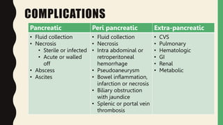
This document provides information on acute pancreatitis including:
1. The epidemiology, causes, pathophysiology, clinical presentation, investigations, management, and complications of acute pancreatitis are summarized. Gallstones and alcohol are the most common causes.
2. Laboratory markers like lipase and amylase are used to diagnose, while CT, MRI, and ultrasound can identify complications like fluid collections and necrosis. Treatment involves fluid resuscitation, pain management, and treating any organ dysfunction.
3. Complications include pancreatic and extra-pancreatic complications like fluid collections, necrosis, infection, and vascular or bowel issues. Infected necrosis requires antibiotics while severe cases may require drainage procedures or surgery.
Acute cholangitis is an infection of the bile ducts caused by obstruction and bacterial overgrowth. It presents with fever, jaundice, and right upper quadrant pain (Charcot's triad). Obstruction leads to increased pressure and bacterial growth in the bile ducts. Diagnosis involves blood tests, imaging like ultrasound or CT, and testing bile if drained. Treatment is antibiotics, hydration, and relieving obstruction endoscopically or surgically. Antibiotics are continued until obstruction is fully resolved to prevent recurrence.
The document provides information on acute pancreatitis including its definition, causes, pathogenesis, clinical manifestations, complications, diagnostic criteria, imaging, and severity scoring systems. Key points include:
- Acute pancreatitis is defined as pancreatic inflammation initiated by pancreatic injury and activation of enzymes.
- Common causes are gallstones, alcohol use, and other structural/metabolic issues.
- Diagnosis requires abdominal pain plus elevated pancreatic enzymes or imaging findings. Severity is evaluated using Ranson or Glasgow criteria.
- Complications can include pancreatic necrosis, fluid collections, vascular issues, and multi-organ failure. Management involves treatment of the underlying cause, pain control, and monitoring for complications.
This document provides an overview of the management of acute pancreatitis. It defines acute pancreatitis as the inflammation of the pancreas often associated with pancreatic duct dilation. It discusses the epidemiology, etiology, pathogenesis, clinical forms, investigations, risk assessment, treatment and prognosis of acute pancreatitis. The management involves resuscitation, assessing severity, treating any underlying causes, and monitoring for complications which can include pancreatic necrosis, infection and multi-organ failure. Severity is assessed using scoring systems like Ranson's criteria or CT severity index to determine prognosis and guide management.
This document provides an overview of pancreatitis, including its epidemiology, pathophysiology, etiology, clinical presentation, workup, severity scoring systems, treatment, prognosis, and complications. It defines acute and chronic pancreatitis and describes the reversible inflammation of the pancreas that occurs in acute pancreatitis. Key points include that the annual incidence is 13-45 per 100,000 people, the pathophysiology involves premature activation of digestive enzymes within the pancreas rather than the intestines, and treatment depends on the severity but generally involves IV rehydration and pain management for mild cases and more aggressive monitoring and support in an ICU for severe cases.
The document discusses pancreatitis, including its anatomy, physiology, classification, signs and symptoms, diagnosis, and management. It addresses both acute and chronic pancreatitis. Acute pancreatitis is commonly caused by gallstones or alcohol and can be mild, moderately severe, or severe based on organ dysfunction. It presents with abdominal pain and elevated pancreatic enzymes. Chronic pancreatitis is usually due to alcohol abuse and causes pain, digestive issues, and diabetes over time. Management involves treating the underlying cause, supportive care, and surgery for complications.
Chronic pancreatitis is a progressive inflammatory condition of the pancreas characterized by irreversible morphological changes and loss of function. It is most commonly caused by long term heavy alcohol use. Symptoms include recurrent abdominal pain, steatorrhea due to exocrine insufficiency, and diabetes mellitus due to endocrine insufficiency. Diagnosis involves functional tests like fecal elastase and imaging modalities like CT, MRI, ERCP and EUS which demonstrate findings of pancreatic duct abnormalities, parenchymal changes and calcifications.
This document defines acute kidney injury (AKI), formerly known as acute renal failure (ARF), and discusses its causes, diagnosis, and management. AKI is defined based on increases in serum creatinine and decreases in urine output. The main causes of AKI are pre-renal (decreased renal blood flow), renal (intrinsic kidney injury), and post-renal (urinary tract obstruction). Common etiologies include acute tubular necrosis, glomerulonephritis, and acute interstitial nephritis. Diagnosis involves laboratory and imaging tests. Management focuses on treating the underlying cause, fluid management, and potentially renal replacement therapy. Prognosis depends on the severity and reversibility of the kidney injury
The document provides information on acute abdomen including its definition, epidemiology, physiology, differential diagnosis by location, history and physical examination findings, important investigations, management principles, and criteria for surgical consultation. Acute abdomen is defined as sudden severe abdominal pain lasting less than 24 hours that often requires urgent diagnosis and some causes need surgical treatment. The differential diagnosis considers location of pain and includes conditions like appendicitis, diverticulitis, bowel obstruction, pancreatitis and others. Key aspects of evaluation involve history, physical exam, labs, imaging and identifying high-risk patients who may require emergent surgery.
This document provides an overview of the management of obstructive jaundice. It begins with definitions and classifications of jaundice. Obstructive jaundice can be intrahepatic or extrahepatic in origin. Common causes of intrahepatic cholestasis include viral hepatitis, alcoholic hepatitis, and drug toxicity. Extrahepatic obstructions are often due to choledocholithiasis (gallstones in the common bile duct), tumors, or strictures. Diagnostic imaging includes ultrasound, MRCP, ERCP, and intraoperative cholangiography. Treatment depends on whether the obstruction is pre-operative or discovered during cholecystectomy, and may involve ERCP, laparoscopic or open CBD exploration, or
Acute Pancreatitis (According to American College of Gastroenterology 2013 gu...Jibran Mohsin
This Presentation focuses on definition, new classification, different scoring systems for severity, rationale for radiological signs and new management recommendations as per 2013 American College of Gastroenterology guidelines
1. The document summarizes guidelines for the management of acute pancreatitis, including diagnosis, etiology, risk stratification, initial management, role of ERCP, antibiotics, nutrition, and surgery.
2. Key points include diagnosing based on abdominal pain and elevated serum amylase/lipase, identifying gallstones and alcohol as common causes, and aggressively hydrating patients while considering nutrition via enteral feeding in severe cases.
3. Surgery is only recommended for gallstone pancreatitis patients without ongoing inflammation or fluid collections in order to prevent recurrence.
This document provides an overview of acute and chronic pancreatitis, including anatomy, pathophysiology, clinical presentation, diagnosis, management, and case examples. It describes the pancreas' exocrine and endocrine functions. Acute pancreatitis results from pancreatic cell damage causing trypsinogen activation and inflammation. Risk factors include gallstones, alcohol use, and hypertriglyceridemia. Diagnosis involves abdominal pain, lipase elevation, and imaging findings. Chronic pancreatitis is characterized by irreversible fibrosis from recurrent acute injury. It can cause exocrine and endocrine insufficiency over time. Diagnosis combines clinical history with secretin stimulation tests, fecal elastase, and imaging. Management focuses on pain control, nutrition
Gallstones:
Most common biliary pathology
Asymptomatic in majority of cases (>80%)
Approx. 1–2% of asymptomatic patients develop symptoms requiring cholecystectomy per year.
Ulcerative colitis is a chronic inflammatory bowel disease that affects the colon. It involves diffuse inflammation and ulceration of the colonic mucosa. The cause is unknown but likely related to genetic and immune factors. Symptoms include bloody diarrhea. Diagnosis involves colonoscopy and biopsy. Treatment involves medications to induce and maintain remission such as mesalamine, corticosteroids, immunomodulators, and biologics. Surgery may be required for severe cases or cancer prevention. Long-term monitoring is needed due to cancer risk.
This document summarizes acute pancreatitis, including its definition, causes, symptoms, pathogenesis, complications, diagnostic tests, severity scoring systems, and management. Acute pancreatitis is characterized by inflammation of the pancreas and is most commonly caused by gallstones or alcoholism. It presents with severe epigastric pain and other gastrointestinal symptoms. The pathogenesis involves premature activation of digestive enzymes within the pancreas that can lead to autodigestion. Complications include pseudocysts, abscesses, necrosis, and systemic complications like shock. Diagnosis involves blood tests showing elevated pancreatic enzymes and imaging tests. Severity is assessed using scoring systems like Ranson criteria, APACHE II, and CT severity index. Treatment focuses on fluid
Pancreatitis is an inflammation of the pancreas that can be acute or chronic. Acute pancreatitis involves reversible injury to the pancreas and can range from mild to severe, with severe cases involving organ failure. Chronic pancreatitis is characterized by irreversible damage to the pancreas that typically causes pain and loss of pancreatic function over time. Treatment for acute pancreatitis depends on severity and may involve hospitalization, IV fluids, monitoring for organ failure, and antibiotics for severe cases. Treatment for chronic pancreatitis focuses on pain management, treating complications, and sometimes surgical interventions.
Peptic ulcer disease is caused by an imbalance between aggressive gastric factors like acid and pepsin and protective mucosal defenses. H. pylori infection plays a key role in most peptic ulcers by damaging the mucosal layer. Treatment involves eradicating H. pylori with triple therapy using a PPI and two antibiotics for 2 weeks, and continuing PPI therapy for an additional 2 weeks to aid ulcer healing. Adherence to the full treatment course is important for successful eradication.
This document discusses pancreatitis, including its anatomy, physiology, etiology, clinical presentation, diagnosis, prognosis, management, and complications. Pancreatitis is defined as inflammation of the pancreas and can be acute or chronic. Acute pancreatitis is commonly caused by gallstones or alcohol and may range from mild to severe, with severe cases involving pancreatic necrosis and multi-organ failure. Diagnosis involves blood tests measuring amylase and lipase along with imaging like CT. Management depends on severity but generally involves hospitalization, IV fluids, pain control, and monitoring for complications.
- Places fingers over the lower ribs on the left side
- Asks patient to take a deep breath
You:
- Percuss over the assistant's fingers
- Dullness indicates splenic enlargement crossing
the midline
Positive Nixon's sign suggests splenomegaly.
Obstructive jaundice is one of the important surgical topics. In this playlist I have discussed the introduction, choledocholithiasis, Carcinoma Pancreas and biliary atresia. If you watch all these videos together you will become confident in Managing obstructive jaundice.
This document provides an overview of approaches to evaluating and treating a patient presenting with jaundice. There are three main types of jaundice discussed: hemolytic, hepatic, and obstructive. For each type, the document outlines relevant clinical findings, laboratory investigations, and potential etiologies. Treatment options are also reviewed for obstructive jaundice, the most common cause being choledocholithiasis, which can be addressed through open or laparoscopic exploration/stone extraction or endoscopic papillotomy. Periampullary carcinoma is another potential etiology that may require curative surgery like the Whipple procedure or palliative interventions.
The document discusses peptic ulcers, including that the prevalence in India is estimated at 4-10 per 1000 people aged 30-60, with males at higher risk. Common causes are H. pylori infection, smoking, alcohol, NSAIDs, and stress. Symptoms include abdominal pain relieved by food as well as bleeding. Diagnosis involves tests like endoscopy. Treatment focuses on eliminating H. pylori, reducing acid secretion, and lifestyle changes. Complications can include hemorrhage, perforation, and obstruction.
Acute pancreatitis is an inflammatory condition of the pancreas caused by the early activation of digestive enzymes within the pancreas. It can range from mild to severe, and in severe cases, it can lead to organ failure. The most common causes are gallstones, alcohol use, and viral infections. Symptoms include severe abdominal pain, nausea, vomiting, and fever. Laboratory tests show elevated levels of pancreatic enzymes in the blood. Severity is assessed using the Ranson score or APACHE II score. Treatment involves intravenous fluids, bowel rest, pain medications, and treating the underlying cause. Complications can include pancreatic pseudocysts, abscesses, and necrosis.
The gallbladder is a hollow organ located beneath the liver that stores and concentrates bile. Cholecystitis is inflammation of the gallbladder, usually caused by gallstones blocking the cystic duct. Symptoms include pain in the upper right abdomen and fever. Ultrasound is often used to diagnose cholecystitis by detecting gallstones or thickening of the gallbladder wall. Treatment typically involves surgical removal of the gallbladder via laparoscopy.
This document discusses acute pancreatitis, including its anatomy, etiology, diagnosis, assessment of severity, treatment, complications, and management guidelines. It covers the key roles of the pancreas in enzyme and electrolyte secretion. Common causes of pancreatitis like gallstones and alcohol are described. Diagnosis involves serum markers, imaging, and severity scores. Treatment focuses on hydration, nutrition, and managing complications. Local complications like pseudocysts and necrosis are defined and approaches to their management are provided. Surgical debridement indications and timing are outlined.
This document provides an overview of acute pancreatitis, including:
- The epidemiology, with highest rates in the US and among males related to alcohol use.
- The pathophysiology, involving premature activation of digestive enzymes within the pancreas.
- Diagnosis is based on abdominal pain plus elevated pancreatic enzymes or imaging findings. Severity is assessed using scores like Ranson's criteria or CT severity index.
- Treatment involves fluid resuscitation, nutritional support, pain management, and antibiotics only for proven or suspected infected pancreatic necrosis. The goals are to prevent complications and infections.
This document provides an overview of acute pancreatitis including its anatomy, etiology, pathophysiology, diagnosis, severity assessment, treatment, and complications. Some key points:
- The pancreas is located in the retroperitoneum and has a head, neck, body and tail supplied by various arteries and veins.
- Acute pancreatitis is defined as inflammation of the pancreas with abdominal pain and elevated pancreatic enzymes. Common causes include gallstones, alcohol use, and hyperlipidemia.
- Inflammation occurs when pancreatic enzymes prematurely activate within the pancreas, causing injury. Systemic complications can develop depending on severity.
- Diagnosis involves history, exam, and lab tests
The document describes the pancreas, pancreatitis, and pancreatic tumors. It discusses the anatomy and function of the pancreas, including that it produces digestive enzymes and hormones. Pancreatitis can be acute or chronic and is defined as inflammation of the pancreas. Acute pancreatitis causes severe abdominal pain and its severity ranges from mild to severe based on organ dysfunction. Chronic pancreatitis is progressive destruction of the pancreas due to recurrent inflammation, causing severe pain and pancreatic insufficiency over time. The document also outlines evaluation and management of pancreatic disorders.
This document provides tips for using a PowerPoint presentation on acute pancreatitis. It recommends:
1. Freely editing and modifying the slides to add your own name.
2. Not worrying about the number of slides, as many are blank except for the title to facilitate active learning sessions.
3. Showing blank slides first to elicit what students already know, then showing the content slide.
4. Repeating this process of blank slide then content slide at the end for review.
5. This format allows for active learning through three revisions of content.
This document provides information on acute pancreatitis including:
- Etiology is often gallstones or alcohol use. Other causes include drugs, genetics, obesity, and diabetes.
- Diagnosis requires abdominal pain consistent with pancreatitis plus serum lipase or amylase 3x upper limit and findings on imaging. CT scan is useful for assessing severity.
- Treatment involves aggressive IV fluids, monitoring for complications, enteral nutrition, and antibiotics only if infection is present. Severe cases may require endoscopic or minimally invasive drainage of pancreatic fluid and necrosis.
- Complications include pancreatic pseudocysts, abscesses, and sterile or infected pancreatic necrosis. Severity is classified based on organ failure
1) Acute pancreatitis has many causes including gallstones, alcohol use, genetic mutations, drugs, hypertriglyceridemia and trauma. It is diagnosed when a patient has abdominal pain consistent with pancreatitis along with elevated pancreatic enzymes or imaging findings of pancreatitis.
2) Severity is classified based on the presence of organ failure and local complications. Predictors of severe acute pancreatitis include age over 60, comorbidities, obesity, and long term heavy alcohol use.
3) Management involves fluid resuscitation, nutritional support either enterally or parenterally, use of antibiotics only if infection is suspected, and minimally invasive techniques to treat fluid collections and necrosis when indicated. Recurrence can
Acute pancreatitis is a common cause of hospitalization. It has various causes including gallstones and alcohol use. It involves inflammation of the pancreas that can range from mild to severe. In severe cases, it has a mortality rate of up to 80% if not properly managed in the first 24 hours. Key aspects of management include IV fluids, pain relief, considering antibiotics in certain situations, early nutritional support preferably through enteral feeding, and evaluating any complications through imaging such as CT scan. Scoring systems can help determine prognosis.
This document discusses acute pancreatitis, including:
- The pathophysiology of acute pancreatitis involving autodigestion of the pancreas from digestive enzymes.
- Common causes like gallstones, alcohol use, and hypertriglyceridemia.
- Diagnosis using clinical signs/symptoms confirmed with blood tests like serum amylase and lipase and imaging like CT scans.
- Determining severity using criteria like Ranson score, APACHE II score, and evidence of organ failure or complications on CT scan.
- Treatment focuses on pain control, IV fluids, nutritional support like enteral feeding, and antibiotics only for infected pancreatic necrosis.
Surgical management of chronic pancreatitis.PritamMandal18
Chronic pancreatitis is characterized by irreversible damage to the pancreas from chronic inflammation and scarring that results in loss of exocrine and endocrine function. Risk factors include alcohol use, genetics, and recurrent acute pancreatitis. Diagnosis involves history, labs, imaging, and function tests. Management is initially conservative but may include endoscopic or surgical interventions for pain relief, complications, or disease progression. Surgical options include drainage procedures or resections, with randomized trials showing various techniques have comparable outcomes.
Acute pancreatitis is an inflammatory process of the pancreas that is usually painful and self-limited. The most common causes are gallstones and alcohol abuse. In mild cases, patients can resume oral intake after symptoms improve. In severe cases, supportive care including fluid resuscitation and nutritional support via enteral feeding is important. Enteral nutrition is preferred over total parenteral nutrition due to lower risks of infection and lower costs with similar or better outcomes. The development of pancreatic necrosis is a marker of severe disease and risk of complications.
This document provides information on acute pancreatitis including its definition, causes, pathogenesis, clinical presentation, diagnosis, severity scoring systems, treatment goals, and approaches to nutrition. The key points are:
1. Acute pancreatitis is an inflammatory process of the pancreas that is usually painful and self-limited, with pancreatic function and morphology returning to normal after attacks. Gallstones and alcohol abuse are the most common causes.
2. The pathogenesis involves premature activation of digestive enzymes within the pancreas, leading to autodigestion and systemic complications in severe cases. Scoring systems like Ranson criteria and CT severity index are used to predict severity and guide management.
3. Treatment goals are to limit systemic
The document discusses acute pancreatitis, including:
1. It provides an overview of the normal anatomy and physiology of the pancreas, as well as the exocrine and endocrine functions.
2. It examines the causes, pathogenesis, clinical presentation, diagnosis, and severity scoring systems used to evaluate acute pancreatitis such as Ranson criteria.
3. It outlines the goals and approaches to treatment for both mild and severe acute pancreatitis, including supportive care, nutritional support, role of ERCP, and antibiotics for infected necrosis.
This document provides an overview of acute pancreatitis including:
- The pathophysiology of premature activation of digestive enzymes within the pancreas leading to autodigestion.
- Common etiologies like gallstones, alcohol use, and hypertriglyceridemia.
- Diagnosis through clinical signs/symptoms confirmed with lab tests like serum amylase and lipase levels or imaging like CT.
- Determining severity using criteria like Ranson score, APACHE II score, or evidence of organ failure/necrosis on CT.
- Goals of treatment include halting disease progression, preventing organ failure through aggressive hydration and nutritional support, adequate pain control, and ERCP for severe gall
This document provides an overview of pancreatitis and pancreatic pseudocysts. It defines acute and chronic pancreatitis, describes the pathogenesis involving premature activation of pancreatic enzymes, and lists common causes like gallstones. Signs and symptoms include abdominal pain while complications involve local issues like pseudocysts or systemic problems. Diagnosis involves blood tests, imaging, and assessing severity with tools like BISAP score. Management focuses on supportive care, treating underlying causes, and draining complications surgically or minimally invasively. Pseudocysts are pancreatic fluid collections that often resolve on their own but sometimes require intervention.
This document provides an overview of acute pancreatitis, including its definition, epidemiology, causes, signs and symptoms, diagnostic tests, treatment, and complications. It notes that acute pancreatitis results from inflammation of the pancreas that can range from mild to severe. Diagnostic testing includes blood tests, imaging like ultrasound, CT, and MRI to determine severity. Treatment involves supportive care, pain management, fluid resuscitation, and treating any underlying causes or complications like infection if they develop.
Approach to maternal collapse and cardiac arrest.pptxKTD Priyadarshani
This is a case based discussion on approach to maternal collapse and cardiac arrest. It includes a detailed account on ERC ALS guideline on maternal cardiac arrest and post resuscitation care.
Pelvic Fracture managemnt- Case based discussion .pptxKTD Priyadarshani
A case based approach on the management of a pelvic fracture. it is based on ATLS guideline. A brief account on anaesthetic and orthopedic point of view also included.
This presentation describes cardiac physiology and classification of antiarrhythmics. It also includes a brief account of main drugs of each group including latest drugs like ranolazine, ivabradine and vernakalent.
This presentation describes updated management of thyroid related emergencies. Anaesthetic considerations of myxoedema coma and thyrotoxic crisis is highlighted.
This presentation is based on JBDS and BSPDE guidelines in adult and Paediatric DKA management. A comparison of adult vs paediatric management is included.
This is about emergency approach to a patient presenting with acute severe hemolysis. It mainly describes general approach and how to choose investigations appropriately. In depth discussion about the management of autoimmune haemolytic anaemia with warm antibody, cold antibody, all-immune antibody, drug induced, microangiopathic syndromes- TTP, HUS, DIC, Macrovascular hemolysis, sickle cell disease, thalassemia, G6PD deficiency, Hereditary spherocytosis and paroxysmal nocturnal hemoglobinuria is included.
This presentation describes the epidemiology, initial assessment, investigation and emergency department management of a patient with atrial fibrillation. Some new research evidences are also discussed to answer some dilemmas.
A brief account on major toxidrome and an explanation about how the clinical features occur. anticholinergic, cholinergic, sympathomimetic, opiate, sedative toxidrome and serotonin syndrome and neuroleptic malignant syndrome are explained with the management.
Toxic alcohol includes Methanol, Ethylene Glycol, Isopropyl alcohol. The toxicokinetics, clinical features are explained separately. Pathophysiology of toxic alcohols explained using diagrams. diagnosis can be done using HAGMA, High osmolar gap, UFR and ECG. Management is determined by block metabolism, correct pH and eliminate toxic metabolites.
This presentation includes overview of Sri cyclic antidepressants, its toxicokinetics, toxic mechanism and clinical features. The management is explained in detailed according Resus- RSI- DEAD steps. Main steps includes resuscitation, risk assessment, investigations, supportive therapy, decontamination, antidote- sodium bicarbonate, lipid emulsion and elimination methods.
This document provides information on snake identification and management of snake bites in Sri Lanka. It discusses the clinical features, epidemiology and management of bites from common snakes in Sri Lanka including Russell's viper, cobra, krait, saw-scaled viper, hump-nosed pit viper and green pit viper. For each snake, it outlines signs of local envenoming, coagulopathy and neurotoxicity. The document provides guidance on airway management, IV fluids, antivenom administration and monitoring for anaphylaxis or other reactions to the antivenom.
this presentation contains epidemiology of propane poisoning, kinetics in overdose and clinical effects. in the management mainly focused on methhemoglobinemia.
The presentation includes epidemiology of poisonous plants, classification based upon latest publication from poison centre- Sri Lanka. It goes in to details of Jatropha circus, Glorriosa superba, Thevetia Peruviana, Datura stramonium, Tabernaemantona dichotomy, Strychnus nut vomica toxicity.
This presentation explains about epidemiology of organophosphate poisoning, the toxic mechanism and pathophysiological basis of clinical features. It briefly outlines diagnosis in an emergency situation and management.
ECG is widely available, non invasive investigation in emergency setting. This presentation describes utility of ECG as a screening tool in an unknown poisoning. It includes toxidromic approach in interpreting ECG to narrow down your differential diagnosis based on published articles.
Presentation includes an account on overview about oral anti diabetics, toxicity of sulfonylureas, biguanides, alpha-glucosidase inhibitors, DPP4 inhibitor, SGLT2 inhibitor and its managemnt.
This presentation includes approach to a patient admitting with calcium channel or beta blocker overdose. Toxic mechanism and clinical features are explained. Management is subdivided to Resuscitation, Risk assessment, supportive care & monitoring, investigations, decontamination, antidote and disposition. Antidotes explained are HEIT therapy, vasopressors, glucagon and lipid emulsion
A brief account on use of bicarbonate in toxicology. It includes use in sodium channel blocker poisoning, serum alkalisation, urine alkalisation and toxic alcohol. Details in to doses and routes also included.
A brief account on Organophosphate poisoning and management practised in Sri Lanka. It includes a description of toxic mechanism, clinical features and management with atropinisation and pralidoxime.
This is based on approach to a patient presenting to emergency department complaining of right hypochondriac pain. It includes anatomy, pathophysiology, epidemiology, clinical assessment, investigation, management, complication and disposition of a biliary infection.
- Video recording of this lecture in English language: https://youtu.be/Pt1nA32sdHQ
- Video recording of this lecture in Arabic language: https://youtu.be/uFdc9F0rlP0
- Link to download the book free: https://nephrotube.blogspot.com/p/nephrotube-nephrology-books.html
- Link to NephroTube website: www.NephroTube.com
- Link to NephroTube social media accounts: https://nephrotube.blogspot.com/p/join-nephrotube-on-social-media.html
Histololgy of Female Reproductive System.pptxAyeshaZaid1
Dive into an in-depth exploration of the histological structure of female reproductive system with this comprehensive lecture. Presented by Dr. Ayesha Irfan, Assistant Professor of Anatomy, this presentation covers the Gross anatomy and functional histology of the female reproductive organs. Ideal for students, educators, and anyone interested in medical science, this lecture provides clear explanations, detailed diagrams, and valuable insights into female reproductive system. Enhance your knowledge and understanding of this essential aspect of human biology.
The skin is the largest organ and its health plays a vital role among the other sense organs. The skin concerns like acne breakout, psoriasis, or anything similar along the lines, finding a qualified and experienced dermatologist becomes paramount.
Breast cancer: Post menopausal endocrine therapyDr. Sumit KUMAR
Breast cancer in postmenopausal women with hormone receptor-positive (HR+) status is a common and complex condition that necessitates a multifaceted approach to management. HR+ breast cancer means that the cancer cells grow in response to hormones such as estrogen and progesterone. This subtype is prevalent among postmenopausal women and typically exhibits a more indolent course compared to other forms of breast cancer, which allows for a variety of treatment options.
Diagnosis and Staging
The diagnosis of HR+ breast cancer begins with clinical evaluation, imaging, and biopsy. Imaging modalities such as mammography, ultrasound, and MRI help in assessing the extent of the disease. Histopathological examination and immunohistochemical staining of the biopsy sample confirm the diagnosis and hormone receptor status by identifying the presence of estrogen receptors (ER) and progesterone receptors (PR) on the tumor cells.
Staging involves determining the size of the tumor (T), the involvement of regional lymph nodes (N), and the presence of distant metastasis (M). The American Joint Committee on Cancer (AJCC) staging system is commonly used. Accurate staging is critical as it guides treatment decisions.
Treatment Options
Endocrine Therapy
Endocrine therapy is the cornerstone of treatment for HR+ breast cancer in postmenopausal women. The primary goal is to reduce the levels of estrogen or block its effects on cancer cells. Commonly used agents include:
Selective Estrogen Receptor Modulators (SERMs): Tamoxifen is a SERM that binds to estrogen receptors, blocking estrogen from stimulating breast cancer cells. It is effective but may have side effects such as increased risk of endometrial cancer and thromboembolic events.
Aromatase Inhibitors (AIs): These drugs, including anastrozole, letrozole, and exemestane, lower estrogen levels by inhibiting the aromatase enzyme, which converts androgens to estrogen in peripheral tissues. AIs are generally preferred in postmenopausal women due to their efficacy and safety profile compared to tamoxifen.
Selective Estrogen Receptor Downregulators (SERDs): Fulvestrant is a SERD that degrades estrogen receptors and is used in cases where resistance to other endocrine therapies develops.
Combination Therapies
Combining endocrine therapy with other treatments enhances efficacy. Examples include:
Endocrine Therapy with CDK4/6 Inhibitors: Palbociclib, ribociclib, and abemaciclib are CDK4/6 inhibitors that, when combined with endocrine therapy, significantly improve progression-free survival in advanced HR+ breast cancer.
Endocrine Therapy with mTOR Inhibitors: Everolimus, an mTOR inhibitor, can be added to endocrine therapy for patients who have developed resistance to aromatase inhibitors.
Chemotherapy
Chemotherapy is generally reserved for patients with high-risk features, such as large tumor size, high-grade histology, or extensive lymph node involvement. Regimens often include anthracyclines and taxanes.
STUDIES IN SUPPORT OF SPECIAL POPULATIONS: GERIATRICS E7shruti jagirdar
Unit 4: MRA 103T Regulatory affairs
This guideline is directed principally toward new Molecular Entities that are
likely to have significant use in the elderly, either because the disease intended
to be treated is characteristically a disease of aging ( e.g., Alzheimer's disease) or
because the population to be treated is known to include substantial numbers of
geriatric patients (e.g., hypertension).
Lecture 6 -- Memory 2015.pptlearning occurs when a stimulus (unconditioned st...AyushGadhvi1
learning occurs when a stimulus (unconditioned stimulus) eliciting a response (unconditioned response) • is paired with another stimulus (conditioned stimulus)
DECLARATION OF HELSINKI - History and principlesanaghabharat01
This SlideShare presentation provides a comprehensive overview of the Declaration of Helsinki, a foundational document outlining ethical guidelines for conducting medical research involving human subjects.
Test bank for karp s cell and molecular biology 9th edition by gerald karp.pdfrightmanforbloodline
Test bank for karp s cell and molecular biology 9th edition by gerald karp.pdf
Osvaldo Bernardo Muchanga-GASTROINTESTINAL INFECTIONS AND GASTRITIS-2024.pdfOsvaldo Bernardo Muchanga
GASTROINTESTINAL INFECTIONS AND GASTRITIS
Osvaldo Bernardo Muchanga
Gastrointestinal Infections
GASTROINTESTINAL INFECTIONS result from the ingestion of pathogens that cause infections at the level of this tract, generally being transmitted by food, water and hands contaminated by microorganisms such as E. coli, Salmonella, Shigella, Vibrio cholerae, Campylobacter, Staphylococcus, Rotavirus among others that are generally contained in feces, thus configuring a FECAL-ORAL type of transmission.
Among the factors that lead to the occurrence of gastrointestinal infections are the hygienic and sanitary deficiencies that characterize our markets and other places where raw or cooked food is sold, poor environmental sanitation in communities, deficiencies in water treatment (or in the process of its plumbing), risky hygienic-sanitary habits (not washing hands after major and/or minor needs), among others.
These are generally consequences (signs and symptoms) resulting from gastrointestinal infections: diarrhea, vomiting, fever and malaise, among others.
The treatment consists of replacing lost liquids and electrolytes (drinking drinking water and other recommended liquids, including consumption of juicy fruits such as papayas, apples, pears, among others that contain water in their composition).
To prevent this, it is necessary to promote health education, improve the hygienic-sanitary conditions of markets and communities in general as a way of promoting, preserving and prolonging PUBLIC HEALTH.
Gastritis and Gastric Health
Gastric Health is one of the most relevant concerns in human health, with gastrointestinal infections being among the main illnesses that affect humans.
Among gastric problems, we have GASTRITIS AND GASTRIC ULCERS as the main public health problems. Gastritis and gastric ulcers normally result from inflammation and corrosion of the walls of the stomach (gastric mucosa) and are generally associated (caused) by the bacterium Helicobacter pylor, which, according to the literature, this bacterium settles on these walls (of the stomach) and starts to release urease that ends up altering the normal pH of the stomach (acid), which leads to inflammation and corrosion of the mucous membranes and consequent gastritis or ulcers, respectively.
In addition to bacterial infections, gastritis and gastric ulcers are associated with several factors, with emphasis on prolonged fasting, chemical substances including drugs, alcohol, foods with strong seasonings including chilli, which ends up causing inflammation of the stomach walls and/or corrosion. of the same, resulting in the appearance of wounds and consequent gastritis or ulcers, respectively.
Among patients with gastritis and/or ulcers, one of the dilemmas is associated with the foods to consume in order to minimize the sensation of pain and discomfort.
Travel vaccination in Manchester offers comprehensive immunization services for individuals planning international trips. Expert healthcare providers administer vaccines tailored to your destination, ensuring you stay protected against various diseases. Conveniently located clinics and flexible appointment options make it easy to get the necessary shots before your journey. Stay healthy and travel with confidence by getting vaccinated in Manchester. Visit us: www.nxhealthcare.co.uk
5-hydroxytryptamine or 5-HT or Serotonin is a neurotransmitter that serves a range of roles in the human body. It is sometimes referred to as the happy chemical since it promotes overall well-being and happiness.
It is mostly found in the brain, intestines, and blood platelets.
5-HT is utilised to transport messages between nerve cells, is known to be involved in smooth muscle contraction, and adds to overall well-being and pleasure, among other benefits. 5-HT regulates the body's sleep-wake cycles and internal clock by acting as a precursor to melatonin.
It is hypothesised to regulate hunger, emotions, motor, cognitive, and autonomic processes.
5. EPIDEMIOLOGY
• Incidence*
– Acute pancreatitis 13-45 per 100,000
• Most have only one episode
• 15-30% at least one recurrence
• 5-25% chronic pancreatitis*
– Chronic pancreatitis 5-12 per 100,000
• Gender
– Acute pancreatitis- both genders equal
– Chronic pancreatitis- More common among men than women
• Age
– Acute- increases with age
– Chronic- primarily affects middle aged patients
• Yadav D, Lowenfels AB. The epidemiology of pancreatitis & pancreatic cancer. Gastroenterology 2013
• Tintinal’s Emergency Medicine 9th edition
6. • Mortality among all admissions 1%
– 15% in pancreatic necrosis
– 30% in infected pancreatic necrosis
• Up to 15% will develop necrosis
Heckler, M., Hackert, T., Hu, K. et al. Severe acute pancreatitis: surgical indications and treatment. Langenbecks Arch Surg 406, 521–535 (2021)
7. CAUSES
1. Gall stones (40-50%)
2. Alcohol (20%)
3. Hypertriglyceridemia ( triglycerides >1000 mg/dL/ 11.30 mmol/L)
4. Medication ( over 500 drugs- but <2% cases)
5. Infection
6. Metabolic
7. Autoimmune
8. Iatrogenic (post ERCP- 5% with in 30 days)
9. Hereditary
10. Malignancy
10-25% of acute pancreatitis cases the etiology is unclear despite workup*
Idiopathic pancreatitis, >40yrs or with prolonged or recurrent disease- consider pancreatic malignancy *
*Waller A, Long B, Koyfman A, Gottlieb M. Acute pancreatitis :updates for emergency clinicians. J Emerg Med. 2018
* Tanner S, Baillie J, DeWitt J, Vege SS: American college of Gastroenterology. American college of gastroenterology guideline: Management of acute
pancreatitis- 2014
9. PATHOPHYSIOLOGY
• An inflammatory process caused by pancreatic autodigestion
– Cell damage
– Activate trypsinogen & inflammatory mediators
– Potentiate feedback loop
• Chronic pancreatitis-
– progressive inflammatory changes
– Leads to structural damage
– Impaired endocrine & exocrine function
– significant fibrosis- enzymes are not elevated
10. CLINICAL PRESENTATION
• Persistent epigastric upper abdominal pain
– Often radiates to back, chest or flanks
– Associated with nausea & vomiting (90%)
– Worsen with lying supine & improve with sitting up with the knees flexed.
• May have anorexia- pain worsen with oral
• Risk factors
– Alcohol
– Past hx of pancreatitis
– Medications
– Positive family history
11. • Abnormal vital signs
– Tachycardia
– Tachypnea
– Fever
– Hypotension
• Abdominal examination
– Guarding
– Reduced bowel sounds +/-
– Occasionally- jaundice, pale, sweaty
• Grey Turner’s sign (0.96%), Cullen’s sign(0.77%) & erythematous skin nodules from
focal subcutaneous fat necrosis and purtscher retinopathy are rare & occur very late in
the disease*
* Wallwe A, Long B, Koyfman A, Gottlieb M. Acute pancreatitis :updates for emergency clinicians. J Emerg Med. 2018
12.
13. DIAGNOSIS
• 2/3 Atlanta criteria (1992-2012)
1. Upper abdominal pain consistent with pancreatitis
2. Serum lipase or amylase at more than 3 time ULN
3. Imaging shows pancreatic inflammation on CT with IV contrast, MRI or USS
14. Severity
• Mild acute- no local or systemic complications
• Moderate acute-
– local or systemic complications or
– transient (<48h) organ failure
• Severe acute-
– persistent organ failure (>48h),
– pancreatic necrosis,
– persistent systemic inflammatory response syndrome
• Pulse >90
• RR >20 or partial pressure of CO2 <32mmHg
• Temperature >38 or <36
• WBC >12,000 or <4000
• Chronic-
– classic pain without laboratory abnormalities,
– accompanied by fibrotic changes or calcification on imaging
15. INVESTIGATIONS
Laboratory studies
• Amylase (3* ULN) sensitivity 70%- 80% PPV (15%-
72%)
– Rise with in few hours, peak with in 48 hrs,
normalizes in 3-5 days
– 20% (alcohol & Hypertriglyceridemia related
disease)- Normal amylase
– Elevate in multiple non pancreatic related disease
(Mnemonic: AMyLASE)
• Aortic aneurysm, appendicitis
• Macroamylasemia
• LOBE tumors (Lung, Ovary, Breast, Esophagus
cancers)
• Acidosis (DKA), Acute renal failure
• Salivary gland disease
• Ectopic rupture, Esophageal perforation
Test Rise Peak
Return
to
baseline
Lipase
4-6
hours
48
8-14
days
Amylase
2-4
hours
24-48 5-7 days
16. • Lipase (3* ULN) 100% Sensitive 99% Specific
– Remains elevated for longer
– Elevated in DM, Renal disease- less associated than amylase
– Mnemonic: LIPASE
• Lipasemia (Macrolipasemia)
• Inflammatory bowel disease
• Perforated duodenum
• Acute cholecystitis
• Embolism (Fat), Extrahepatic biliary obstruction
– More sensitive in delayed presentation & pancreatitis associated with alcohol &
hypertriglyceridemia
• Limitations
– Amylase *3ULN & Lipase *2 ULN
– Any elevation above normal is consistent with diagnosis
– Cutoffs for older with co-morbidities
– Amylase or lipase- level does not correlate severity or prognosis
17. • Urine trypsinogen 2 dipstick (sensitivity 82% & specificity 94%)
– Rapid, non invasive test
– Not widely available
• Alanine aminotransferase >150 U/L within the first 48 hrs
– Predict gallstone pancreatitis (>85% PPV)
• CRP may be useful in predicting disease severity*
• Procalcitonin may be a better marker of pancreatic necrosis than CRP at 24 h
• BUN- a prognostic indicator in acute pancreatitis- initial value & change within 24 hrs
can predict mortality*
• Chemokine monocyte chemotactic protein-1 (MCP-1) polymorphism- predict severity
• Majidi S, Golembioski A, Wilson SL, Thompson EC, Acute pancreatitis; Etiology, pathology, diagnosis & treatment. South Med J 2017
• Wu BU, Hwang JQ, Gardner TH et al; Lactated ringer’s solution reduces syatemic inflammation compared with saline in patients with acute
pancreatitis, Clin Gastroenterol Hepatol 2011
18. • Assess other organ involvement
– Renal
– Liver
– Electrolytes
– Glucose
– WBC
– Hb/HCT
19. Imaging-
• TAS
– Look for gallstones
• Endoscopic USS*
– In both acute & chronic pancreatitis
– Useful information regarding
pancreatic function, micro lithiasis
& periampullary lesions
– But needs highly skilled
gastroenterologist
*Tanner S, Baillie J, DeWitt J, Vege SS: American college of Gastroenterology. American college of gastroenterology guideline: Management of acute
pancreatitis- 2014
20. Computed Tomography
• Routine CT with IV/O contrast- not recommended
– Most have uncomplicated disease
– No evidence that early CT improves clinical outcome- probably CT findings are delayed &
may underestimate disease severity
– Early detection of peripancreatic fluid collection or pancreatic necrosis with in first few days-
require no Rx
– Complete extent of local complications is usually not appreciated until at least 3 days after
onset of symptoms (false negative in too early disease)
– IV contrast- allergic reactions, nephrotoxicity & worsening of pancreatitis
• If clinical diagnosis is in doubt- consider further evaluation with IV contrast abdominal
CT
– Pancreatic parenchymal inflammation +/- peripancreatic fat inflammation
– Pancreatic parenchymal necrosis or peripancreatic necrosis
– Peripancreatic fluid collection
– Pancreatic pseudocyst
21.
22. • Non contrast MRI (MRCP)
– Can identify complications of pancreatitis & choledocholithiasis
– Alternative for renal failure, allergic for IV contrast, pregnancy
23. SCORING!
• Scoring systems- all have high false positive rates
1. Ranson’s criteria
2. Acute physiology & chronic health examination –II (APACHE)
3. Modified Glasgow score
4. Bedside index for severity in acute pancreatitis (BISAP)
5. Balthazar CT severity index
24. RANSON’S CRITERIA
• Require both admission & 48 h data
• >2 at presentation- severe illness require
admission
• Predict severity & mortality
25. GLASGOW SCORE
• Prognostic score
• >3- severe pancreatitis likely
• Refer to HDU/ICU
• <3- severe pancreatitis
unlikely
26. BISAP
• More sensitive & specific
than Ranson’s criteria &
APACHE II
• Allows for early
identification of patients at
increased risk for in hospital
mortality
28. MANAGEMENT
• Goals
1. Limit the severity of pancreatic inflammation & necrosis
2. Provide supportive treatment
29. TREATMENT
1. Aggressive crystalloid therapy ( decreases morbidity & mortality)
– Vomiting, third spacing of fluids
– Large volumes of NS- hyperchloremic metabolic acidosis- further activate trypsinogen, AKI
& immune dysfunction*
– RL- reduces SIRS but contains Ca
– 20ml/kg bolus over 30 min, then 1.5-3 ml/kg/h for 12-24 hrs
2. Monitor
– Vital signs
– Pulse oximetry
3. Pain-
– IV Opioids
– Ketorolac may induce or worsen existing pancreatitis*
*de- Madaria E, Herrera- Marante I, Gonzalez- Camacho V et al. fluid resuscitayion with lactated Ringer’s solution vs normal saline in acute pancreatitis: a
triple blind, randomized, controlled trial 2018
*Majidi S, Golembioski A, Wilson SL, Thompson EC, Acute pancreatitis; Etiology, pathology, diagnosis & treatment. South Med J 2017
30. 1. Nausea /Vomiting
– Antiemetics
– NBM
– No benefit to NG tube ( exception ileus)
– Early initiation of a low fat diet is beneficial (24-48 hr) vs TPN
2. Electrolyte imbalance –
– Low Ca, Mg,
– Hyperglycemia
3. Antibiotics
– No prophylaxis
– If known or strongly suspect an infection only
– For septic patients give antibiotics until infected pancreatic necrosis excluded*
• Imipenem/cilastatin 500mg 6h
• Naser JY, Papachiristou GI, Early fluid resuscitation I acute pancreatitis: a lot more than just fluids. Clin Gastroenterol Hepatol 2011
• Waller A, Long B, Koyfman A, Gottlieb M. Acute pancreatitis :updates for emergency clinicians. J Emerg Med. 2018
31. 1. Other organ support
– CVS
– Respi
– Renal
2. Surgical management
1. GE referral if
• Liver dysfunction
• Cholangitis
• Dilated CBD
2. Biliary obstruction/ choledocholithiasis- urgent ERCP + sphincterotomy
3. Gallstone pancreatitis- Cholecystectomy
3. Preventive measures
– Counselling & cessation of alcohol*
*Yadav D, Lowenfels AB. The epidemiology of pancreatitis & pancreatic cancer. Gastroenterology 2013
32. ERCP
• ERCP + Sphincterotomy and stone extraction
– Reduce length of hospital stay
– Reduce complication rate
– Reduce mortality
• Biliary pancreatitis + dilated obstructed CBD + Elevated plasma bilirubin level
– ERCP is warranted with in first 72 hrs
33. COMPLICATIONS
Pancreatic Peri pancreatic Extra-pancreatic
• Fluid collection
• Necrosis
• Sterile or infected
• Acute or walled
off
• Abscess
• Ascites
• Fluid collection
• Necrosis
• Intra abdominal or
retroperitoneal
hemorrhage
• Pseudoaneurysm
• Bowel inflammation,
infarction or necrosis
• Biliary obstruction
with jaundice
• Splenic or portal vein
thrombosis
• CVS
• Pulmonary
• Hematologic
• GI
• Renal
• Metabolic
34. PERI PANCREATIC FLUID
• 30-57% of patient
– Resolve
– Progress to encapsulation of fluid ( pseudocyst) 4 wks
– Necrotic tissue ( walled off necrosis)- 4-6 wks
• Sterile vs infected- clinical challenge
• Intervention
– Infected pancreatic necrosis
– 33%*
– Mortality rate 15%
* Van Dijk SM, Hallenslebn NDL, van Santvoort HC, et al. Acute pancreatitis, recent advances through randomized trials 2017
35. • Infection can be confirmed by*
– Gas within a necrotic collection on imaging
– Positive result of fine needle aspiration
– Clinical suspicion based on signs of infection or new or persistent organ failure
• Antibiotics
– E coli 26%
– Pseudomonas species 16%
– Staphylococcus species 15%
• Delay intervention until walled off necrosis occurs*
– Follows a step up approach
• Catheter drainage first, then necrosectomy only when indicated
• Minimally invasive techniques are preferred over open surgery
*Van Dijk SM, Hallenslebn NDL, van Santvoort HC, et al. Acute pancreatitis, recent advances through randomized trials 2017
36. CHRONIC PANCREATITIS
• Classic triad- pancreatic calcifications, steatorrhea, DM
• Not seen until the patient develops very advanced disease*
• Amylase & lipase are often normal
• Home pain control- amitriptyline, pregabalin & chronic opiates
• Symptoms can be controlled by
– Alcohol cessation
– Small- low fat meals
– Smoking cessation
– Enzyme supplements
37. • Pseudocysts
– (10% ) Of chronic pancreatitis
• Often asymptomatic
• Depend on size & location- pain, duodenal or biliary obstruction, fistula formation, vascular
occlusion
38. DISPOSITION & FOLLOW-UP
• Discharge- young, non biliary pancreatitis, pain controlled, tolerate oral, no evidence
of gallstones
– Refer for follow-up to prevent recurrence
• Admission
– 1st bout of acute pancreatitis, biliary pancreatitis
– Pain not controlled
– Not tolerating oral
– Persistent abnormal vital signs
– Signs of other organ insufficiency
39. • Consider ICU if
– Elevated BUN
– >2 SIRS criteria
– Sign of persistent organ insufficiency
– BISAP score >2
– Abnormal vitals
40. REFERENCES
• Tintinali’s Emergency Medicine 9th
• Yadav D, Lowenfels AB. The epidemiology of pancreatitis & pancreatic cancer. Gastroenterology 2013
• Waller A, Long B, Koyfman A, Gottlieb M. Acute pancreatitis :updates for emergency clinicians. J Emerg Med.
2018
• Tanner S, Baillie J, DeWitt J, Vege SS: American college of Gastroenterology. American college of
gastroenterology guideline: Management of acute pancreatitis- 2014
• Szucs A, Marjai T, Szentesi A et al; chronic pancreatitis: multicenter prospective data collection and analysis
by the Hungarian pancreatic study group. PLoS One 2017
• Majidi S, Golembioski A, Wilson SL, Thompson EC, Acute pancreatitis; Etiology, pathology, diagnosis &
treatment. South Med J 2017
• de- Madaria E, Herrera- Marante I, Gonzalez- Camacho V et al. fluid resuscitayion with lactated Ringer’s
solution vs normal saline in acute pancreatitis: a triple blind, randomized, controlled trial 2018
• Van Dijk SM, Hallenslebn NDL, van Santvoort HC, et al. Acute pancreatitis, recent advances through
randomized trials 2017
Editor's Notes
Gallstones- increase intrapancreatic duct pressure & acid reflux in to pancreas, activate trypsin
Alcohol- sensitizes pancreatic cells to cholecystokinin- increase trypsin production
Free fatty acids when lipase act on triglyceride & initiate inflammatory cascade
Grey turner- reddish brown discoloration along the flanks- retroperitoneal blood or extravasation of pancreatic exudate
Cullen’s sign- bluish discoloration around the umbilicus signifying hemoperitoneum
Imaging- look for cause & complications
Magnitude of morphologic change on imaging studies does not necessariy correlate with disease severity
Supportive & symptom based
Fluid resuscitation- increased macro & micro circulatory support of the pancreas
Antibiotics if cholangitis, UTI, Pneumonia or infected pancreatic necrosis only