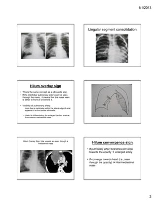
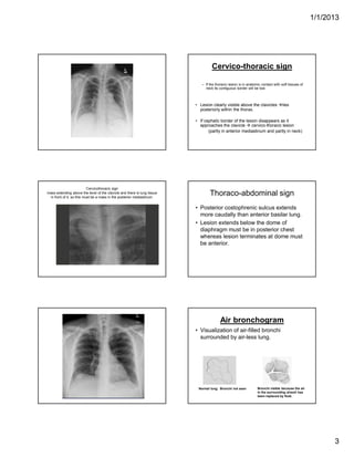
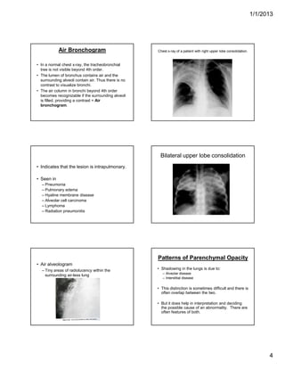
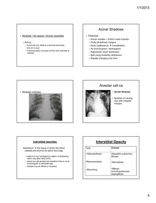
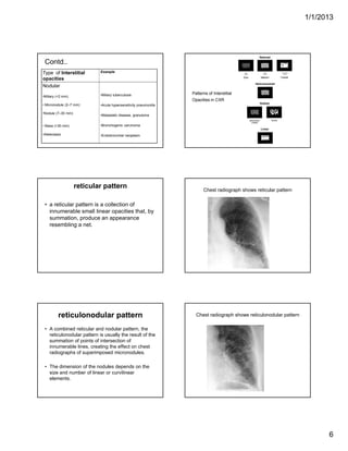
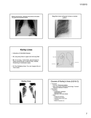
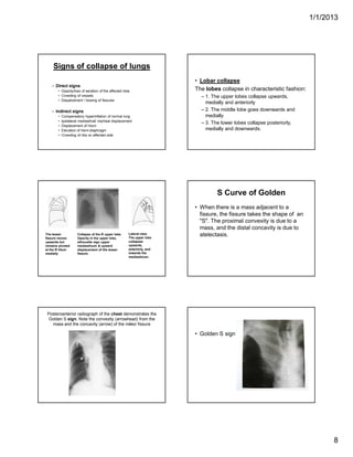
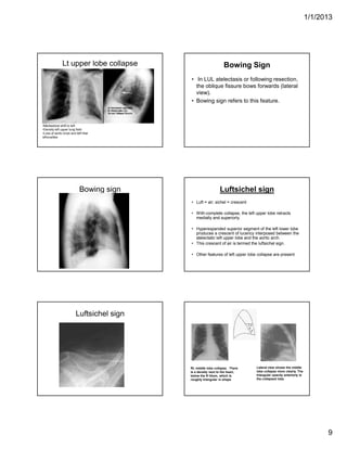
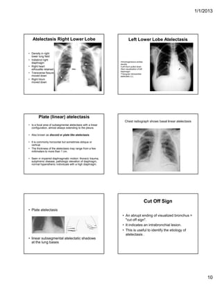
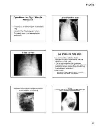
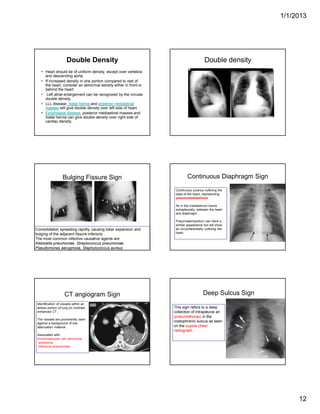
SlideShare a Scribd company logo
1/1/2013
1
Abnormal Sign in Chest X-
ray
Silhouette sign:
Density difference  delineation of the outline.
There are four basic densities in x-ray images:
gas
fat
water / soft tissue
bone / calcium
Loss of density difference of the adjacent
structures  loss of silhouette.
Silhouette sign contd…
• The silhouette sign is the absence of depiction
of an anatomic soft-tissue border resulting
from the juxtaposition of structures of similar
radiographic attenuation.
• The sign actually refers to the absence of a
silhouette.
• If the borders are retained 
– Not adjacent to each other or
– Of different radiographic densities.
Silhouette Sign
• Cardiac margins are clearly seen because there
is contrast between the fluid density of the heart
and the adjacent air filled alveoli.
• If the adjacent lung is devoid of air, the clarity of
the silhouette will be lost.
• Pleura encircles the lung and diseases of the
pleura can also obliterate silhouettes. The same
is true for mediastinal masses.
Silhouette Adjacent
Lobe/Segment
Right diaphragm RLL/Basal segments
Right heart margin RML/Medial segment
Ascending aorta RUL/Anterior segment
Aortic knob LUL/Apicoposterior segment
Left heart margin Lingula/Inferior segment
Descending aorta LLL/Superior and medial
segments
Left diaphragm LLL/Basal segments
Chest radiograph shows silhouette sign, with
obscuration of right border of heart (arrows).
1/1/2013
2
Lingular segment consolidation
Hilum overlay sign
• This is the same concept as a silhouette sign.
• If the interlobar pulmonary artery can be seen
through the mass, it means that the mass seen
is either in front of or behind it.
• Visibility of pulmonary artery
more than a centimeter within the lateral edge of what
appears to be the cardiac silhouette.
– Useful in differentiating the enlarged cardiac shadow
from anterior mediastinal mass.
Hilum Overlay Sign: hilar vessels are seen through a
mediastinal mass Hilum convergence sign
• If pulmonary artery branches converge
towards the opacity  enlarged artery
• If converge towards heart (i.e., seen
through the opacity)  hilar/mediastinal
mass
1/1/2013
3
Cervico-thoracic sign
– If the thoracic lesion is in anatomic contact with soft tissues of
neck its contiguous border will be lost.
• Lesion clearly visible above the clavicles lies
posteriorly within the thorax.
• If cephalic border of the lesion disappears as it
approaches the clavicle  cervico-thoracic lesion
(partly in anterior mediastinum and partly in neck)
Cervicothoracic sign
mass extending above the level of the clavicle and there is lung tissue
in front of it, so this must be a mass in the posterior mediastinum.
Thoraco-abdominal sign
• Posterior costophrenic sulcus extends
more caudally than anterior basilar lung.
• Lesion extends below the dome of
diaphragm must be in posterior chest
whereas lesion terminates at dome must
be anterior.
Air bronchogram
• Visualization of air-filled bronchi
surrounded by air-less lung.
Normal lung. Bronchi not seen Bronchi visible because the air
in the surrounding alveoli has
been replaced by fluid.
1/1/2013
4
Air Bronchogram
• In a normal chest x-ray, the tracheobronchial
tree is not visible beyond 4th order.
• The lumen of bronchus contains air and the
surrounding alveoli contain air. Thus there is no
contrast to visualize bronchi.
• The air column in bronchi beyond 4th order
becomes recognizable if the surrounding alveoli
is filled, providing a contrast = Air
bronchogram.
Chest x-ray of a patient with right upper lobe consolidation.
• Indicates that the lesion is intrapulmonary.
• Seen in
– Pneumonia
– Pulmonary edema
– Hyaline membrane disease
– Alveolar cell carcinoma
– Lymphoma
– Radiation pneumonitis
Bilateral upper lobe consolidation
• Air alveologram
– Tiny areas of radiolucency within the
surrounding air-less lung
Patterns of Parenchymal Opacity
• Shadowing in the lungs is due to:
– Alveolar disease
– Interstitial disease
• This distinction is sometimes difficult and there is
often overlap between the two.
• But it does help in interpretation and deciding
the possible cause of an abnormality. There are
often features of both.
1/1/2013
5
• Alveolar / Air-space / Acinar opacities
– Acinus
• functional unit, distal to a terminal bronchiole
• 6-8 mm in size
• Communication via pores of Kohn and channels of
Lambert
Acinar Shadows
• Features
– Acinar nodules – 6-8mm sized nodules
– Fluffy (ill-defined) margins
– Early coalescence  consolidation
– Air bronchogram / alveologram
– Segmental / lobar distribution
– Bat's wing (butterfly) distribution
– Rapidly changing over time
• Alveolar nodules
Alveolar cell ca
• Acinar Nodules
• Nodules of varying
size with irregular
margins
Interstitial opacities
Interstitium  the tissue in which the blood
vessels and bronchi lie within the lungs.
• leads to a non homogenous pattern of shadowing
which may take many forms.
• alveoli are still aerated and therefore there is no air
bronchogram or silhouette sign.
• disease may be diffuse or localised.
Interstitial Opacity
Type Example
• Reticular/linear
•Reticulonodular
• Branching
•Idiopathic pulmonary
fibrosis
•Sarcoidosis
•Allergic
bronchopulmonary
aspergillosis
1/1/2013
6
Contd..
Type of Interstitial
opacities
Example
Nodular
•Miliary (<2 mm)
• Micronodule (2–7 mm)
•Nodule (7–30 mm)
• Mass (>30 mm)
•Atelectasis
•Miliary tuberculosis
•Acute hypersensitivity pneumonitis
•Metastatic disease, granuloma
•Bronchogenic carcinoma
•Endobronchial neoplasm
Patterns of Interstitial
Opacities in CXR
reticular pattern
• a reticular pattern is a collection of
innumerable small linear opacities that, by
summation, produce an appearance
resembling a net.
Chest radiograph shows reticular pattern
reticulonodular pattern
• A combined reticular and nodular pattern, the
reticulonodular pattern is usually the result of the
summation of points of intersection of
innumerable lines, creating the effect on chest
radiographs of superimposed micronodules.
• The dimension of the nodules depends on the
size and number of linear or curvilinear
elements.
Chest radiograph shows reticulonodular pattern
1/1/2013
7
Bilateral reticulonodular shadowing most obvious at the bases.
This patient had rheumatoid arthritis.
Magnified chest radiograph shows a nodular
pattern.
Kerley Lines
• Indicative of interstitial disease.
• A: Long wavy lines in upper and mid lung field
• B: 2-3 cm long, 1-2mm thick, pleural based in
bases perpendicular to lateral chest; reliable,
thickening of interlobular septa.
• C: Fine fibrillatory lines: You can imagine this on
most cases
Kerley C lines
Kerley A lines
Kerley B lines
Kerley lines Causes of Kerley’s lines (A,B & C)
• Transient
– Common: Pulmonary edema
– Rare: Pneumonia, pulmonary hemorrhage, Transient
respiratory distress of newborn
• Persistent
– Common:
• Lymphangitic metastases
• Pneumoconiosis
• Rheumatic mitral valve disease
– Rare:
• Alveolar cell carcinoma
• Congenital heart disease
• Interstitial fibrosis of any cause
• Lymphoma
• Mineral oil aspiration etc.
1/1/2013
8
Signs of collapse of lungs
– Direct signs
• Opacity/loss of aeration of the affected lobe
• Crowding of vessels
• Dispalcement / bowing of fissures
– Indirect signs
• Compensatory hyperinflation of normal lung
• Ipsilateral mediastinal/ tracheal displacement
• Displacement of hilum
• Elevation of hemi-diaphragm
• Crowding of ribs on affected side
• Lobar collapse
The lobes collapse in characteristic fashion:
– 1. The upper lobes collapse upwards,
medially and anteriorly
– 2. The middle lobe goes downwards and
medially
– 3. The lower lobes collapse posteriorly,
medially and downwards.
The lesser
fissure moves
upwards but
remains pivoted
at the R hilum
medially
Lateral view.
The upper lobe
collapses
upwards,
anteriorly, and
towards the
mediastinum.
Collapse of the R upper lobe.
Opacity in the upper lobe,
silhouette sign upper
mediastinum & upward
displacement of the lesser
fissure.
S Curve of Golden
• When there is a mass adjacent to a
fissure, the fissure takes the shape of an
"S". The proximal convexity is due to a
mass, and the distal concavity is due to
atelectasis.
Posteroanterior radiograph of the chest demonstrates the
Golden S sign. Note the convexity (arrowhead) from the
mass and the concavity (arrow) of the minor fissure
• Golden S sign
1/1/2013
9
Lt upper lobe collapse
•Mediastinal shift to left
•Density left upper lung field
•Loss of aortic knob and left hilar
silhouettes
Bowing Sign
• In LUL atelectasis or following resection,
the oblique fissure bows forwards (lateral
view).
• Bowing sign refers to this feature.
Bowing sign Luftsichel sign
• Luft = air; sichel = crescent
• With complete collapse, the left upper lobe retracts
medially and superiorly.
• Hyperexpanded superior segment of the left lower lobe
produces a crescent of lucency interposed between the
atelectatic left upper lobe and the aortic arch.
• This crescent of air is termed the luftsichel sign.
• Other features of left upper lobe collapse are present
Luftsichel sign
Rt. middle lobe collapse. There
is a density next to the heart,
below the R hilum, which is
roughly triangular in shape
Lateral view shows the middle
lobe collapse more clearly. The
triangular opacity anteriorly is
the collapsed lobe
1/1/2013
10
Atelectasis Right Lower Lobe
• Density in right
lower lung field
• Indistinct right
diaphragm
• Right heart
silhouette retained
• Transverse fissure
moved down
• Right hilum
moved down
Left Lower Lobe Atelectasis
•Inhomogeneous cardiac
density
•Left hilum pulled down
•Non-visualization of left
diaphragm
•Triangular retrocardiac
atelectatic LLL
Plate (linear) atelectasis
• Is a focal area of subsegmental atelectasis with a linear
configuration, almost always extending to the pleura.
• Also known as discoid or plate like atelectasis.
• It is commonly horizontal but sometimes oblique or
vertical.
• The thickness of the atelectasis may range from a few
millimeters to more than 1 cm.
• Seen in impaired diaphragmatic motion: thoracic trauma,
subphrenic disease, pathologic elevation of diaphragm,
normal hypersthenic individuals with a high diaphragm.
Chest radiograph shows basal linear atelectasis
• Plate atelectasis
• linear subsegmental atelectatic shadows
at the lung bases
Cut Off Sign
• An abrupt ending of visualized bronchus =
"cut off sign".
• It indicates an intrabronchial lesion.
• This is useful to identify the etiology of
atelectasis .
1/1/2013
11
Open Bronchus Sign / Alveolar
Atelectasis
• Presence of air bronchogram in atelectatic
lung.
• indicates that the airways are patent.
• Commonly seen in adhesive alveolar
atelectasis.
Open bronchus sign
Close up view
Air crescent/ halo sign
• An air crescent is a collection of air in a
crescentic shape that separates the wall of a
cavity from an inner mass.
• The air crescent sign is often considered
characteristic of either Aspergillus colonization of
preexisting cavities or retraction of infarcted lung
in angioinvasive aspergillosis.
• other causes:
– tuberculosis, Wegener granulomatosis, intracavitary
hemorrhage, lung cancer, lung gangrene
Magnified chest radiograph shows air crescent
(arrows) adjacent to mycetoma
Halo Sign
In a cavity with fungus ball there is a crescentic lucent space along the upper
portion of a density giving the appearance of a halo.
1/1/2013
12
Double Density
• Heart should be of uniform density, except over vertebra
and descending aorta.
• If increased density in one portion compared to rest of
the heart, consider an abnormal density either in front or
behind the heart.
• Left atrial enlargement can be recognized by the circular
double density.
• LLL disease, hiatal hernia and posterior mediastinal
masses will give double density over left side of heart.
• Esophageal disease, posterior mediastinal masses and
hiatal hernia can give double density over right side of
cardiac density.
Double density
Bulging Fissure Sign
Consolidation spreading rapidly, causing lobar expansion and
bulging of the adjacent fissure inferiorly .
The most common infective causative agents are
Klebsiella pneumoniae, Streptococcus pneumoniae,
Pseudomonas aeruginosa, Staphylococcus aureus
Continuous Diaphragm Sign
Continuous lucency outlining the
base of the heart, representing
pneumomediastinum .
Air in the mediastinum tracks
extrapleurally, between the heart
and diaphragm .
Pneumopericardium can have a
similar appearance but will show
air circumferentially outlining the
heart.
CT angiogram Sign
Identification of vessels within an
airless portion of lung on contrast-
enhanced CT .
The vessels are prominently seen
against a background of low-
attenuation material .
Associated with:
bronchoalveolar cell carcinoma
lymphoma
infectious pneumonias.
Deep Sulcus Sign
This sign refers to a deep
collection of intrapleural air
(pneumothorax) in the
costophrenic sulcus as seen
on the supine chest
radiograph .
1/1/2013
13
Fallen Lung Sign
This sign refers to the appearance
of the collapsed lung occurring
with a fractured bronchus .
The bronchial fracture results in
the lung to fall away from the
hilum, either inferiorly and laterally
in an upright patient or posteriorly,
as seen on CT in a supine patient.
DD:
Pneumothorax causes a lung to
collapse inward toward the hilum.
Finger in Glove Sign
In allergic bronchopulmonary aspergillosis.
The impacted bronchi appear radiographically as opacities with distinctive
shapes.
Hampton Hump Sign
Pulmonary infarction secondary to pulmonary embolism produces
an abnormal area of opacification on the chest radiograph, which
is always in contact with the pleural surface.
Juxtaphrenic Peak Sign
This sign refers to a small triangular shadow that obscures the dome of the
diaphragm secondary to upper lobe atelectasis . The shadow is caused by
traction on the lower end of the major fissure, the inferior accessory fissure, or
the inferior pulmonary ligament.
THANK YOU
QUESTIONS
• Silhouette Sign.
• Hilum overlay sign.
• Air bronchogram.
• Patterns of parenchymal opacities.
• Causes of kerely’s line.
• Signs of collapse of lung.
• Luftsichel sign.
• Air crescent sign.
• Continous diaphragm sign.

More Related Content

What's hot

Radiographic Presentation of Congenital Heart Disease
Radiographic Presentation of Congenital Heart DiseaseRadiographic Presentation of Congenital Heart Disease
Radiographic Presentation of Congenital Heart Disease
Tarique Ajij
 
Segmental anatomy of lungs , anatomy of mediastinum and secondary lobule
Segmental anatomy of lungs , anatomy of mediastinum and secondary lobuleSegmental anatomy of lungs , anatomy of mediastinum and secondary lobule
Segmental anatomy of lungs , anatomy of mediastinum and secondary lobuleGamal Agmy
 
Presentation1.pptx, radiological imaging of pulmonary infection.
Presentation1.pptx, radiological imaging of pulmonary infection.Presentation1.pptx, radiological imaging of pulmonary infection.
Presentation1.pptx, radiological imaging of pulmonary infection.Abdellah Nazeer
 
Interstitial lung diseases radiology
Interstitial lung diseases radiologyInterstitial lung diseases radiology
Interstitial lung diseases radiology
Shrikant Nagare
 
Radiological signs in chest medicine part 2
Radiological signs in chest medicine part 2Radiological signs in chest medicine part 2
Radiological signs in chest medicine part 2Gamal Agmy
 
Radiology signs
Radiology signsRadiology signs
Radiology signs
Nitin Jain
 
Ascites and Pleural Effusion
 Ascites and Pleural Effusion Ascites and Pleural Effusion
Ascites and Pleural Effusion
Media Genie
 
Radiological features of Lung cancer Dr. Muhammad Bin Zulfiqar
Radiological features of Lung cancer Dr. Muhammad Bin ZulfiqarRadiological features of Lung cancer Dr. Muhammad Bin Zulfiqar
Radiological features of Lung cancer Dr. Muhammad Bin Zulfiqar
Dr. Muhammad Bin Zulfiqar
 
Lines & mediastinal stripes 01
Lines & mediastinal stripes 01Lines & mediastinal stripes 01
Signs in Chest Xray
Signs in Chest Xray Signs in Chest Xray
Signs in Chest Xray
Archana Koshy
 
Mediastinum-RADIOLOGY
Mediastinum-RADIOLOGYMediastinum-RADIOLOGY
Mediastinum-RADIOLOGYNavdeep Shah
 
Hrct
HrctHrct
Diagnostic Imaging of Pulmonary infections
Diagnostic Imaging of Pulmonary infectionsDiagnostic Imaging of Pulmonary infections
Diagnostic Imaging of Pulmonary infections
Mohamed M.A. Zaitoun
 
HRCT Chest
HRCT ChestHRCT Chest
HRCT Chest
Manjit Tendolkar
 
Diagnostic Imaging of Diffuse Lung Lesions
Diagnostic Imaging of Diffuse Lung LesionsDiagnostic Imaging of Diffuse Lung Lesions
Diagnostic Imaging of Diffuse Lung Lesions
Mohamed M.A. Zaitoun
 
Interstitial lung diseases- HRCT
Interstitial lung diseases- HRCTInterstitial lung diseases- HRCT
Interstitial lung diseases- HRCTNavdeep Shah
 
Chest x ray interpretation
Chest x ray interpretationChest x ray interpretation
Chest x ray interpretation
Kamal Sharma
 
Chest Xray
Chest XrayChest Xray
Chest Xray
Hassan Tarig
 
11 cavitary lesions of the lungs
11 cavitary lesions of the lungs11 cavitary lesions of the lungs
11 cavitary lesions of the lungs
Dr. Muhammad Bin Zulfiqar
 

What's hot (20)

Radiographic Presentation of Congenital Heart Disease
Radiographic Presentation of Congenital Heart DiseaseRadiographic Presentation of Congenital Heart Disease
Radiographic Presentation of Congenital Heart Disease
 
Segmental anatomy of lungs , anatomy of mediastinum and secondary lobule
Segmental anatomy of lungs , anatomy of mediastinum and secondary lobuleSegmental anatomy of lungs , anatomy of mediastinum and secondary lobule
Segmental anatomy of lungs , anatomy of mediastinum and secondary lobule
 
Presentation1.pptx, radiological imaging of pulmonary infection.
Presentation1.pptx, radiological imaging of pulmonary infection.Presentation1.pptx, radiological imaging of pulmonary infection.
Presentation1.pptx, radiological imaging of pulmonary infection.
 
Interstitial lung diseases radiology
Interstitial lung diseases radiologyInterstitial lung diseases radiology
Interstitial lung diseases radiology
 
Radiological signs in chest medicine part 2
Radiological signs in chest medicine part 2Radiological signs in chest medicine part 2
Radiological signs in chest medicine part 2
 
Radiology signs
Radiology signsRadiology signs
Radiology signs
 
Ascites and Pleural Effusion
 Ascites and Pleural Effusion Ascites and Pleural Effusion
Ascites and Pleural Effusion
 
Radiological features of Lung cancer Dr. Muhammad Bin Zulfiqar
Radiological features of Lung cancer Dr. Muhammad Bin ZulfiqarRadiological features of Lung cancer Dr. Muhammad Bin Zulfiqar
Radiological features of Lung cancer Dr. Muhammad Bin Zulfiqar
 
Lines & mediastinal stripes 01
Lines & mediastinal stripes 01Lines & mediastinal stripes 01
Lines & mediastinal stripes 01
 
Signs in Chest Xray
Signs in Chest Xray Signs in Chest Xray
Signs in Chest Xray
 
Mediastinum-RADIOLOGY
Mediastinum-RADIOLOGYMediastinum-RADIOLOGY
Mediastinum-RADIOLOGY
 
CXR - Cavity in Lung
CXR - Cavity in LungCXR - Cavity in Lung
CXR - Cavity in Lung
 
Hrct
HrctHrct
Hrct
 
Diagnostic Imaging of Pulmonary infections
Diagnostic Imaging of Pulmonary infectionsDiagnostic Imaging of Pulmonary infections
Diagnostic Imaging of Pulmonary infections
 
HRCT Chest
HRCT ChestHRCT Chest
HRCT Chest
 
Diagnostic Imaging of Diffuse Lung Lesions
Diagnostic Imaging of Diffuse Lung LesionsDiagnostic Imaging of Diffuse Lung Lesions
Diagnostic Imaging of Diffuse Lung Lesions
 
Interstitial lung diseases- HRCT
Interstitial lung diseases- HRCTInterstitial lung diseases- HRCT
Interstitial lung diseases- HRCT
 
Chest x ray interpretation
Chest x ray interpretationChest x ray interpretation
Chest x ray interpretation
 
Chest Xray
Chest XrayChest Xray
Chest Xray
 
11 cavitary lesions of the lungs
11 cavitary lesions of the lungs11 cavitary lesions of the lungs
11 cavitary lesions of the lungs
 

Similar to Abnormal sign in chest X- Ray

Abnormal signs in chest x ray
Abnormal signs in chest x rayAbnormal signs in chest x ray
Abnormal signs in chest x ray
Milan Silwal
 
Helpful radiological signs in cxr25 11-91
Helpful radiological signs in cxr25 11-91Helpful radiological signs in cxr25 11-91
Helpful radiological signs in cxr25 11-91
aalmasi1970
 
Chest radiology part 1
Chest radiology part 1Chest radiology part 1
Chest radiology part 1Gamal Agmy
 
Radiological presentation of chest diseases gamal agmy
Radiological presentation of chest diseases  gamal agmyRadiological presentation of chest diseases  gamal agmy
Radiological presentation of chest diseases gamal agmyGamal Agmy
 
Radiological Presentation of Chest Diseases
Radiological Presentation of Chest Diseases  Radiological Presentation of Chest Diseases
Radiological Presentation of Chest Diseases Gamal Agmy
 
Reading chest-x-rays
Reading chest-x-rays Reading chest-x-rays
Reading chest-x-rays
Samriddha Pokharel
 
Normal Chest X-Rays & Its Systemic Approach- Anatomy
Normal Chest X-Rays & Its Systemic Approach- AnatomyNormal Chest X-Rays & Its Systemic Approach- Anatomy
Normal Chest X-Rays & Its Systemic Approach- Anatomy
Dr.Bijay Yadav
 
Collapse consolidation
Collapse consolidationCollapse consolidation
Collapse consolidation
airwave12
 
Radiological Signs in Chest Medicine
Radiological Signs in Chest MedicineRadiological Signs in Chest Medicine
Radiological Signs in Chest MedicineGamal Agmy
 
Radiological signs in chest medicine
Radiological signs in chest medicineRadiological signs in chest medicine
Radiological signs in chest medicineGamal Agmy
 
Patttern of lung disease on chest x ray
Patttern of lung disease on   chest x rayPatttern of lung disease on   chest x ray
Patttern of lung disease on chest x ray
Vrishit Saraswat
 
Interactive radiology case presentation
Interactive radiology case presentationInteractive radiology case presentation
Interactive radiology case presentationGamal Agmy
 
Chest x ray review.
Chest x ray review.Chest x ray review.
Chest x ray review.
Muhammad Ali
 
Chest imaging
Chest imagingChest imaging
Chest imaging
JANARDANCNMC
 
Abnormal Chest X rays
Abnormal Chest X raysAbnormal Chest X rays
Abnormal Chest X rays
Dr. Tom Mishael J
 
XRAY
XRAYXRAY
XRAY
Sufindc
 
CHEST XRAYS RJJ.pptx
CHEST XRAYS RJJ.pptxCHEST XRAYS RJJ.pptx
CHEST XRAYS RJJ.pptx
rohanjohnjacob
 
X-ray images of lung diseases (pneumonia, tuberculosis, and tumors)..pptx
X-ray images of lung diseases (pneumonia, tuberculosis, and tumors)..pptxX-ray images of lung diseases (pneumonia, tuberculosis, and tumors)..pptx
X-ray images of lung diseases (pneumonia, tuberculosis, and tumors)..pptx
Aavani13
 
Chest x. ray interpretation and teaching
Chest x. ray interpretation and teachingChest x. ray interpretation and teaching
Chest x. ray interpretation and teaching
samirelansary
 

Similar to Abnormal sign in chest X- Ray (20)

Abnormal signs in chest x ray
Abnormal signs in chest x rayAbnormal signs in chest x ray
Abnormal signs in chest x ray
 
Helpful radiological signs in cxr25 11-91
Helpful radiological signs in cxr25 11-91Helpful radiological signs in cxr25 11-91
Helpful radiological signs in cxr25 11-91
 
Chest radiology part 1
Chest radiology part 1Chest radiology part 1
Chest radiology part 1
 
Radiological presentation of chest diseases gamal agmy
Radiological presentation of chest diseases  gamal agmyRadiological presentation of chest diseases  gamal agmy
Radiological presentation of chest diseases gamal agmy
 
Radiological Presentation of Chest Diseases
Radiological Presentation of Chest Diseases  Radiological Presentation of Chest Diseases
Radiological Presentation of Chest Diseases
 
Reading chest-x-rays
Reading chest-x-rays Reading chest-x-rays
Reading chest-x-rays
 
Normal Chest X-Rays & Its Systemic Approach- Anatomy
Normal Chest X-Rays & Its Systemic Approach- AnatomyNormal Chest X-Rays & Its Systemic Approach- Anatomy
Normal Chest X-Rays & Its Systemic Approach- Anatomy
 
Collapse consolidation
Collapse consolidationCollapse consolidation
Collapse consolidation
 
Chest x rays
Chest x raysChest x rays
Chest x rays
 
Radiological Signs in Chest Medicine
Radiological Signs in Chest MedicineRadiological Signs in Chest Medicine
Radiological Signs in Chest Medicine
 
Radiological signs in chest medicine
Radiological signs in chest medicineRadiological signs in chest medicine
Radiological signs in chest medicine
 
Patttern of lung disease on chest x ray
Patttern of lung disease on   chest x rayPatttern of lung disease on   chest x ray
Patttern of lung disease on chest x ray
 
Interactive radiology case presentation
Interactive radiology case presentationInteractive radiology case presentation
Interactive radiology case presentation
 
Chest x ray review.
Chest x ray review.Chest x ray review.
Chest x ray review.
 
Chest imaging
Chest imagingChest imaging
Chest imaging
 
Abnormal Chest X rays
Abnormal Chest X raysAbnormal Chest X rays
Abnormal Chest X rays
 
XRAY
XRAYXRAY
XRAY
 
CHEST XRAYS RJJ.pptx
CHEST XRAYS RJJ.pptxCHEST XRAYS RJJ.pptx
CHEST XRAYS RJJ.pptx
 
X-ray images of lung diseases (pneumonia, tuberculosis, and tumors)..pptx
X-ray images of lung diseases (pneumonia, tuberculosis, and tumors)..pptxX-ray images of lung diseases (pneumonia, tuberculosis, and tumors)..pptx
X-ray images of lung diseases (pneumonia, tuberculosis, and tumors)..pptx
 
Chest x. ray interpretation and teaching
Chest x. ray interpretation and teachingChest x. ray interpretation and teaching
Chest x. ray interpretation and teaching
 

More from Upakar Paudel

Radiology Contrast media and its Types
Radiology Contrast media and its TypesRadiology Contrast media and its Types
Radiology Contrast media and its Types
Upakar Paudel
 
Measurement of Radiation (Thimble Ionization Chamber, Free air Ionization Cha...
Measurement of Radiation (Thimble Ionization Chamber, Free air Ionization Cha...Measurement of Radiation (Thimble Ionization Chamber, Free air Ionization Cha...
Measurement of Radiation (Thimble Ionization Chamber, Free air Ionization Cha...
Upakar Paudel
 
CT Procedure OF Abdomen & Pelvis
CT Procedure OF Abdomen & Pelvis CT Procedure OF Abdomen & Pelvis
CT Procedure OF Abdomen & Pelvis
Upakar Paudel
 
Digital Radiography
Digital RadiographyDigital Radiography
Digital Radiography
Upakar Paudel
 
HSG DCS and Sailography
HSG DCS  and SailographyHSG DCS  and Sailography
HSG DCS and Sailography
Upakar Paudel
 
Sterilization & Disinfection
Sterilization &  DisinfectionSterilization &  Disinfection
Sterilization & Disinfection
Upakar Paudel
 
Basic Pulse Sequences In MRI
Basic Pulse Sequences In MRIBasic Pulse Sequences In MRI
Basic Pulse Sequences In MRI
Upakar Paudel
 
Basic principle of gamma camera
Basic principle of gamma cameraBasic principle of gamma camera
Basic principle of gamma camera
Upakar Paudel
 
Sensitometry or characteristic curve
Sensitometry or characteristic curve   Sensitometry or characteristic curve
Sensitometry or characteristic curve
Upakar Paudel
 
CT Procedure of Thorax (CT Chest)
CT Procedure of Thorax (CT Chest)CT Procedure of Thorax (CT Chest)
CT Procedure of Thorax (CT Chest)
Upakar Paudel
 
Recent Advances In MRI
Recent Advances In MRIRecent Advances In MRI
Recent Advances In MRI
Upakar Paudel
 
How to write a thesis
How to write a thesisHow to write a thesis
How to write a thesis
Upakar Paudel
 
ERCP PROCEDURE
ERCP PROCEDURE ERCP PROCEDURE
ERCP PROCEDURE
Upakar Paudel
 
CT Generation (Generation of CT)
CT Generation (Generation of CT)CT Generation (Generation of CT)
CT Generation (Generation of CT)
Upakar Paudel
 

More from Upakar Paudel (14)

Radiology Contrast media and its Types
Radiology Contrast media and its TypesRadiology Contrast media and its Types
Radiology Contrast media and its Types
 
Measurement of Radiation (Thimble Ionization Chamber, Free air Ionization Cha...
Measurement of Radiation (Thimble Ionization Chamber, Free air Ionization Cha...Measurement of Radiation (Thimble Ionization Chamber, Free air Ionization Cha...
Measurement of Radiation (Thimble Ionization Chamber, Free air Ionization Cha...
 
CT Procedure OF Abdomen & Pelvis
CT Procedure OF Abdomen & Pelvis CT Procedure OF Abdomen & Pelvis
CT Procedure OF Abdomen & Pelvis
 
Digital Radiography
Digital RadiographyDigital Radiography
Digital Radiography
 
HSG DCS and Sailography
HSG DCS  and SailographyHSG DCS  and Sailography
HSG DCS and Sailography
 
Sterilization & Disinfection
Sterilization &  DisinfectionSterilization &  Disinfection
Sterilization & Disinfection
 
Basic Pulse Sequences In MRI
Basic Pulse Sequences In MRIBasic Pulse Sequences In MRI
Basic Pulse Sequences In MRI
 
Basic principle of gamma camera
Basic principle of gamma cameraBasic principle of gamma camera
Basic principle of gamma camera
 
Sensitometry or characteristic curve
Sensitometry or characteristic curve   Sensitometry or characteristic curve
Sensitometry or characteristic curve
 
CT Procedure of Thorax (CT Chest)
CT Procedure of Thorax (CT Chest)CT Procedure of Thorax (CT Chest)
CT Procedure of Thorax (CT Chest)
 
Recent Advances In MRI
Recent Advances In MRIRecent Advances In MRI
Recent Advances In MRI
 
How to write a thesis
How to write a thesisHow to write a thesis
How to write a thesis
 
ERCP PROCEDURE
ERCP PROCEDURE ERCP PROCEDURE
ERCP PROCEDURE
 
CT Generation (Generation of CT)
CT Generation (Generation of CT)CT Generation (Generation of CT)
CT Generation (Generation of CT)
 

Recently uploaded

Report Back from SGO 2024: What’s the Latest in Cervical Cancer?
Report Back from SGO 2024: What’s the Latest in Cervical Cancer?Report Back from SGO 2024: What’s the Latest in Cervical Cancer?
Report Back from SGO 2024: What’s the Latest in Cervical Cancer?
bkling
 
BENIGN PROSTATIC HYPERPLASIA.BPH. BPHpdf
BENIGN PROSTATIC HYPERPLASIA.BPH. BPHpdfBENIGN PROSTATIC HYPERPLASIA.BPH. BPHpdf
BENIGN PROSTATIC HYPERPLASIA.BPH. BPHpdf
DR SETH JOTHAM
 
Couples presenting to the infertility clinic- Do they really have infertility...
Couples presenting to the infertility clinic- Do they really have infertility...Couples presenting to the infertility clinic- Do they really have infertility...
Couples presenting to the infertility clinic- Do they really have infertility...
Sujoy Dasgupta
 
Non-respiratory Functions of the Lungs.pdf
Non-respiratory Functions of the Lungs.pdfNon-respiratory Functions of the Lungs.pdf
Non-respiratory Functions of the Lungs.pdf
MedicoseAcademics
 
Physiology of Special Chemical Sensation of Taste
Physiology of Special Chemical Sensation of TastePhysiology of Special Chemical Sensation of Taste
Physiology of Special Chemical Sensation of Taste
MedicoseAcademics
 
Flu Vaccine Alert in Bangalore Karnataka
Flu Vaccine Alert in Bangalore KarnatakaFlu Vaccine Alert in Bangalore Karnataka
Flu Vaccine Alert in Bangalore Karnataka
addon Scans
 
TEST BANK for Operations Management, 14th Edition by William J. Stevenson, Ve...
TEST BANK for Operations Management, 14th Edition by William J. Stevenson, Ve...TEST BANK for Operations Management, 14th Edition by William J. Stevenson, Ve...
TEST BANK for Operations Management, 14th Edition by William J. Stevenson, Ve...
kevinkariuki227
 
Surat @ℂall @Girls ꧁❤8527049040❤꧂@ℂall @Girls Service Vip Top Model Safe
Surat @ℂall @Girls ꧁❤8527049040❤꧂@ℂall @Girls Service Vip Top Model SafeSurat @ℂall @Girls ꧁❤8527049040❤꧂@ℂall @Girls Service Vip Top Model Safe
Surat @ℂall @Girls ꧁❤8527049040❤꧂@ℂall @Girls Service Vip Top Model Safe
Savita Shen $i11
 
BRACHYTHERAPY OVERVIEW AND APPLICATORS
BRACHYTHERAPY OVERVIEW  AND  APPLICATORSBRACHYTHERAPY OVERVIEW  AND  APPLICATORS
BRACHYTHERAPY OVERVIEW AND APPLICATORS
Krishan Murari
 
Phone Us ❤85270-49040❤ #ℂall #gIRLS In Surat By Surat @ℂall @Girls Hotel With...
Phone Us ❤85270-49040❤ #ℂall #gIRLS In Surat By Surat @ℂall @Girls Hotel With...Phone Us ❤85270-49040❤ #ℂall #gIRLS In Surat By Surat @ℂall @Girls Hotel With...
Phone Us ❤85270-49040❤ #ℂall #gIRLS In Surat By Surat @ℂall @Girls Hotel With...
Savita Shen $i11
 
THOA 2.ppt Human Organ Transplantation Act
THOA 2.ppt Human Organ Transplantation ActTHOA 2.ppt Human Organ Transplantation Act
THOA 2.ppt Human Organ Transplantation Act
DrSathishMS1
 
Surgical Site Infections, pathophysiology, and prevention.pptx
Surgical Site Infections, pathophysiology, and prevention.pptxSurgical Site Infections, pathophysiology, and prevention.pptx
Surgical Site Infections, pathophysiology, and prevention.pptx
jval Landero
 
KDIGO 2024 guidelines for diabetologists
KDIGO 2024 guidelines for diabetologistsKDIGO 2024 guidelines for diabetologists
KDIGO 2024 guidelines for diabetologists
د.محمود نجيب
 
Ocular injury ppt Upendra pal optometrist upums saifai etawah
Ocular injury  ppt  Upendra pal  optometrist upums saifai etawahOcular injury  ppt  Upendra pal  optometrist upums saifai etawah
Ocular injury ppt Upendra pal optometrist upums saifai etawah
pal078100
 
Cervical & Brachial Plexus By Dr. RIG.pptx
Cervical & Brachial Plexus By Dr. RIG.pptxCervical & Brachial Plexus By Dr. RIG.pptx
Cervical & Brachial Plexus By Dr. RIG.pptx
Dr. Rabia Inam Gandapore
 
MANAGEMENT OF ATRIOVENTRICULAR CONDUCTION BLOCK.pdf
MANAGEMENT OF ATRIOVENTRICULAR CONDUCTION BLOCK.pdfMANAGEMENT OF ATRIOVENTRICULAR CONDUCTION BLOCK.pdf
MANAGEMENT OF ATRIOVENTRICULAR CONDUCTION BLOCK.pdf
Jim Jacob Roy
 
Maxilla, Mandible & Hyoid Bone & Clinical Correlations by Dr. RIG.pptx
Maxilla, Mandible & Hyoid Bone & Clinical Correlations by Dr. RIG.pptxMaxilla, Mandible & Hyoid Bone & Clinical Correlations by Dr. RIG.pptx
Maxilla, Mandible & Hyoid Bone & Clinical Correlations by Dr. RIG.pptx
Dr. Rabia Inam Gandapore
 
Hemodialysis: Chapter 3, Dialysis Water Unit - Dr.Gawad
Hemodialysis: Chapter 3, Dialysis Water Unit - Dr.GawadHemodialysis: Chapter 3, Dialysis Water Unit - Dr.Gawad
Hemodialysis: Chapter 3, Dialysis Water Unit - Dr.Gawad
NephroTube - Dr.Gawad
 
HOT NEW PRODUCT! BIG SALES FAST SHIPPING NOW FROM CHINA!! EU KU DB BK substit...
HOT NEW PRODUCT! BIG SALES FAST SHIPPING NOW FROM CHINA!! EU KU DB BK substit...HOT NEW PRODUCT! BIG SALES FAST SHIPPING NOW FROM CHINA!! EU KU DB BK substit...
HOT NEW PRODUCT! BIG SALES FAST SHIPPING NOW FROM CHINA!! EU KU DB BK substit...
GL Anaacs
 
New Directions in Targeted Therapeutic Approaches for Older Adults With Mantl...
New Directions in Targeted Therapeutic Approaches for Older Adults With Mantl...New Directions in Targeted Therapeutic Approaches for Older Adults With Mantl...
New Directions in Targeted Therapeutic Approaches for Older Adults With Mantl...
i3 Health
 

Recently uploaded (20)

Report Back from SGO 2024: What’s the Latest in Cervical Cancer?
Report Back from SGO 2024: What’s the Latest in Cervical Cancer?Report Back from SGO 2024: What’s the Latest in Cervical Cancer?
Report Back from SGO 2024: What’s the Latest in Cervical Cancer?
 
BENIGN PROSTATIC HYPERPLASIA.BPH. BPHpdf
BENIGN PROSTATIC HYPERPLASIA.BPH. BPHpdfBENIGN PROSTATIC HYPERPLASIA.BPH. BPHpdf
BENIGN PROSTATIC HYPERPLASIA.BPH. BPHpdf
 
Couples presenting to the infertility clinic- Do they really have infertility...
Couples presenting to the infertility clinic- Do they really have infertility...Couples presenting to the infertility clinic- Do they really have infertility...
Couples presenting to the infertility clinic- Do they really have infertility...
 
Non-respiratory Functions of the Lungs.pdf
Non-respiratory Functions of the Lungs.pdfNon-respiratory Functions of the Lungs.pdf
Non-respiratory Functions of the Lungs.pdf
 
Physiology of Special Chemical Sensation of Taste
Physiology of Special Chemical Sensation of TastePhysiology of Special Chemical Sensation of Taste
Physiology of Special Chemical Sensation of Taste
 
Flu Vaccine Alert in Bangalore Karnataka
Flu Vaccine Alert in Bangalore KarnatakaFlu Vaccine Alert in Bangalore Karnataka
Flu Vaccine Alert in Bangalore Karnataka
 
TEST BANK for Operations Management, 14th Edition by William J. Stevenson, Ve...
TEST BANK for Operations Management, 14th Edition by William J. Stevenson, Ve...TEST BANK for Operations Management, 14th Edition by William J. Stevenson, Ve...
TEST BANK for Operations Management, 14th Edition by William J. Stevenson, Ve...
 
Surat @ℂall @Girls ꧁❤8527049040❤꧂@ℂall @Girls Service Vip Top Model Safe
Surat @ℂall @Girls ꧁❤8527049040❤꧂@ℂall @Girls Service Vip Top Model SafeSurat @ℂall @Girls ꧁❤8527049040❤꧂@ℂall @Girls Service Vip Top Model Safe
Surat @ℂall @Girls ꧁❤8527049040❤꧂@ℂall @Girls Service Vip Top Model Safe
 
BRACHYTHERAPY OVERVIEW AND APPLICATORS
BRACHYTHERAPY OVERVIEW  AND  APPLICATORSBRACHYTHERAPY OVERVIEW  AND  APPLICATORS
BRACHYTHERAPY OVERVIEW AND APPLICATORS
 
Phone Us ❤85270-49040❤ #ℂall #gIRLS In Surat By Surat @ℂall @Girls Hotel With...
Phone Us ❤85270-49040❤ #ℂall #gIRLS In Surat By Surat @ℂall @Girls Hotel With...Phone Us ❤85270-49040❤ #ℂall #gIRLS In Surat By Surat @ℂall @Girls Hotel With...
Phone Us ❤85270-49040❤ #ℂall #gIRLS In Surat By Surat @ℂall @Girls Hotel With...
 
THOA 2.ppt Human Organ Transplantation Act
THOA 2.ppt Human Organ Transplantation ActTHOA 2.ppt Human Organ Transplantation Act
THOA 2.ppt Human Organ Transplantation Act
 
Surgical Site Infections, pathophysiology, and prevention.pptx
Surgical Site Infections, pathophysiology, and prevention.pptxSurgical Site Infections, pathophysiology, and prevention.pptx
Surgical Site Infections, pathophysiology, and prevention.pptx
 
KDIGO 2024 guidelines for diabetologists
KDIGO 2024 guidelines for diabetologistsKDIGO 2024 guidelines for diabetologists
KDIGO 2024 guidelines for diabetologists
 
Ocular injury ppt Upendra pal optometrist upums saifai etawah
Ocular injury  ppt  Upendra pal  optometrist upums saifai etawahOcular injury  ppt  Upendra pal  optometrist upums saifai etawah
Ocular injury ppt Upendra pal optometrist upums saifai etawah
 
Cervical & Brachial Plexus By Dr. RIG.pptx
Cervical & Brachial Plexus By Dr. RIG.pptxCervical & Brachial Plexus By Dr. RIG.pptx
Cervical & Brachial Plexus By Dr. RIG.pptx
 
MANAGEMENT OF ATRIOVENTRICULAR CONDUCTION BLOCK.pdf
MANAGEMENT OF ATRIOVENTRICULAR CONDUCTION BLOCK.pdfMANAGEMENT OF ATRIOVENTRICULAR CONDUCTION BLOCK.pdf
MANAGEMENT OF ATRIOVENTRICULAR CONDUCTION BLOCK.pdf
 
Maxilla, Mandible & Hyoid Bone & Clinical Correlations by Dr. RIG.pptx
Maxilla, Mandible & Hyoid Bone & Clinical Correlations by Dr. RIG.pptxMaxilla, Mandible & Hyoid Bone & Clinical Correlations by Dr. RIG.pptx
Maxilla, Mandible & Hyoid Bone & Clinical Correlations by Dr. RIG.pptx
 
Hemodialysis: Chapter 3, Dialysis Water Unit - Dr.Gawad
Hemodialysis: Chapter 3, Dialysis Water Unit - Dr.GawadHemodialysis: Chapter 3, Dialysis Water Unit - Dr.Gawad
Hemodialysis: Chapter 3, Dialysis Water Unit - Dr.Gawad
 
HOT NEW PRODUCT! BIG SALES FAST SHIPPING NOW FROM CHINA!! EU KU DB BK substit...
HOT NEW PRODUCT! BIG SALES FAST SHIPPING NOW FROM CHINA!! EU KU DB BK substit...HOT NEW PRODUCT! BIG SALES FAST SHIPPING NOW FROM CHINA!! EU KU DB BK substit...
HOT NEW PRODUCT! BIG SALES FAST SHIPPING NOW FROM CHINA!! EU KU DB BK substit...
 
New Directions in Targeted Therapeutic Approaches for Older Adults With Mantl...
New Directions in Targeted Therapeutic Approaches for Older Adults With Mantl...New Directions in Targeted Therapeutic Approaches for Older Adults With Mantl...
New Directions in Targeted Therapeutic Approaches for Older Adults With Mantl...
 

Abnormal sign in chest X- Ray

  • 1. 1/1/2013 1 Abnormal Sign in Chest X- ray Silhouette sign: Density difference  delineation of the outline. There are four basic densities in x-ray images: gas fat water / soft tissue bone / calcium Loss of density difference of the adjacent structures  loss of silhouette. Silhouette sign contd… • The silhouette sign is the absence of depiction of an anatomic soft-tissue border resulting from the juxtaposition of structures of similar radiographic attenuation. • The sign actually refers to the absence of a silhouette. • If the borders are retained  – Not adjacent to each other or – Of different radiographic densities. Silhouette Sign • Cardiac margins are clearly seen because there is contrast between the fluid density of the heart and the adjacent air filled alveoli. • If the adjacent lung is devoid of air, the clarity of the silhouette will be lost. • Pleura encircles the lung and diseases of the pleura can also obliterate silhouettes. The same is true for mediastinal masses. Silhouette Adjacent Lobe/Segment Right diaphragm RLL/Basal segments Right heart margin RML/Medial segment Ascending aorta RUL/Anterior segment Aortic knob LUL/Apicoposterior segment Left heart margin Lingula/Inferior segment Descending aorta LLL/Superior and medial segments Left diaphragm LLL/Basal segments Chest radiograph shows silhouette sign, with obscuration of right border of heart (arrows).
  • 2. 1/1/2013 2 Lingular segment consolidation Hilum overlay sign • This is the same concept as a silhouette sign. • If the interlobar pulmonary artery can be seen through the mass, it means that the mass seen is either in front of or behind it. • Visibility of pulmonary artery more than a centimeter within the lateral edge of what appears to be the cardiac silhouette. – Useful in differentiating the enlarged cardiac shadow from anterior mediastinal mass. Hilum Overlay Sign: hilar vessels are seen through a mediastinal mass Hilum convergence sign • If pulmonary artery branches converge towards the opacity  enlarged artery • If converge towards heart (i.e., seen through the opacity)  hilar/mediastinal mass
  • 3. 1/1/2013 3 Cervico-thoracic sign – If the thoracic lesion is in anatomic contact with soft tissues of neck its contiguous border will be lost. • Lesion clearly visible above the clavicles lies posteriorly within the thorax. • If cephalic border of the lesion disappears as it approaches the clavicle  cervico-thoracic lesion (partly in anterior mediastinum and partly in neck) Cervicothoracic sign mass extending above the level of the clavicle and there is lung tissue in front of it, so this must be a mass in the posterior mediastinum. Thoraco-abdominal sign • Posterior costophrenic sulcus extends more caudally than anterior basilar lung. • Lesion extends below the dome of diaphragm must be in posterior chest whereas lesion terminates at dome must be anterior. Air bronchogram • Visualization of air-filled bronchi surrounded by air-less lung. Normal lung. Bronchi not seen Bronchi visible because the air in the surrounding alveoli has been replaced by fluid.
  • 4. 1/1/2013 4 Air Bronchogram • In a normal chest x-ray, the tracheobronchial tree is not visible beyond 4th order. • The lumen of bronchus contains air and the surrounding alveoli contain air. Thus there is no contrast to visualize bronchi. • The air column in bronchi beyond 4th order becomes recognizable if the surrounding alveoli is filled, providing a contrast = Air bronchogram. Chest x-ray of a patient with right upper lobe consolidation. • Indicates that the lesion is intrapulmonary. • Seen in – Pneumonia – Pulmonary edema – Hyaline membrane disease – Alveolar cell carcinoma – Lymphoma – Radiation pneumonitis Bilateral upper lobe consolidation • Air alveologram – Tiny areas of radiolucency within the surrounding air-less lung Patterns of Parenchymal Opacity • Shadowing in the lungs is due to: – Alveolar disease – Interstitial disease • This distinction is sometimes difficult and there is often overlap between the two. • But it does help in interpretation and deciding the possible cause of an abnormality. There are often features of both.
  • 5. 1/1/2013 5 • Alveolar / Air-space / Acinar opacities – Acinus • functional unit, distal to a terminal bronchiole • 6-8 mm in size • Communication via pores of Kohn and channels of Lambert Acinar Shadows • Features – Acinar nodules – 6-8mm sized nodules – Fluffy (ill-defined) margins – Early coalescence  consolidation – Air bronchogram / alveologram – Segmental / lobar distribution – Bat's wing (butterfly) distribution – Rapidly changing over time • Alveolar nodules Alveolar cell ca • Acinar Nodules • Nodules of varying size with irregular margins Interstitial opacities Interstitium  the tissue in which the blood vessels and bronchi lie within the lungs. • leads to a non homogenous pattern of shadowing which may take many forms. • alveoli are still aerated and therefore there is no air bronchogram or silhouette sign. • disease may be diffuse or localised. Interstitial Opacity Type Example • Reticular/linear •Reticulonodular • Branching •Idiopathic pulmonary fibrosis •Sarcoidosis •Allergic bronchopulmonary aspergillosis
  • 6. 1/1/2013 6 Contd.. Type of Interstitial opacities Example Nodular •Miliary (<2 mm) • Micronodule (2–7 mm) •Nodule (7–30 mm) • Mass (>30 mm) •Atelectasis •Miliary tuberculosis •Acute hypersensitivity pneumonitis •Metastatic disease, granuloma •Bronchogenic carcinoma •Endobronchial neoplasm Patterns of Interstitial Opacities in CXR reticular pattern • a reticular pattern is a collection of innumerable small linear opacities that, by summation, produce an appearance resembling a net. Chest radiograph shows reticular pattern reticulonodular pattern • A combined reticular and nodular pattern, the reticulonodular pattern is usually the result of the summation of points of intersection of innumerable lines, creating the effect on chest radiographs of superimposed micronodules. • The dimension of the nodules depends on the size and number of linear or curvilinear elements. Chest radiograph shows reticulonodular pattern
  • 7. 1/1/2013 7 Bilateral reticulonodular shadowing most obvious at the bases. This patient had rheumatoid arthritis. Magnified chest radiograph shows a nodular pattern. Kerley Lines • Indicative of interstitial disease. • A: Long wavy lines in upper and mid lung field • B: 2-3 cm long, 1-2mm thick, pleural based in bases perpendicular to lateral chest; reliable, thickening of interlobular septa. • C: Fine fibrillatory lines: You can imagine this on most cases Kerley C lines Kerley A lines Kerley B lines Kerley lines Causes of Kerley’s lines (A,B & C) • Transient – Common: Pulmonary edema – Rare: Pneumonia, pulmonary hemorrhage, Transient respiratory distress of newborn • Persistent – Common: • Lymphangitic metastases • Pneumoconiosis • Rheumatic mitral valve disease – Rare: • Alveolar cell carcinoma • Congenital heart disease • Interstitial fibrosis of any cause • Lymphoma • Mineral oil aspiration etc.
  • 8. 1/1/2013 8 Signs of collapse of lungs – Direct signs • Opacity/loss of aeration of the affected lobe • Crowding of vessels • Dispalcement / bowing of fissures – Indirect signs • Compensatory hyperinflation of normal lung • Ipsilateral mediastinal/ tracheal displacement • Displacement of hilum • Elevation of hemi-diaphragm • Crowding of ribs on affected side • Lobar collapse The lobes collapse in characteristic fashion: – 1. The upper lobes collapse upwards, medially and anteriorly – 2. The middle lobe goes downwards and medially – 3. The lower lobes collapse posteriorly, medially and downwards. The lesser fissure moves upwards but remains pivoted at the R hilum medially Lateral view. The upper lobe collapses upwards, anteriorly, and towards the mediastinum. Collapse of the R upper lobe. Opacity in the upper lobe, silhouette sign upper mediastinum & upward displacement of the lesser fissure. S Curve of Golden • When there is a mass adjacent to a fissure, the fissure takes the shape of an "S". The proximal convexity is due to a mass, and the distal concavity is due to atelectasis. Posteroanterior radiograph of the chest demonstrates the Golden S sign. Note the convexity (arrowhead) from the mass and the concavity (arrow) of the minor fissure • Golden S sign
  • 9. 1/1/2013 9 Lt upper lobe collapse •Mediastinal shift to left •Density left upper lung field •Loss of aortic knob and left hilar silhouettes Bowing Sign • In LUL atelectasis or following resection, the oblique fissure bows forwards (lateral view). • Bowing sign refers to this feature. Bowing sign Luftsichel sign • Luft = air; sichel = crescent • With complete collapse, the left upper lobe retracts medially and superiorly. • Hyperexpanded superior segment of the left lower lobe produces a crescent of lucency interposed between the atelectatic left upper lobe and the aortic arch. • This crescent of air is termed the luftsichel sign. • Other features of left upper lobe collapse are present Luftsichel sign Rt. middle lobe collapse. There is a density next to the heart, below the R hilum, which is roughly triangular in shape Lateral view shows the middle lobe collapse more clearly. The triangular opacity anteriorly is the collapsed lobe
  • 10. 1/1/2013 10 Atelectasis Right Lower Lobe • Density in right lower lung field • Indistinct right diaphragm • Right heart silhouette retained • Transverse fissure moved down • Right hilum moved down Left Lower Lobe Atelectasis •Inhomogeneous cardiac density •Left hilum pulled down •Non-visualization of left diaphragm •Triangular retrocardiac atelectatic LLL Plate (linear) atelectasis • Is a focal area of subsegmental atelectasis with a linear configuration, almost always extending to the pleura. • Also known as discoid or plate like atelectasis. • It is commonly horizontal but sometimes oblique or vertical. • The thickness of the atelectasis may range from a few millimeters to more than 1 cm. • Seen in impaired diaphragmatic motion: thoracic trauma, subphrenic disease, pathologic elevation of diaphragm, normal hypersthenic individuals with a high diaphragm. Chest radiograph shows basal linear atelectasis • Plate atelectasis • linear subsegmental atelectatic shadows at the lung bases Cut Off Sign • An abrupt ending of visualized bronchus = "cut off sign". • It indicates an intrabronchial lesion. • This is useful to identify the etiology of atelectasis .
  • 11. 1/1/2013 11 Open Bronchus Sign / Alveolar Atelectasis • Presence of air bronchogram in atelectatic lung. • indicates that the airways are patent. • Commonly seen in adhesive alveolar atelectasis. Open bronchus sign Close up view Air crescent/ halo sign • An air crescent is a collection of air in a crescentic shape that separates the wall of a cavity from an inner mass. • The air crescent sign is often considered characteristic of either Aspergillus colonization of preexisting cavities or retraction of infarcted lung in angioinvasive aspergillosis. • other causes: – tuberculosis, Wegener granulomatosis, intracavitary hemorrhage, lung cancer, lung gangrene Magnified chest radiograph shows air crescent (arrows) adjacent to mycetoma Halo Sign In a cavity with fungus ball there is a crescentic lucent space along the upper portion of a density giving the appearance of a halo.
  • 12. 1/1/2013 12 Double Density • Heart should be of uniform density, except over vertebra and descending aorta. • If increased density in one portion compared to rest of the heart, consider an abnormal density either in front or behind the heart. • Left atrial enlargement can be recognized by the circular double density. • LLL disease, hiatal hernia and posterior mediastinal masses will give double density over left side of heart. • Esophageal disease, posterior mediastinal masses and hiatal hernia can give double density over right side of cardiac density. Double density Bulging Fissure Sign Consolidation spreading rapidly, causing lobar expansion and bulging of the adjacent fissure inferiorly . The most common infective causative agents are Klebsiella pneumoniae, Streptococcus pneumoniae, Pseudomonas aeruginosa, Staphylococcus aureus Continuous Diaphragm Sign Continuous lucency outlining the base of the heart, representing pneumomediastinum . Air in the mediastinum tracks extrapleurally, between the heart and diaphragm . Pneumopericardium can have a similar appearance but will show air circumferentially outlining the heart. CT angiogram Sign Identification of vessels within an airless portion of lung on contrast- enhanced CT . The vessels are prominently seen against a background of low- attenuation material . Associated with: bronchoalveolar cell carcinoma lymphoma infectious pneumonias. Deep Sulcus Sign This sign refers to a deep collection of intrapleural air (pneumothorax) in the costophrenic sulcus as seen on the supine chest radiograph .
  • 13. 1/1/2013 13 Fallen Lung Sign This sign refers to the appearance of the collapsed lung occurring with a fractured bronchus . The bronchial fracture results in the lung to fall away from the hilum, either inferiorly and laterally in an upright patient or posteriorly, as seen on CT in a supine patient. DD: Pneumothorax causes a lung to collapse inward toward the hilum. Finger in Glove Sign In allergic bronchopulmonary aspergillosis. The impacted bronchi appear radiographically as opacities with distinctive shapes. Hampton Hump Sign Pulmonary infarction secondary to pulmonary embolism produces an abnormal area of opacification on the chest radiograph, which is always in contact with the pleural surface. Juxtaphrenic Peak Sign This sign refers to a small triangular shadow that obscures the dome of the diaphragm secondary to upper lobe atelectasis . The shadow is caused by traction on the lower end of the major fissure, the inferior accessory fissure, or the inferior pulmonary ligament. THANK YOU QUESTIONS • Silhouette Sign. • Hilum overlay sign. • Air bronchogram. • Patterns of parenchymal opacities. • Causes of kerely’s line. • Signs of collapse of lung. • Luftsichel sign. • Air crescent sign. • Continous diaphragm sign.